

Review
doi: 10.1136/bcr.09.2010.3304.
Bilateral infarction of paramedian thalami: a report of two cases of artery of Percheron occlusion and review of the literature
Affiliations
- PMID: 22715252
- PMCID: PMC3062066
- DOI: 10.1136/bcr.09.2010.3304
Item in Clipboard
Review
Bilateral infarction of paramedian thalami: a report of two cases of artery of Percheron occlusion and review of the literature
BMJ Case Rep.
.
Abstract
Artery of Percheron is a normal variant of the paramedian branches of posterior cerebral artery. This artery supplies the paramedian areas of the thalami and upper midbrain. Occlusion of this artery is rare and results in a multitude of neurological signs and symptoms, which might prompt the physician think of an aetiology other than vascular insults, and therefore change the management plan. The authors report two ischaemic strokes, which developed because of this arterial occlusion; their presentation differed from each other.
Conflict of interest statement
Figures
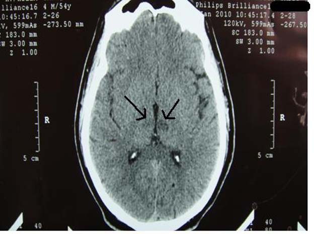
Non-contrast brain CT of the patient in case number one, which was interpreted as normal by the emergency physician. Note the bilateral paramedian thalamic hypodensities (black arrows).
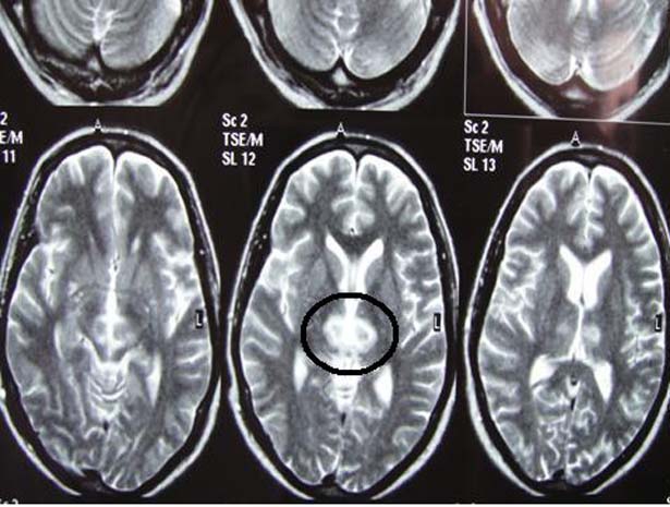
Axial T2-weighted brain MRI of patient number one. There are hyperintense signal abnormalities in the paramedian thalami.
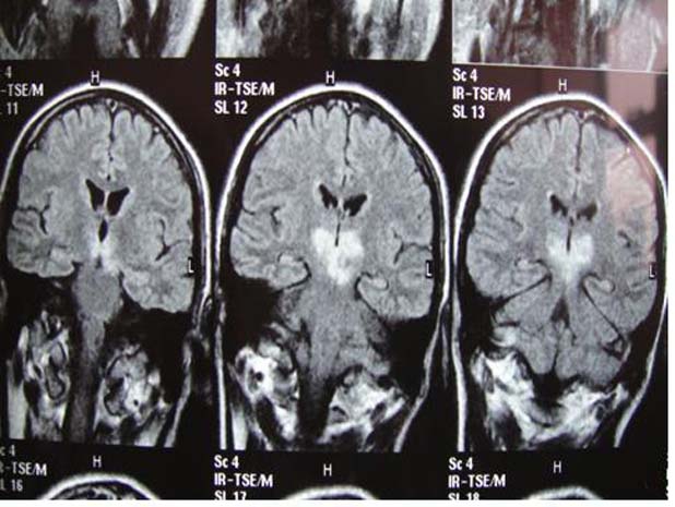
Coronal T2 FLAIR brain MRI of our first patient. Note the persistent hyperintense signal abnormality (which did not suppress on the FLAIR film) at the paramedian thalami and the left side of the rostral midbrain. The patient presented with acute thalamopeduncular syndrome.

Brain CT scan with no contrast in patient number two at the time of admission. It was unremarkable.
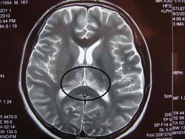
Axial T2-weighted brain MRI film of patient number two. Note the hyperintense signals at both paramedian thalami (within the black circle).
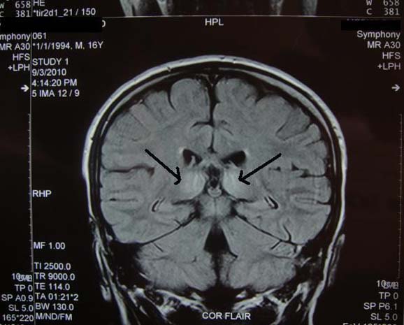
Coronal T2 FLAIR brain MRI image of the patient in case number two. The hypersignal abnormalities are localised to both paramedian thalami; these signals were not suppressed on the FLAIR film (black arrows).
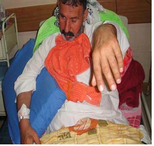
In response to our request, ‘raise your arms,’ the patient was able to lift up his left upper limb only. Note the left-sided complete ptosis.
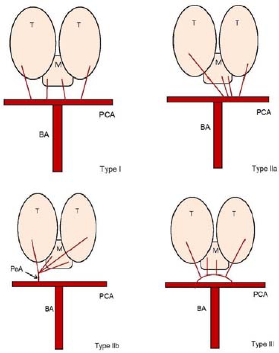
A diagram illustrating the four variants of the arterial supply to the human thalami and upper mesencephalon. Note that Percheron artery represents type IIb (T, thalamus; M, midbrain; PCA, posterior cerebral artery; BA, basilar artery; PeA, Percheron artery).
References
-
- Percheron G. The anatomy of the arterial supply of the human thalamus and its use for the interpretation of the thalamic vascular pathology. Z Neurol 1973;205:1–13 - PubMed
-
- Percheron G. Arteries of the human thalamus. I. Artery and polar thalamic territory of the posterior communicating artery. Rev Neurol (Paris) 1976;132:297–307 - PubMed
-
- Percheron G. Arteries of the human thalamus. II. Arteries and paramedian thalamic territory of the communicating basilar artery. Rev Neurol (Paris) 1976;132:309–24 - PubMed
-
- Bogousslavsky J, Van Melle G, Regli F. The Lausanne Stroke Registry: analysis of 1,000 consecutive patients with first stroke. Stroke 1988;19:1083–92 - PubMed
-
- Uz A. Variations in the origin of the thalamoperforating arteries. J Clin Neurosci 2007;14:134–7 - PubMed
Publication types
MeSH terms
LinkOut - more resources
Full Text Sources