. 2012 Aug;85(4):768-81.
doi: 10.1111/j.1365-2958.2012.08138.x. Epub 2012 Jul 11.

A conformational switch controls cell wall-remodelling enzymes required for bacterial cell division

Affiliations

A conformational switch controls cell wall-remodelling enzymes required for bacterial cell division

Desirée C Yang et al. Mol Microbiol. 2012 Aug.

Abstract

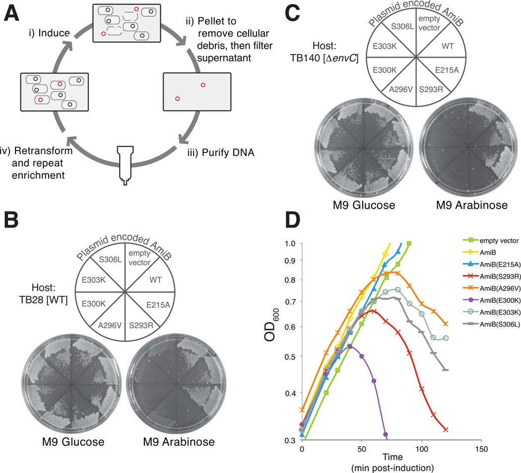
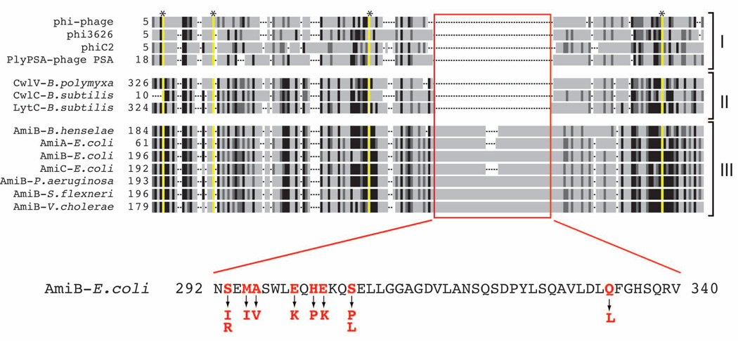
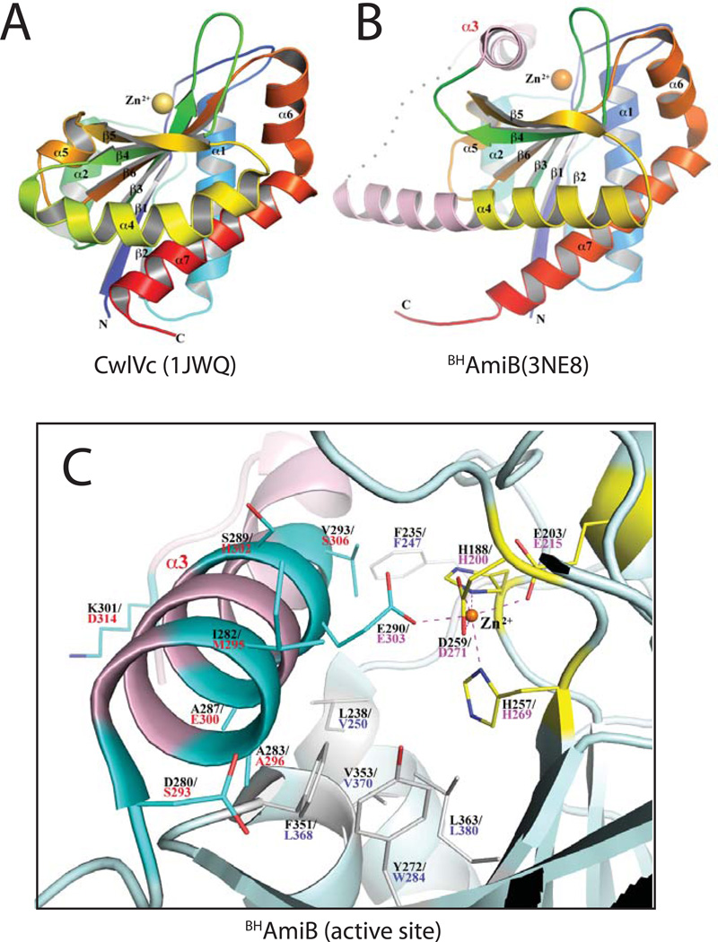
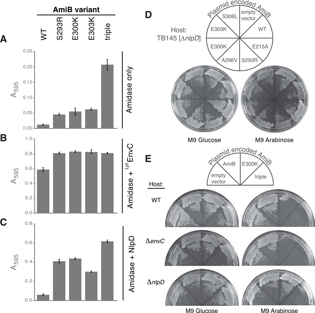
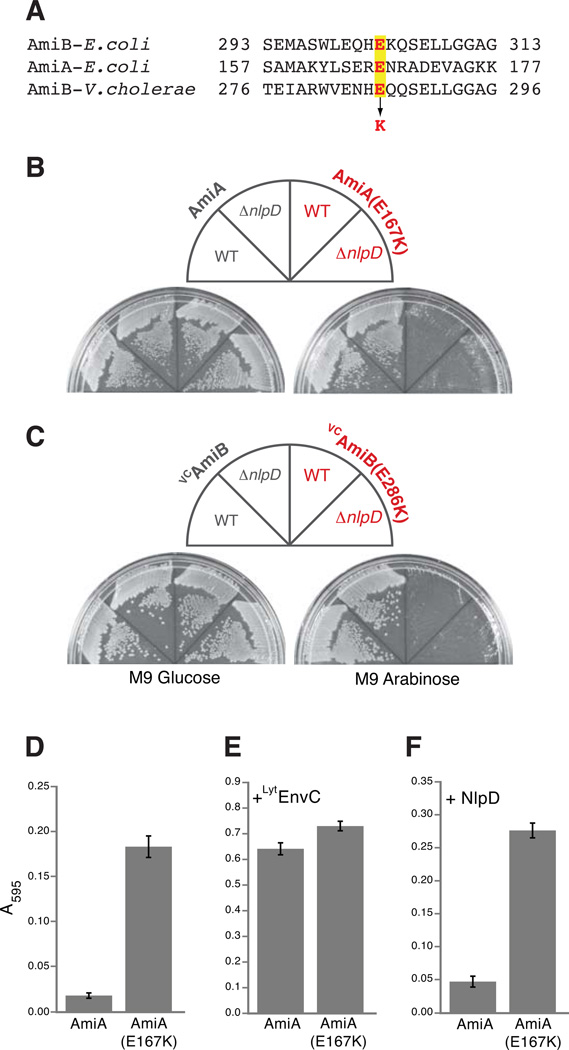
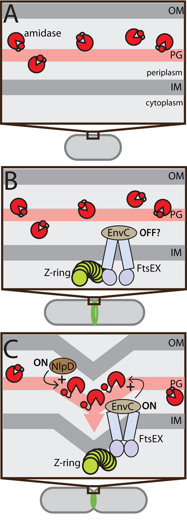
Remodelling of the peptidoglycan (PG) exoskeleton is intimately tied to the growth and division of bacteria. Enzymes that hydrolyse PG are critical for these processes, but their activities must be tightly regulated to prevent the generation of lethal breaches in the PG matrix. Despite their importance, the mechanisms regulating PG hydrolase activity have remained elusive. Here we investigate the control of cell division hydrolases called amidases (AmiA, AmiB and AmiC) required for Escherichia coli cell division. Poorly regulated amiB mutants were isolated encoding lytic AmiB variants with elevated basal PG hydrolase activities in vitro. The structure of an AmiB orthologue was also solved, revealing that the active site of AmiB is occluded by a conserved alpha helix. Strikingly, most of the amino acid substitutions in the lytic AmiB variants mapped to this domain and are predicted to disrupt its interaction with the active site. Our results therefore support a model in which cell separation is stimulated by the reversible relief of amidase autoinhibition governed by conserved subcomplexes within the cytokinetic ring. Analogous conformational control mechanisms are likely to be part of a general strategy used to control PG hydrolases present within multienzyme PG-remodelling machines.


Figures

Figure 1
Figure 1. Plasmid release enrichment strategy for the isolation of lytic amiB (lytamiB) mutants
(A) Overview of the plasmid release enrichment protocol. (i) Cells carrying mutagenized amiB-containing plasmid (black and red circles) are induced with 0.2% arabinose. (ii) Plasmids encoding rare lytamiB mutants (red circles) promote cell lysis and are released into the culture supernatant. Cells and cell debris are removed by centrifugation and filtration. (iii) Plasmid DNA is purified from the culture supernatants using Qiagen columns. (iv) Purified DNA can then be retransformed into the parental strain to repeat the enrichment protocol for as many rounds as necessary. (B) TB28 [WT] cells carrying an empty vector or expression constructs encoding AmiB (WT), AmiB(E215A) or the indicated LytAmiB variants were spread on M9-casamino acid (M9-CAA) agar containing either 0.2% glucose or 0.2% arabinose supplemented with 10 µg/ml chloramphenicol (Cm10). Plates were incubated overnight at 37°C and photographed. The AmiB(E215A) variant is a catalytically dead mutant used as a control (Uehara et al, 2010). (C) Same as (B) except the host strain was TB140 [ΔenvC]. (D) TB170 [ΔamiB] carrying an empty vector or expression constructs encoding AmiB (WT), AmiB(E215A) or the indicated LytAmiB variants were were grown at 37°C in M9-CAA-Cm10 supplemented with 0.2% maltose to an OD600 of about 0.3. At t = 0, arabinose was added to a final concentration of 0.2% and growth was monitored by following culture OD600. Samples for immunoblot analysis were taken just prior to lysis at t = 50 (see Fig. S1). Growth of cells harboring the empty vector or the AmiB (WT) or AmiB(E215A) control constructs continued without observable lysis.
Figure 2
Figure 2. Amino acid substitutions in the LytAmiB variants map to a potential regulatory domain
Shown is a schematic representation of a multiple sequence alignment generated using amidase sequences from bacteria and phages. Identities and similarities are indicated by the black and dark grey regions, respectively. Residues essential for catalysis are highlighted in yellow and with an asterisk at the top of the alignment. Gaps in the alignment are indicated as dashed lines. The amidases are grouped into the following categories: (I) phage endolysins, (II) bacterial amidases involved in mother cell lysis following sporulation, and (III) cell separation amidases. The red box highlights what appears to be a ~50 amino acid insertion region found only in the cell separation amidases. The sequence of the insertion from E. coli AmiB is shown below the alignment. Residues in red were altered in LytAmiB variants. The identity of the substitutions are indicated by the arrows. See Figure S2 for the complete sequence alignment.
Figure 3
Figure 3. Crystal structure of Bartonella henselae AmiB
(A–B) Shown are rainbow-colored cartoon diagrams of the CwlVc (A) and BHAmiB (B) structures with the N-termini in blue and the C-termini in red. The catalytic Zn2+ ion is drawn as a sphere. All secondary structures are labeled. In numbering α-helices, α3 is omitted from the numbering of CwlVc so that all common elements are numbered identically for CwlVc and BHAmiB. The regulatory region unique to cell separation amidases is colored pink in the BHAmiB structure with the structurally disordered loop region (E303-T308) represented by a dotted line. The unique α3 helix is labeled in pink. (C) Close-up view of the BHAmiB active site and regulatory helix. All residues are drawn in stick format and the Zn2+ ion is represented as an orange sphere. The Zn2+ ion is coordinated by H188, E203, H257, D259 (bidentate) and E290 with their carbon atoms drawn in yellow. Residue numbers for E.coli AmiB are provided in red underneath those for BHAmiB. Positions in the structure corresponding to amino acid substitutions in the LytAmiB variants are drawn with their carbon atoms in cyan. The residue corresponding to Q333 of E.coli AmiB (T315 of BHAmiB) is located in the N-terminal region of the α4 helix and they are out of view in this figure. Hydrophobic residues on the floor of binding cleft are also displayed in white.
Figure 4
Figure 4. LytAmiB variants have elevated basal PG hydrolase activity
(A) Dye-release assays measuring basal PG hydrolase activity of the LytAmiB variants relative to AmiB(WT). The variant referred to as “triple” is AmiB(S293R, E300K, E303K). The indicated proteins (2 µM) were incubated with RBB-labeled PG at 37°C for 30 min. Undigested PG was pelleted by centrifugation and the absorbance of the supernatants was measured at 595 nm. Reactions were performed in triplicate and the error bars indicate the standard deviation. (B–C) Same as in (A) except LytEnvC (2 µM) or NlpD (2 µM) were combined with the amidases. Supernatants from control reactions containing LytEnvC only (2 µM), NlpD only (2 µM), lysozyme (4 µM) or buffer alone had the following A595 readings (average +/− std. dev.): 0.02 +/− 0.002, 0.04 +/− 0.008, 0.56 +/− 0.022 and 0.007 +/− 0.002. The A595 reading for the lysozyme reaction is a typical reading for a reaction that has gone to completion. (D) TB145 [ΔnlpD] cells carrying an empty vector or expression constructs encoding AmiB (WT), AmiB(E215A) or the indicated LytAmiB variants were spread on M9-CAA-Cm10 agar containing either 0.2% glucose or 0.2% arabinose. Plates were incubated overnight at 37°C and photographed. (E) TB28[WT], TB140[ΔenvC], or TB145[ΔnlpD] carrying an empty vector or expression constructs encoding AmiB (WT), AmiB(E300K), or AmiB(triple) were plated and photographed as described in (D).
Figure 5
Figure 5. Lytic and PG hydrolase activities of other amidases with substitutions in their regulatory domains
(A) Sequence alignment of a portion of the regulatory domains of E. coli AmiB, E. coli AmiA, and V. cholerae AmiB (VCAmiB). The highlighted glutamic acid (E) residues in red were changed to lysines (K) in AmiA and VCAmiB to yield AmiA(E167K) and VCAmiB(E286K), respectively. (B–C) As indicated, TB28[WT] or TB145[ΔnlpD] cells carrying AmiA, AmiA(E167K), VCAmiB, or VCAmiB(E286K) were spread on M9-CAA-Cm10 agar containing either 0.2% glucose or 0.2% arabinose. Plates were incubated overnight at 37°C and photographed. (D) Dye-release assay measuring basal PG hydrolase activity for AmiA(E167K) relative to AmiA (WT). Assays were performed as described in Fig. 4 except that reactions contained 1µM amidase with or without an additional 1µM of purified LytM factor as indicated.
Figure 6
Figure 6. Conformational control of amidase activity during the cell cycle
Shown is a schematic diagram illustrating the activation status of cell separation amidases through the cell cycle. (A) At early stages in the cell cycle prior to the formation of the Z-ring, periplasmic amidases (red pac-men) are likely to be largely inhibited by their regulatory helices (red circles). (B) FtsEX and EnvC are early recruits to the Z-ring, arriving well before the initiation of constriction (Peters et al, 2011). It is not known if the FtsEX-EnvC system is capable of amidase activation immediately following its recruitment, or if it requires further septal ring maturation. Even if it is active at this stage, it is unlikely to stimulate a high level of amidase activity because the amidase are not concentrated at midcell before the onset of cell constriction (Peters et al, 2011). (C) Once constriction is initiated NlpD, AmiB, and AmiC are recruited to the septal ring and both amidase activation systems are presumably activated to stimulate amidase activity (Peters et al, 2011). Our results indicate that amidase activation proceeds via the release of the regulatory helix from their active site. For simplicity, the three different amidases are not individually identified in the figure.

References

    1. Baba T, Schneewind O. Targeting of muralytic enzymes to the cell division site of Gram-positive bacteria: repeat domains direct autolysin to the equatorial surface ring of Staphylococcus aureus. EMBO J. 1998;17:4639–4646. - PMC - PubMed
    1. Bendezú FO, Hale CA, Bernhardt TG, de Boer PAJ. RodZ (YfgA) is required for proper assembly of the MreB actin cytoskeleton and cell shape in E. coli. EMBO J. 2009;28:193–204. - PMC - PubMed
    1. Bernhardt TG, de Boer PAJ. The Escherichia coli amidase AmiC is a periplasmic septal ring component exported via the twin-arginine transport pathway. Mol Microbiol. 2003;48:1171–1182. - PMC - PubMed
    1. Bi EF, Lutkenhaus J. FtsZ ring structure associated with division in Escherichia coli. Nature. 1991;354:161–164. - PubMed
    1. Böth D, Schneider G, Schnell R. Peptidoglycan Remodeling in Mycobacterium tuberculosis: Comparison of Structures and Catalytic Activities of RipA and RipB. J Mol Biol. 2011;413:247–260. - PubMed

Publication types

MeSH terms

Substances