. 2012 Jun 20:12:91.
doi: 10.1186/1471-2148-12-91.

Evolutionary genomics of mycovirus-related dsRNA viruses reveals cross-family horizontal gene transfer and evolution of diverse viral lineages

Affiliations

Evolutionary genomics of mycovirus-related dsRNA viruses reveals cross-family horizontal gene transfer and evolution of diverse viral lineages

Huiquan Liu et al. BMC Evol Biol. .

Abstract

Background: Double-stranded (ds) RNA fungal viruses are typically isometric single-shelled particles that are classified into three families, Totiviridae, Partitiviridae and Chrysoviridae, the members of which possess monopartite, bipartite and quadripartite genomes, respectively. Recent findings revealed that mycovirus-related dsRNA viruses are more diverse than previously recognized. Although an increasing number of viral complete genomic sequences have become available, the evolution of these diverse dsRNA viruses remains to be clarified. This is particularly so since there is little evidence for horizontal gene transfer (HGT) among dsRNA viruses.

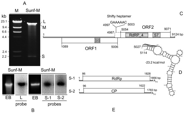
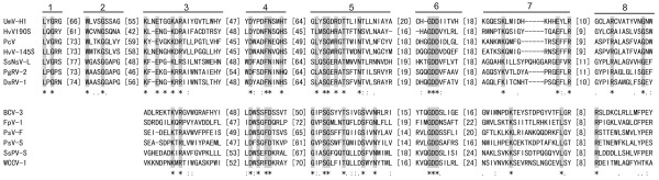
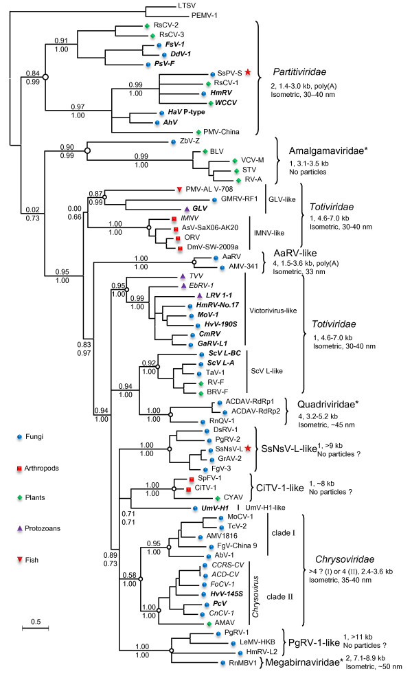
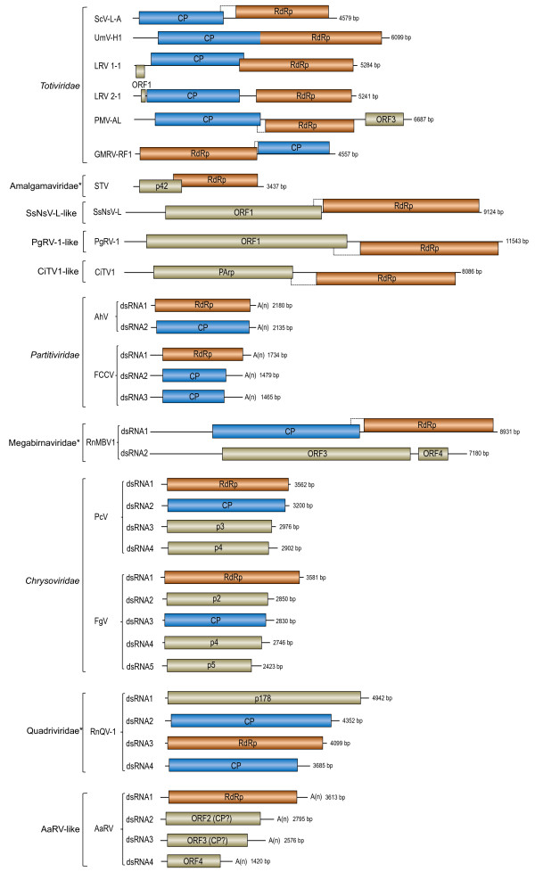
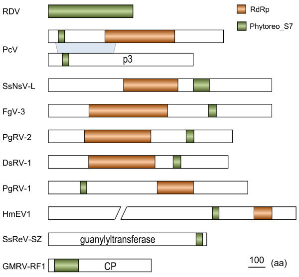
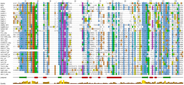
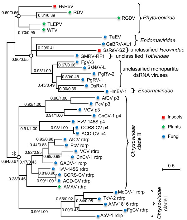
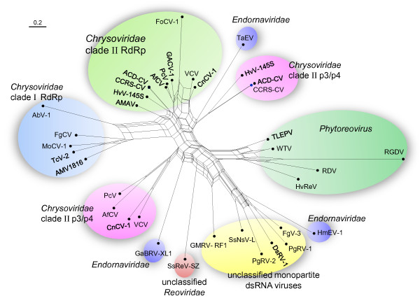
Results: In this study, we report the molecular properties of two novel dsRNA mycoviruses that were isolated from a field strain of Sclerotinia sclerotiorum, Sunf-M: one is a large monopartite virus representing a distinct evolutionary lineage of dsRNA viruses; the other is a new member of the family Partitiviridae. Comprehensive phylogenetic analysis and genome comparison revealed that there are at least ten monopartite, three bipartite, one tripartite and three quadripartite lineages in the known dsRNA mycoviruses and that the multipartite lineages have possibly evolved from different monopartite dsRNA viruses. Moreover, we found that homologs of the S7 Domain, characteristic of members of the genus phytoreovirus in family Reoviridae are widely distributed in diverse dsRNA viral lineages, including chrysoviruses, endornaviruses and some unclassified dsRNA mycoviruses. We further provided evidence that multiple HGT events may have occurred among these dsRNA viruses from different families.

Conclusions: Our study provides an insight into the phylogeny and evolution of mycovirus-related dsRNA viruses and reveals that the occurrence of HGT between different virus species and the development of multipartite genomes during evolution are important macroevolutionary mechanisms in dsRNA viruses.


Figures

Figure 1
Figure 1
Molecular characteristics of L- and S-dsRNA inS. sclerotiorumstrain Sunf-M. (A) Agarose gel electrophoresis of dsRNA isolated from mycelial extracts of Sunf-M. The nucleic acid preparation was fractionated on 1.0% agarose gel and stained with ethidium bromide. Lane M, DNA size markers generated by digestion of λDNA with HindIII. (B) Northern hybridization analysis of L- and S-dsRNA. dsRNAs were separated on a 1.0% agarose gel, transferred on to Hybond-N + membrane and hybridized with 32P-labelled probes prepared by random-primer labelling of cloned cDNA to L, S-1 and S-2 dsRNA, respectively. (C) Schematic representation of the genomic organization of L-dsRNA shows the presence of two ORFs. The dotted line box indicates a possible extension of ORF2 by frameshifting. The conserved domains of deduced proteins are shown: SIS, Sugar ISomerase domain; S7, Phytoreovirus S7 protein; RdRP_4, Viral RNA-dependent RNA-polymerase. (D) The pseudoknot structure immediately downstream of the putative frameshift site. The RNA secondary structure was predicted by KnotSeeker program [45] and drawn by VARNAv3-7 program [46]. (E) Schematic representation of predicted genome organization of S dsRNA.
Figure 2
Figure 2
Comparison of the conserved motifs of RdRps of selected dsRNA mycoviruses including the putative RdRps encoded by SsNsV-L and SsPV-S. Numbers 1–8 refer to the eight conserved motifs characteristic of RdRps of RNA viruses. The amino acid positions corresponding to conserved motifs 1 and 2 for the RdRps of viruses in the family Partitiviridae are not well-defined and, therefore, they are not presented. Asterisks, colons and dots indicate identical amino acid residues (gray shaded), conserved amino acid residues and semi-conserved amino acid residues, respectively. Numbers in square brackets correspond to the number of amino acid residues separating the motifs. See Additional file 2: Table S1 and Additional file 1: Figure S3 for abbreviations of virus names and viral protein accession numbers.
Figure 3
Figure 3
Phylogenetic tree of mycovirus-related dsRNA viruses. The tree presented here is the consensus of two trees calculated using phyML-maximum-likelihood (ML) and Bayesian (BI) methods, respectively. Numbers at various nodes indicate, respectively, SH-like approximate likelihood ratio test (SH-aLRT) probabilities (above) and Bayesian posterior probabilities (below). The characteristics (numbers and sizes of genome segments and particle morphology) of different viral lineages are shown. Question mark (?) indicated that characteristics were not determined for all members of this lineage. The viral families that were proposed but have not been recognized by ICTV are indicated by asterisks, and their names are not italicized. The names of the ICTV-recognized or proposed (but not yet recognized) virus species are written in bold italics or italics, respectively. Pentagram indicates the two viruses reported in this study. The host range of viruses was indicated. This tree was rooted with ss(+)RNA viruses. The scale bar corresponds to 0.5 amino acid substitutions per site. See Additional file 2: Table S1 in the supplemental material for abbreviations of virus names and viral protein accession numbers.
Figure 4
Figure 4
Genomic organization and comparison of representative viruses in different dsRNA viral lineages. The Colored boxes and lines represent open reading frames (ORFs) and non-coding sequences, respectively, roughly to scale: orange, RNA-dependent RNA polymerase (RdRp); blue, capsid protein (CP); Brown beige, unknown function. Dotted line boxes indicate possible extension of the downstream ORFs by frameshifting. The viral families that were proposed but have not been recognized by ICTV are indicated by asterisks, and their names are not italicized. See Additional file 2: Table S1 in the supplemental material for abbreviations of virus names.
Figure 5
Figure 5
Locations of Phytoreovirus S7 domain in proteins of different viruses. See Table 1 for virus names and viral protein accession numbers.
Figure 6
Figure 6
Multiple alignments of the Phytoreo_S7 domain homologs from diverse viral lineages. The default color scheme for ClustalW alignment in the Jalview program was used. Jnetpred is the consensus secondary structure prediction: alpha-helices are shown as red rods and beta strands as green arrows. Quality is the quality level for the multiple alignments. See Table 1 for virus names and viral protein accession numbers.
Figure 7
Figure 7
Phylogenetic tree of the Phytoreo_S7 domain homologs from diverse viral lineages. This tree is the consensus tree of two trees calculated using phyML-maximum-likelihood (ML) and Bayesian (BI) methods, respectively. Numbers at various nodes indicate, respectively, SH-like approximate likelihood ratio test (aLRT) probabilities/Bayesian posterior probabilities. The topology of asterisk marked clade was evaluated independently. The host range of viruses was indicated. The scale bars correspond to 0.5 amino acid substitutions per site. See Table 1 for virus names and viral protein accession numbers.
Figure 8
Figure 8
Neighbor-Net analysis of the Phytoreo_S7 domain homologs from diverse viral lineages. The analysis was conducted under the WAG model of substitution. Scale bar corresponds to 0.2 amino acid substitutions per site. The major viral lineages are indicated. The box-like appearance in the basal branches of this phylogeny suggests regions of unresolved branches or conflicting phylogenetic signals. See Table 1 for virus names and viral protein accession numbers.

Similar articles

Cited by

References

    1. Ghabrial SA, Suzuki N. Viruses of plant pathogenic fungi. Annu Rev Phytopathol. 2009;47:353–384. doi: 10.1146/annurev-phyto-080508-081932. - DOI - PubMed
    1. Pearson MN, Beever RE, Boine B, Arthur K. Mycoviruses of filamentous fungi and their relevance to plant pathology. Mol Plant Pathol. 2009;10(1):115–128. doi: 10.1111/j.1364-3703.2008.00503.x. - DOI - PMC - PubMed
    1. Liu HQ, Fu YP, Jiang DH, Li GQ, Xie J, Peng YL, Yi XH, Ghabrial SA. A novel mycovirus that is related to the human pathogen hepatitis E virus and rubi-like viruses. J Virol. 2009;83(4):1981–1991. doi: 10.1128/JVI.01897-08. - DOI - PMC - PubMed
    1. Yu X, Li B, Fu YP, Jiang DH, Ghabrial SA, Li GQ, Peng YL, Xie JT, Cheng JS, Huang JB. et al.A geminivirus-related DNA mycovirus that confers hypovirulence to a plant pathogenic fungus. Proc Natl Acad Sci U S A. 2010;107(18):8387–8392. doi: 10.1073/pnas.0913535107. - DOI - PMC - PubMed
    1. King AMQ, Adams MJ, Carstens EB, Lefkowitz EJ. (Eds): Virus taxonomy: Ninth Report of the International Committee on taxonomy of viruses. New York: Elsevier; 2011.

Publication types

Substances

LinkOut - more resources