

A Bacillus subtilis sensor kinase involved in triggering biofilm formation on the roots of tomato plants
- PMID: 22716461
- PMCID: PMC3518419
- DOI: 10.1111/j.1365-2958.2012.08109.x
A Bacillus subtilis sensor kinase involved in triggering biofilm formation on the roots of tomato plants
Abstract
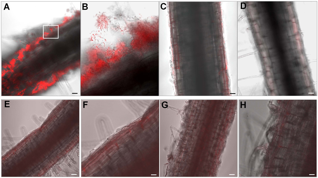
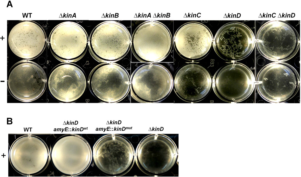
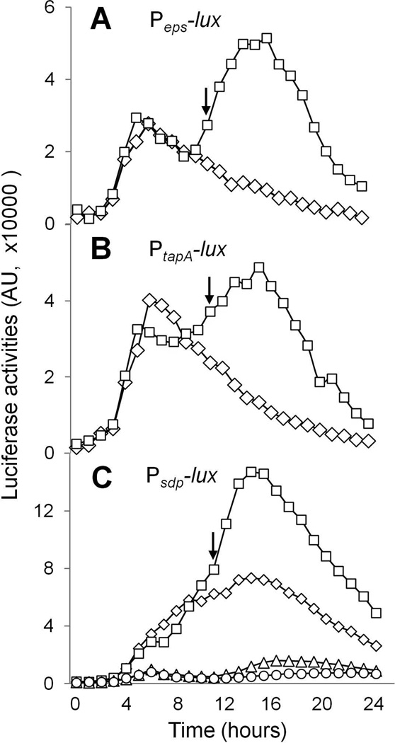
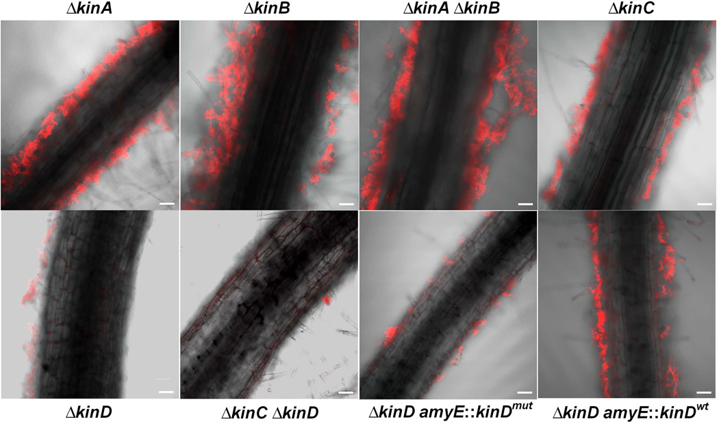
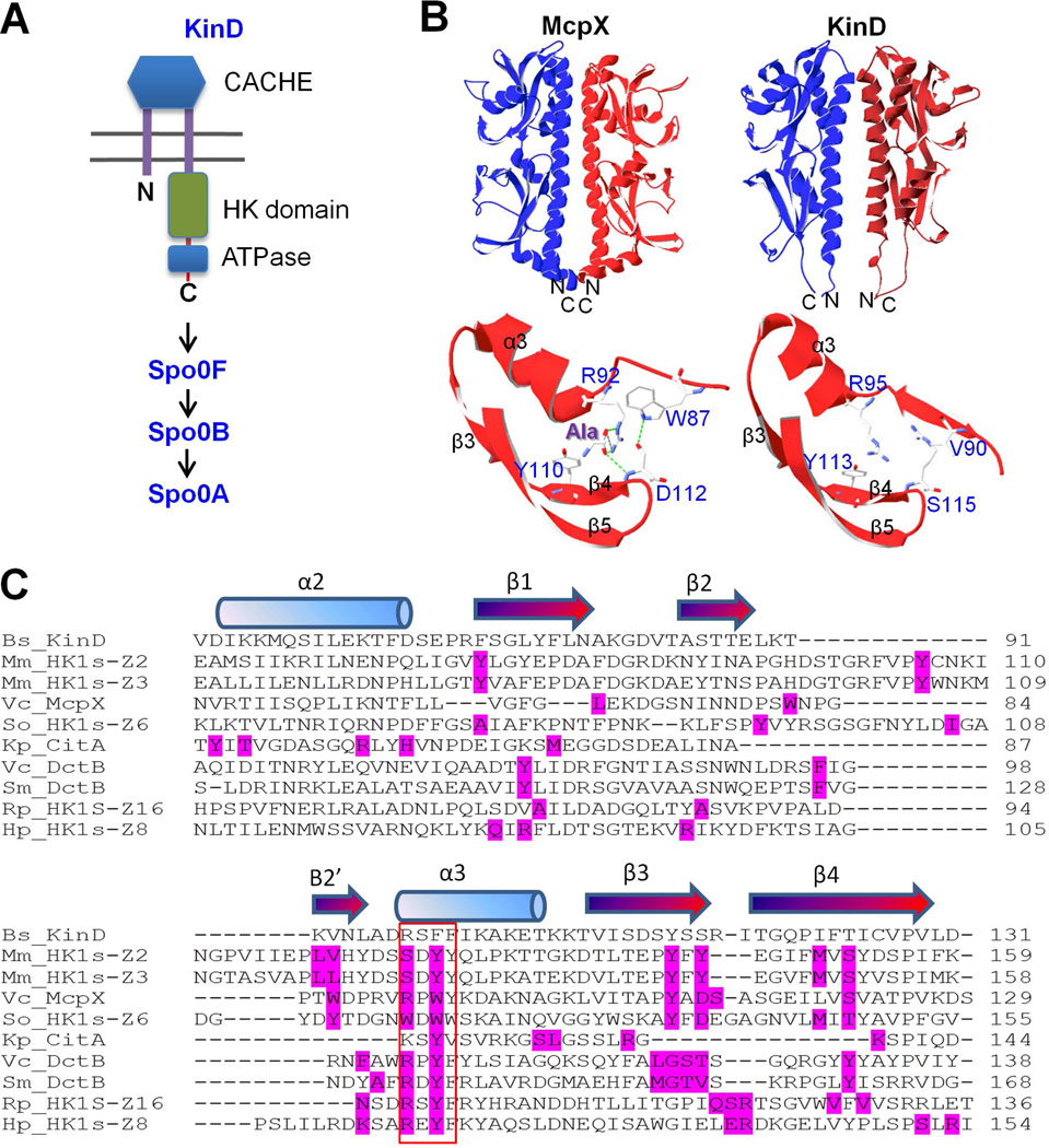
The soil bacterium Bacillus subtilis is widely used in agriculture as a biocontrol agent able to protect plants from a variety of pathogens. Protection is thought to involve the formation of bacterial communities - biofilms - on the roots of the plants. Here we used confocal microscopy to visualize biofilms on the surface of the roots of tomato seedlings and demonstrated that biofilm formation requires genes governing the production of the extracellular matrix that holds cells together. We further show that biofilm formation was dependent on the sensor histidine kinase KinD and in particular on an extracellular CACHE domain implicated in small molecule sensing. Finally, we report that exudates of tomato roots strongly stimulated biofilm formation ex planta and that an abundant small molecule in the exudates, (L) -malic acid, was able to stimulate biofilm formation at high concentrations in a manner that depended on the KinD CACHE domain. We propose that small signalling molecules released by the roots of tomato plants are directly or indirectly recognized by KinD, triggering biofilm formation.
© 2012 Blackwell Publishing Ltd.
Figures
Similar articles
-
Sticking together: building a biofilm the Bacillus subtilis way.Nat Rev Microbiol. 2013 Mar;11(3):157-68. doi: 10.1038/nrmicro2960. Epub 2013 Jan 28. Nat Rev Microbiol. 2013. PMID: 23353768 Free PMC article. Review.
-
Potassium sensing histidine kinase in Bacillus subtilis.Methods Enzymol. 2010;471:229-51. doi: 10.1016/S0076-6879(10)71013-2. Epub 2010 Mar 1. Methods Enzymol. 2010. PMID: 20946851 Free PMC article.
-
Biocontrol of tomato wilt disease by Bacillus subtilis isolates from natural environments depends on conserved genes mediating biofilm formation.Environ Microbiol. 2013 Mar;15(3):848-864. doi: 10.1111/j.1462-2920.2012.02860.x. Epub 2012 Aug 30. Environ Microbiol. 2013. PMID: 22934631 Free PMC article.
-
Inactivation of cysL Inhibits Biofilm Formation by Activating the Disulfide Stress Regulator Spx in Bacillus subtilis.J Bacteriol. 2019 Mar 26;201(8):e00712-18. doi: 10.1128/JB.00712-18. Print 2019 Apr 15. J Bacteriol. 2019. PMID: 30718304 Free PMC article.
-
Inhibition of biofilm formation by Cd2+ on Bacillus subtilis 1JN2 depressed its biocontrol efficiency against Ralstonia wilt on tomato.Microbiol Res. 2018 Oct;215:1-6. doi: 10.1016/j.micres.2018.06.002. Epub 2018 Jun 2. Microbiol Res. 2018. PMID: 30172295
Cited by
-
Kin Discrimination Modifies Strain Distribution, Spatial Segregation, and Incorporation of Extracellular Matrix Polysaccharide Mutants of Bacillus subtilis Strains into Mixed Floating Biofilms.Appl Environ Microbiol. 2022 Sep 22;88(18):e0087122. doi: 10.1128/aem.00871-22. Epub 2022 Sep 12. Appl Environ Microbiol. 2022. PMID: 36094206 Free PMC article.
-
Osmotic pressure can regulate matrix gene expression in Bacillus subtilis.Mol Microbiol. 2012 Oct;86(2):426-36. doi: 10.1111/j.1365-2958.2012.08201.x. Epub 2012 Sep 7. Mol Microbiol. 2012. PMID: 22882172 Free PMC article.
-
Bacillus subtilis biofilm induction by plant polysaccharides.Proc Natl Acad Sci U S A. 2013 Apr 23;110(17):E1621-30. doi: 10.1073/pnas.1218984110. Epub 2013 Apr 8. Proc Natl Acad Sci U S A. 2013. PMID: 23569226 Free PMC article.
-
Sticking together: building a biofilm the Bacillus subtilis way.Nat Rev Microbiol. 2013 Mar;11(3):157-68. doi: 10.1038/nrmicro2960. Epub 2013 Jan 28. Nat Rev Microbiol. 2013. PMID: 23353768 Free PMC article. Review.
-
Disease to dirt: the biology of microbial amyloids.PLoS Pathog. 2013;9(11):e1003740. doi: 10.1371/journal.ppat.1003740. Epub 2013 Nov 21. PLoS Pathog. 2013. PMID: 24278013 Free PMC article. Review. No abstract available.
References
-
- Anantharaman V, Aravind L. CACHE - a signaling domain common to animal Ca2+-channel subunits and a class of prokaryotic chemotaxis receptors. Trends Biochem. Sci. 2000;25:535–537. - PubMed
-
- Berg G, Krechel A, Ditz M, Sikora RA, Ulrich A, Hallmann J. Endophytic and ectophytic potato-associated bacterial communities differ in structure and antagonistic function against plant pathogenic fungi. FEMS Microbiol. Ecol. 2005;51:215–229. - PubMed
Publication types
MeSH terms
Substances
Grants and funding
LinkOut - more resources
Full Text Sources
Molecular Biology Databases
