Review
. 2012 Oct;35(10):607-18.
doi: 10.1016/j.tins.2012.05.003. Epub 2012 Jun 19.

CaMKII regulation in information processing and storage

Affiliations
Review

CaMKII regulation in information processing and storage

Steven J Coultrap et al. Trends Neurosci. 2012 Oct.

Abstract

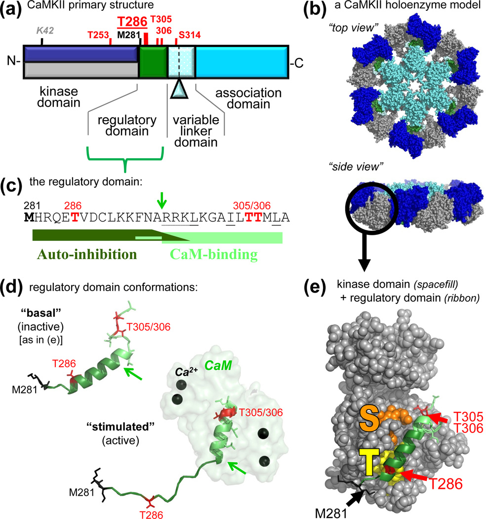
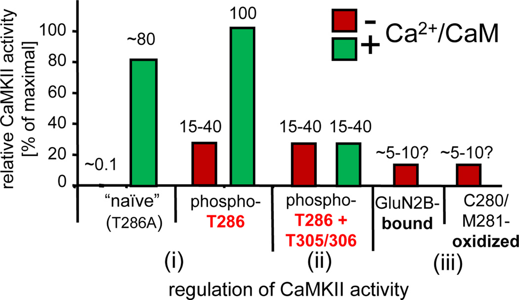
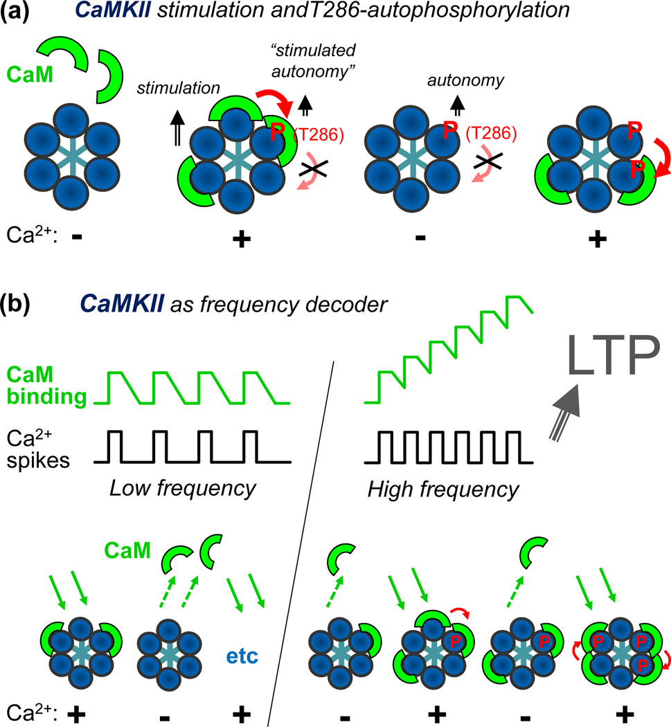
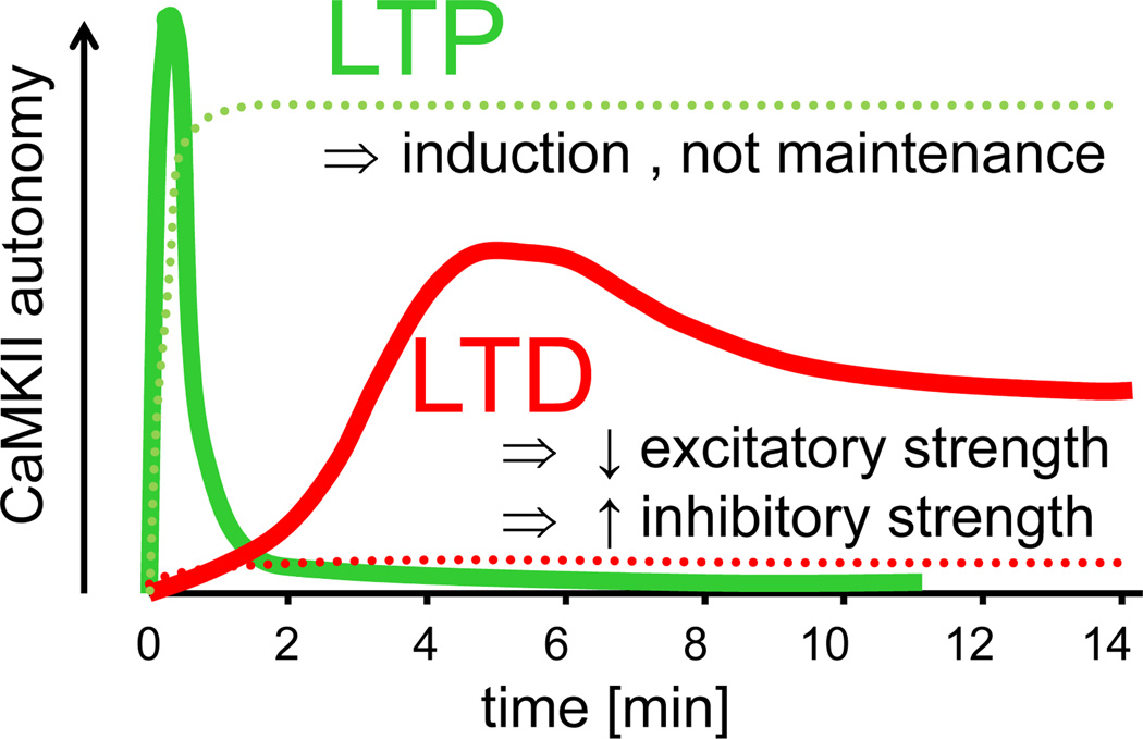
The Ca(2+)/Calmodulin(CaM)-dependent protein kinase II (CaMKII) is activated by Ca(2+)/CaM, but becomes partially autonomous (Ca(2+)-independent) upon autophosphorylation at T286. This hallmark feature of CaMKII regulation provides a form of molecular memory and is indeed important in long-term potentiation (LTP) of excitatory synapse strength and memory formation. However, emerging evidence supports a direct role in information processing, while storage of synaptic information may instead be mediated by regulated interaction of CaMKII with the NMDA receptor (NMDAR) complex. These and other CaMKII regulation mechanisms are discussed here in the context of the kinase structure and their impact on postsynaptic functions. Recent findings also implicate CaMKII in long-term depression (LTD), as well as functional roles at inhibitory synapses, lending renewed emphasis on better understanding the spatiotemporal control of CaMKII regulation.


Figures

Figure 1
Figure 1
CaMKII structure and regulation. (a) Primary structure of CaMKIIα, with an N-terminal kinase domain, followed by a CaM-binding regulatory domain, a variable linker domain (subject to alternative splicing, with the splice insert of αB indicated), and a C-terminal association domain. Phosphorylation sites (red) and other important residues are indicated (see also Table 1). The other isoforms (β, γ,δ) contain an additional Thr after the third residues, and the numbers of their homologous positions are accordingly higher by one. These other isoforms also show more extensive alternative splicing in the variable domain, which is typically longer. (b) A model of a 12meric CaMKII holoenzyme (in two different view angles), based on several partial crystal structures [44, 46, 59], with the association domains forming a central hub [44, 46], and the kinases domain [59] radiating outward. The 12meric nature, the particle size, and the principle arrangement is supported by EM studies [44, 45]; however, the precise positioning of the kinase domains in this holoenzyme conformation is unclear (but compare Fig. 4). The reconstruction was kindly provided by Drs. Chao and Kuriyan, and the image is adapted, with permission, from [35]. (c) Sequence of the regulatory domain. The autoinhibitory and CaM-binding regions are indicated. The arrow indicates the N-terminus of the core CaM-binding region (R296; also indicated by arrows in panel d); Ca2+/CaM trapping on the T286-phosphorylated form of the kinase also involves F293 [2]. Autoinhibition is maintained in a kinase 1-N294 truncation, but additional C-terminal residues participate in the substrate site block [2] (see also panel e). The oxidation (black) and phosphorylation (red) sites that generate autonomous activity (T286) [2, 29, 30] or inhibit CaM-binding (T306/306) [, –72] are marked. (d) The regulatory domain in the basal (inactive) and stimulated (active) conformation, illustrating a structural transition of the different regions upon Ca2+/CaM binding [32]. The positioning relative to the kinase domain is known for the basal state (see panel e) but unclear for the active states. The region shown here for CaMKIIδ is identical in CaMKIIα. (e) Crystal structure of human CaMKIIδ in its basal state, with the regulatory domain (ribbon) held in place in part by interactions with the T286 binding T-site (yellow) and blocking access to the substrate binding S-site (orange). The structural representations in panels d and e are based on Protein Data Bank (PDB) files 2VN9 and 2WEL [32].
Figure 2
Figure 2
Levels of CaMKII activity in response to different forms of regulation. (i) Naïve CaMKII shows only low basal activity (~0.1% of maximum), but is fully activated by Ca2+/CaM-stimulation [2]. This is accompanied by rapid T286-autophosphorylation; when T286-autophosphorylation is prevented by T286A mutation, the activity level remains slightly lower [58]. After T286-autophosphorylation, CaMKII remains partially active even after dissociation of Ca2+/CaM (autonomous), but this autonomous activity is significantly further stimulated by Ca2+/CaM [29]. (ii) T305/306-autophosphorylation of T286-phosphorylated CaMKII prevents Ca2+/CaM binding and thus further stimulation of the autonomous activity. (iii) Autonomous activity can also be induced by GluN2B binding [61] or by C280/M281 oxidation [30]; like T286-autophosphorylation, both mechanisms require an initial Ca2+/CaM-stimulus [30, 61]. The levels of “autonomy” (the ratio of autonomous over maximal CaM-stimulated activity) after GluN2B binding or oxidation are estimated adjustments (based on conditions in the original reports that overestimate autonomy, either by use of an autonomy-favoring substrate [29, 61] or by conditions resulting in unusually low rates of stimulated activity [30]). The activity estimates shown are based on an adapted compilation from [29, 30, 58, 61].
Figure 3
Figure 3
Schematic representations of frequency detection by CaMKII autophosphorylation at T286. (a) Ca2+/CaM-binding separately stimulates each individual subunit of a CaMKII holoenzyme (only 6 of the 12 subunits shown in the schematic model). However, T286 autophosphorylation requires binding of two CaM molecules to two neighboring subunit, one to activate the subunit acting as kinase, the other to expose T286 for phosphorylation (see also Fig. 1e) on the neighboring subunit acting as substrate in this intra-holoenzyme inter-subunit reaction [82, 83]. Phospho-T286 generates Ca2+-independent “autonomous” activity (that can be further stimulated by Ca2+/CaM; see Fig. 2). It can likely also substitute for the kinase-directed function of CaM in further autophosphorylation, but it cannot substitute for the substrate-directed function of CaM [83]. (b) A simple model for frequency detection by T286 autophosphorylation. During submaximal Ca2+-spikes, some CaM molecules bind to some subunits of a CaMKII holoenzyme, and then dissociate during the spike interval, and so on. However, at higher frequencies, with the spike interval in the range of the CaM dissociation time, additional CaM molecules accumulate before all of the initial CaM molecules dissociate. This increases the chance of CaM binding to neighboring subunit and thus autophosphorylation at T286. Such a frequency-dependent response was found in biochemical studies [40, 47], and suggestively correlates with the requirements for LTP induction, which is favored by HFS and has een shown to depend on CaMKII autonomy [26, 27].
Figure 4
Figure 4
Two distinct inactive conformations of the CaMKII holoenzyme: compact and extended. (a) The only current holoenzyme crystal structure (in a compact conformation) with accurate kinase domain positioning (left; PDB1 3SOA) [31] was derived from a monomeric linker-less CaMKIIα with a partial association domain (right; PDB 3SOA) [31], together with the known structure of the association domain assembly [32, 44, 46]. (b) In a compact conformation, the kinase domains fold back to the association domains, leaving the CaM-binding regulatory domain inaccessible to stimulation (as depicted in the crystal structure in panel a) [31]. In an extended conformation, the kinase domains protrude further outward, making the regulatory domains accessible (compare to the model depicted in Fig. 1b). CaM-binding to the extended conformation may, in turn, also favor transition of neighboring subunits to this CaM-binding competent extended conformation [31]. (c) Small-angle X-ray scattering (SAXS) analysis indicates that the I321E mutation in a linker-less CaMKIIα that interferes with the interaction of the kinase domain with the association domain causes a transition from a compact (i) to an extended conformation (ii) [31]. This mutation reduces cooperativity of activation by CaM not only for linker-less but also for full-length CaMKIIα (wild type, iii), at least under molecular crowding conditions that mimic cellular protein concentrations [31]. In SAXS analysis (without molecular crowding) the major CaMKIIα isoform was found in the extended conformation [31]. All panels are adapted, with permission, from [31].
Figure 5
Figure 5
Schematic diagram illustrating the time course of CaMKII autonomy after LTP (green) versus LTD (red) stimuli. The time course of CaMKII autonomy according to recent studies (solid lines) [23, 25, 28, 143] differs from the “traditional” perception (dotted lines) [139], but is consistent with a role of CaMKII T286 phosphorylation in LTP induction rather than maintenance [27], as well as with newly discovered functions in mechanisms induced by excitatory LTD-stimuli in hippocampal pyramidal neurons (depression of excitatory synapses [23] and potentiation of inhibitory synapses [25]). After LTP, the rapid activation of CaMKII in spines was found to decay with two time-constants (6 sec and 45 sec) [28], with the slower time-constant that is consistent with the decay of autonomy found in a previous biochemical study [143] illustrated here. The recent LTD-related studies assessed T286 phosphorylation after mGluR [23] or NMDAR [25] dependent LTD-stimuli, but found very similar time courses (with a peak at 5 min followed by persistence at a lower level, as illustrated here). The illustration represents an adapted compilation of the results of several studies [23, 25, 28, 139, 143]. The similarities between the two more recent studies related to LTP [28, 143] and LTD [23, 25] provide good estimates of the respective time course (as shown), however, a direct comparison between the absolute levels of peak autonomy after LTP versus LTD has not yet been made (and the relative peak levels shown are arbitrary).

References

    1. Schulman H, Greengard P. Stimulation of brain membrane protein phosphorylation by calcium and an endogenous heat-stable protein. Nature. 1978;271:478–479. - PubMed
    1. Hudmon A, Schulman H. Neuronal CA2+/calmodulin-dependent protein kinase II: the role of structure and autoregulation in cellular function. Annu Rev Biochem. 2002;71:473–510. - PubMed
    1. Feng B, et al. Quantitative estimates of the cytoplasmic PSD, and NMDAR-bound pools of CaMKII in dendritic spines. Brain Res. 2011;1419:46–52. - PMC - PubMed
    1. Erondu NE, Kennedy MB. Regional distribution of type II Ca2+/calmodulin-dependent protein kinase in rat brain. J Neurosci. 1985;5:3270–3277. - PMC - PubMed
    1. Malinow R, et al. Inhibition of postsynaptic PKC or CaMKII blocks induction but not expression of LTP. Science. 1989;245:862–866. - PubMed

Publication types

MeSH terms

Substances