Review
. 2013 Feb;228(2):258-66.
doi: 10.1002/jcp.24134.

Rules of engagement for base excision repair in chromatin

Affiliations
Review

Rules of engagement for base excision repair in chromatin

Ian D Odell et al. J Cell Physiol. 2013 Feb.

Abstract

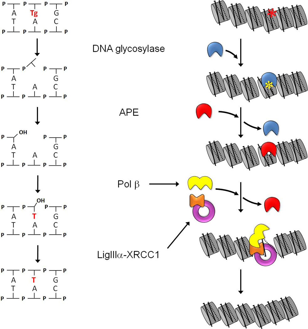
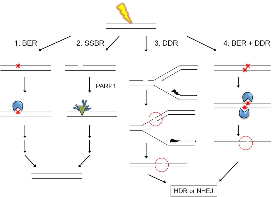
Most of the DNA in eukaryotes is packaged in tandemly arrayed nucleosomes that, together with numerous DNA- and nucleosome-associated enzymes and regulatory factors, make up chromatin. Chromatin modifying and remodeling agents help regulate access to selected DNA segments in chromatin, thereby facilitating transcription and DNA replication and repair. Studies of nucleotide excision repair (NER), single strand break repair (SSBR), and the homology-directed repair (HDR), and non-homologous end-joining (NHEJ) double strand break repair pathways have led to an "access-repair-restore" paradigm, in which chromatin in the vicinity of damaged DNA is disrupted, thereby enabling efficient repair and the subsequent repackaging of DNA into nucleosomes. When damage is extensive, these repair processes are accompanied by cell cycle checkpoint activation, which provides cells with sufficient time to either complete the repair or initiate apoptosis. It is not clear, however, if base excision repair (BER) of the ~20,000 or more oxidative DNA damages that occur daily in each nucleated human cell can be viewed through this same lens. Until recently, we did not know if BER requires or is accompanied by nucleosome disruption, and it is not yet clear that anything short of overwhelming oxidative damage (resulting in the shunting of DNA substrates into other repair pathways) results in checkpoint activation. This review highlights studies of how oxidatively damaged DNA in nucleosomes is discovered and repaired, and offers a working model of events associated with BER in chromatin that we hope will have heuristic value.


Figures

Figure 1
Figure 1. Short-patch base excision repair (BER; left) and a working model of BER in chromatin (right)
The left-hand schematic illustrates the enzymatic steps in short-patch BER, and the right-hand schematic depicts the corresponding steps in chromatin. Repair begins with excision of an oxidized base (in this case, thymine glycol) by a bifunctional DNA glycosylase (blue), followed by cleavage of the phosphodiester bond. APE (red) displaces the product-bound DNA glycosylase and removes the 3’ blocking moiety, leaving a gap that can be filled by DNA polymerase β (yellow) and sealed by DNA ligase IIIα (purple). The scaffolding protein XRCC1 (orange) contains separate binding sites for Pol β and DNA ligase IIIα. The complex of XRCC1 with DNA ligase IIIα disrupts DNA gap- and nick-containing nucleosomes and thus, is able to facilitate both the polymerization and ligation steps. Although not yet documented, the model depicts the re-assembly of the newly repaired DNA into a nucleosome, with the aid of histone chaperones.
Figure 2
Figure 2. Channeling of oxidatively damaged DNA into multiple DNA repair pathways
Reactive oxygen species (ROS), in this case generated by exposure to γ-radiation (yellow lightning), can produce single and multiple clustered base damages (red stars), and single strand DNA breaks (SSBs). As depicted in the left-most diagram and in Figure 1, most oxidatively damaged bases and AP sites are repaired in an error-free manner by short-patch BER (pathway 1). SSBs generated as intermediates during BER appear to remain bound by DNA glycosylases (blue) or APE until displaced by the next enzyme in the BER pathway, and are thus protected from PARP1 (green triangle) binding. Binding of PARP1 to de novo-generated SSBs channels substrates into the single-strand break repair pathway (pathway 2). Pathway 3 depicts the generation of a double strand DNA break (DSB) via replication up to a SSB. DSBs are also produced during the attempted, near simultaneous BER of closely opposed lesions (pathway 4). In either case, DSBs trigger what is now commonly called the DNA damage response (DDR), which encompasses both homology-directed repair (HDR) and non-homologous end joining (NHEJ) pathways.
Figure 3
Figure 3. Dynamic properties of nucleosomes that permit BER
(A) Location of DNA base damages in model nucleosomes used to study BER. The nucleosome structure is adapted from (Davey et al., 2002), pdb 1KX5. Histones H2A, H2B, H3, and H4 are colored red, green, blue, and yellow, respectively. For clarity, only one wrap of the DNA is shown, with outermost DNA on the left. Symbols: dotted line, dyad axis; purple spheres, DNA base lesions; tan arrow, direction of spontaneous, transient partial DNA unwrapping that facilitates processing of sterically-occluded lesions. (B) Transitions between nucleosome states. Spontaneous, partial unwrapping of DNA from the histone octamer transforms the canonical nucleosome (N) to an unwrapped configuration (U) that enables the BER of sterically occluded lesions. Out of register re-wrapping (possibly facilitated by a chromatin remodeling agent) may generate configuration R, containing a DNA loop that can lead to a shift in nucleosome position. Although such shifts have not been observed in in vitro studies of BER on model nucleosomes, they may contribute to lesion exposure and BER in vivo.

References

    1. Anderson JD, Widom J. Sequence and position-dependence of the equilibrium accessibility of nucleosomal DNA target sites. Journal of molecular biology. 2000;296(4):979–987. - PubMed
    1. Apel K, Hirt H. Reactive oxygen species: metabolism, oxidative stress, and signal transduction. Annual review of plant biology. 2004;55:373–399. - PubMed
    1. Banerjee A, Santos WL, Verdine GL. Structure of a DNA glycosylase searching for lesions. Science (New York, NY. 2006;311(5764):1153–1157. - PubMed
    1. Beard BC, Wilson SH, Smerdon MJ. Suppressed catalytic activity of base excision repair enzymes on rotationally positioned uracil in nucleosomes. Proceedings of the National Academy of Sciences of the United States of America. 2003;100(13):7465–7470. - PMC - PubMed
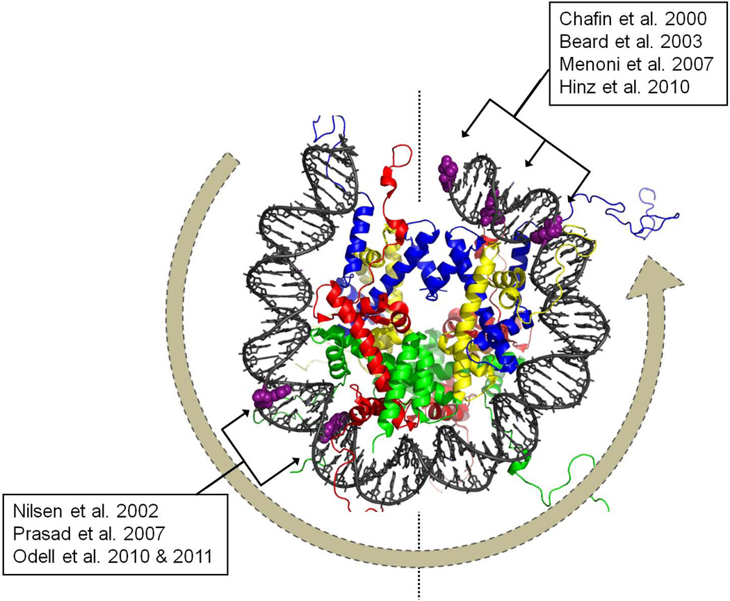
    1. Beard WA, Wilson SH. Structural design of a eukaryotic DNA repair polymerase: DNA polymerase beta. Mutation research. 2000;460(3–4):231–244. - PubMed

Publication types

LinkOut - more resources