

Podocyte detachment and reduced glomerular capillary endothelial fenestration promote kidney disease in type 2 diabetic nephropathy
- PMID: 22718189
- PMCID: PMC3472108
- DOI: 10.1038/ki.2012.234
Podocyte detachment and reduced glomerular capillary endothelial fenestration promote kidney disease in type 2 diabetic nephropathy
Abstract
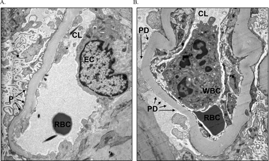
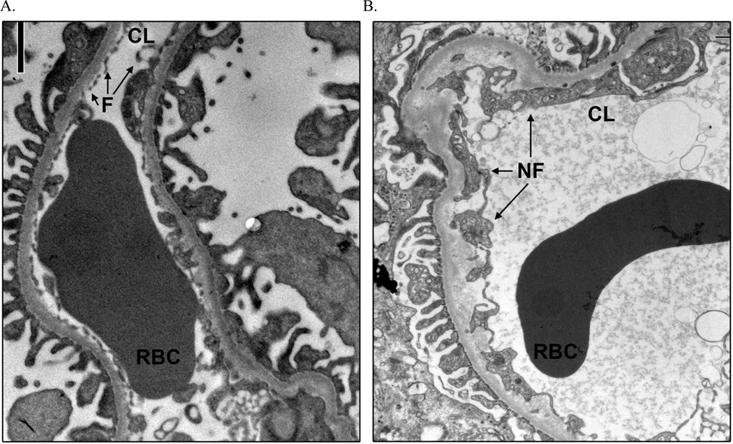
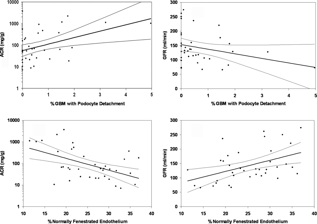
Podocyte detachment and reduced endothelial cell fenestration and relationships between these features and the classic structural changes of diabetic nephropathy have not been described in patients with type 2 diabetes. Here we studied these relationships in 37 Pima Indians with type 2 diabetes of whom 11 had normal albuminuria, 16 had microalbuminuria, and 10 had macroalbuminuria. Biopsies from 10 kidney donors (not American Indians) showed almost undetectable (0.03%) podocyte detachment and 43.5% endothelial cell fenestration. In patients with type 2 diabetes, by comparison, the mean percentage of podocyte detachment was significantly higher in macroalbuminuria (1.48%) than in normal albuminuria (0.41%) or microalbuminuria (0.37%). Podocyte detachment correlated significantly with podocyte number per glomerulus and albuminuria. The mean percentage of endothelial cell fenestration was significantly lower in macroalbuminuria (19.3%) than in normal albuminuria (27.4%) or microalbuminuria (27.2%) and correlated significantly with glomerular basement membrane thickness, albuminuria, fractional mesangial area, and the glomerular filtration rate (iothalamate clearance). Podocyte detachment and diminished endothelial cell fenestration were not correlated, but were related to classic lesions of diabetic nephropathy. Thus, our findings confirm the important role these injuries play in the development and progression of kidney disease in type 2 diabetes, just as they do in type 1 diabetes. Whether podocyte detachment creates conduits for proteins to escape the glomerular circulation and reduced endothelial fenestration lowers glomerular hydraulic permeability requires further study.
Trial registration: ClinicalTrials.gov NCT00340678.
Figures
Comment in
-
The glomerular endothelium emerges as a key player in diabetic nephropathy.Kidney Int. 2012 Nov;82(9):949-51. doi: 10.1038/ki.2012.258. Kidney Int. 2012. PMID: 23064188
-
Is endothelial dysfunction more deleterious than podocyte injury in diabetic nephropathy?Kidney Int. 2013 Jun;83(6):1202-3. doi: 10.1038/ki.2013.36. Kidney Int. 2013. PMID: 23728007 No abstract available.
-
The authors reply.Kidney Int. 2013 Jun;83(6):1203. doi: 10.1038/ki.2013.40. Kidney Int. 2013. PMID: 23728009 Free PMC article. No abstract available.
References
-
- Shankland SJ. The podocyte's response to injury: role in proteinuria and glomerulosclerosis. Kidney Int. 2006;69:2131–2147. - PubMed
-
- Jefferson JA, Shankland SJ, Pichler RH. Proteinuria in diabetic kidney disease: a mechanistic viewpoint. Kidney Int. 2008;74:22–36. - PubMed
-
- Steffes MW, Schmidt D, McCrery R, Basgen JM. International Diabetic Nephropathy Study Group. Glomerular cell number in normal subjects and in type 1 diabetic patients. Kidney Int. 2001;59:2104–2113. - PubMed
-
- White KE, Bilous RW Diabiopsies Study Group. Structural alterations to the podocyte are related to proteinuria in type 2 diabetic patients. Nephrol Dial Transplant. 2004;19:1437–1440. - PubMed
Publication types
MeSH terms
Substances
Associated data
Grants and funding
LinkOut - more resources
Full Text Sources
Other Literature Sources
Medical
