. 2012 Aug 10;287(33):27326-34.
doi: 10.1074/jbc.M112.366658. Epub 2012 Jun 20.

Structure of the nuclear factor κB-inducing kinase (NIK) kinase domain reveals a constitutively active conformation

Affiliations

Structure of the nuclear factor κB-inducing kinase (NIK) kinase domain reveals a constitutively active conformation

Jinsong Liu et al. J Biol Chem. .

Abstract

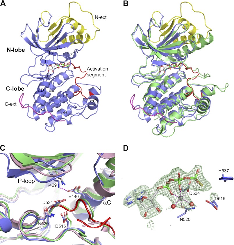
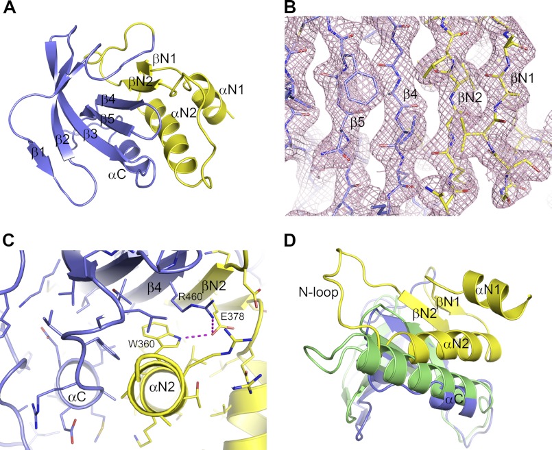
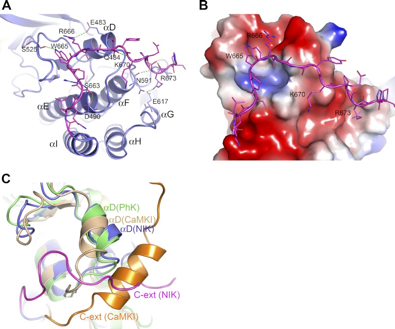
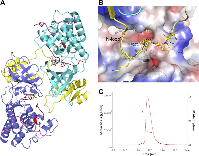
NF-κB-inducing kinase (NIK) is a central component in the non-canonical NF-κB signaling pathway. Excessive NIK activity is implicated in various disorders, such as autoimmune conditions and cancers. Here, we report the first crystal structure of truncated human NIK in complex with adenosine 5'-O-(thiotriphosphate) at a resolution of 2.5 Å. This truncated protein is a catalytically active construct, including an N-terminal extension of 60 residues prior to the kinase domain, the kinase domain, and 20 residues afterward. The structure reveals that the NIK kinase domain assumes an active conformation in the absence of any phosphorylation. Analysis of the structure uncovers a unique role for the N-terminal extension sequence, which stabilizes helix αC in the active orientation and keeps the kinase domain in the catalytically competent conformation. Our findings shed light on the long-standing debate over whether NIK is a constitutively active kinase. They also provide a molecular basis for the recent observation of gain-of-function activity for an N-terminal deletion mutant (ΔN324) of NIK, leading to constitutive non-canonical NF-κB signaling with enhanced B-cell adhesion and apoptosis resistance.


Figures

FIGURE 1.
FIGURE 1.
Identification of catalytically active NIK kinase domain constructs. A, NIK sequence motifs based on literature reports (16, 21). NRD, negative regulatory domain; NCR, non-catalytic region; BR, basic region; PRR, proline-rich repeat. B, schematic of a selected panel of truncated NIK constructs with the kinase domain shown as slate boxes. C, in vitro kinase phosphorylation assays. Left panel, NIK autophosphorylation and IKKα(K43A) phosphorylation by NIK. IKKα(K43A) and NIK construct 329–747(K429A) (329–747-KA) are inactive alanine mutants of the catalytic Lys-43 and Lys-429, respectively. Right panel, immunoblot (IB) analysis of total cell extracts. Positions of molecular mass standards (in kilodaltons) are shown on the left for both panels. IP, immunoprecipitate.
FIGURE 2.
FIGURE 2.
Crystal structure of NIK construct 330–679 in complex with ATPγS. A, overall NIK structure shown in ribbon diagram. The kinase domain is colored in slate with the activation segment highlighted in red. The N-terminal (N-ext) and C-terminal (C-ext) extensions to the kinase domain are colored in yellow and magenta, respectively. ATPγS is shown in stick representation and is atomic color-coded gray for carbon, red for oxygen, blue for nitrogen, yellow for sulfur, and green for phosphorus. N-lobe, N-terminal lobe; C-lobe, C-terminal lobe. B, overall structure comparison of NIK with a constitutively active PhK (Protein Data Bank code 2PHK). NIK is color-coded as described for A, and PhK is colored green. C, ribbon diagram of the NIK kinase domain active site compared with PhK and cAMP-dependent kinase (Protein Data Bank code 1ATP). Residues are shown in atomic color scheme with the carbon color scheme being slate for NIK, green for PhK, and pink for cAMP-dependent kinase. D, the bound nucleotide ATPγS in NIK with omit electron density map contoured at 2.5σ. A Mg2+ ion is shown as a gray sphere, and two water molecules are shown as red spheres.
FIGURE 3.
FIGURE 3.
N-terminal extension to the NIK kinase domain. A, extended N-terminal lobe of the NIK kinase domain. The typical N-terminal lobe of the kinase domain is shown in slate, and the N-terminal extension is shown in yellow. B, packing of strand βN2 of the N-terminal extension with strand β4 in the kinase N-terminal lobe. The pink mesh represents the 2FoFc electron density map. C, packing interactions between the N-terminal extension and the kinase N-terminal lobe. D, position of helix αC in NIK in comparison with that in PhK, shown in green.
FIGURE 4.
FIGURE 4.
C-terminal extension to the NIK kinase domain. A, extensive interactions from the C-terminal extension with the kinase C-terminal lobe. The color scheme is the same as described in the legend to Fig. 2A. B, interactions of the C-terminal extension with the kinase C-terminal lobe, which is shown in electrostatic molecular surface representation. C, superposition of the structures of active PhK and autoinhibited calcium/calmodulin-dependent protein kinase I (CaMKI; Protein Data Bank code 1A06) with NIK in the C-terminal extension (C-ext) area. The position of helix αD in NIK resembles that in the active PhK.
FIGURE 5.
FIGURE 5.
Dimer versus monomer. A, head-to-tail dimer of two independent NIK molecules in the asymmetric unit. The color scheme is the same as described in the legend to Fig. 2, except that the kinase domain of one monomer is colored cyan. B, N-loop binding in the dimer. The N-loop of one monomer is shown in ribbon and stick representation, binding to a region of the P+1 substrate-binding pocket of the dimer partner shown in electrostatic molecular surface representation. C, solution characterization of NIK construct 330–679. SEC-SLS of NIK showed a major peak and a minor peak with measured molecular masses of ∼44 and ∼100 kDa, respectively.

References

    1. Vallabhapurapu S., Karin M. (2009) Regulation and function of NF-κB transcription factors in the immune system. Annu. Rev. Immunol. 27, 693–733 - PubMed
    1. Hayden M. S., Ghosh S. (2008) Shared principles in NF-κB signaling. Cell 132, 344–362 - PubMed
    1. Bonizzi G., Karin M. (2004) The two NF-κB activation pathways and their role in innate and adaptive immunity. Trends Immunol. 25, 280–288 - PubMed
    1. Senftleben U., Cao Y., Xiao G., Greten F. R., Krähn G., Bonizzi G., Chen Y., Hu Y., Fong A., Sun S. C., Karin M. (2001) Activation by IKKα of a second, evolutionary conserved, NF-κB signaling pathway. Science 293, 1495–1499 - PubMed
    1. Pomerantz J. L., Baltimore D. (2002) Two pathways to NF-κB. Mol. Cell 10, 693–695 - PubMed

MeSH terms

Substances

Associated data

LinkOut - more resources