. 2012 Sep;86(17):9361-8.
doi: 10.1128/JVI.01214-12. Epub 2012 Jun 20.

Characterization in vitro and in vivo of pandemic (H1N1) 2009 influenza viruses isolated from patients

Affiliations

Characterization in vitro and in vivo of pandemic (H1N1) 2009 influenza viruses isolated from patients

Tokiko Watanabe et al. J Virol. 2012 Sep.

Abstract

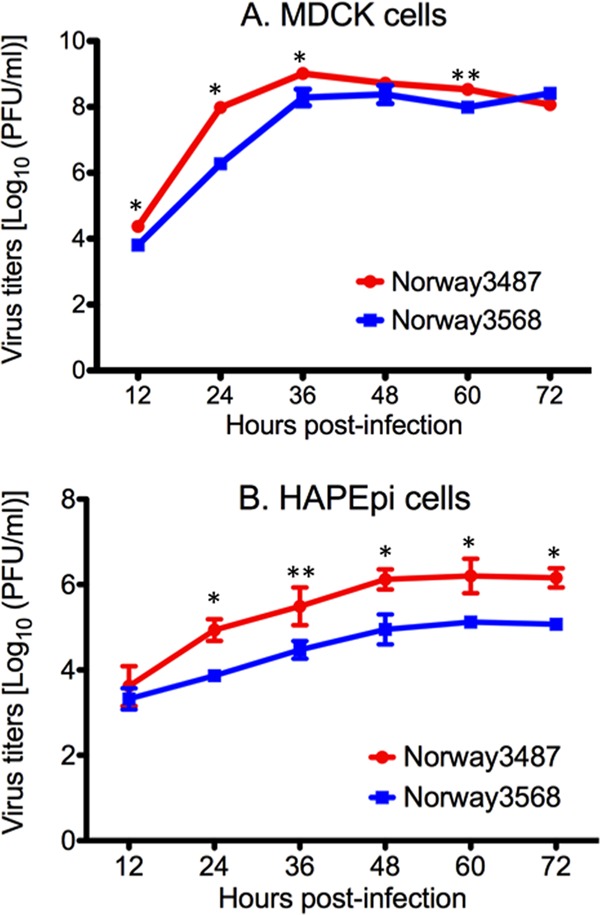
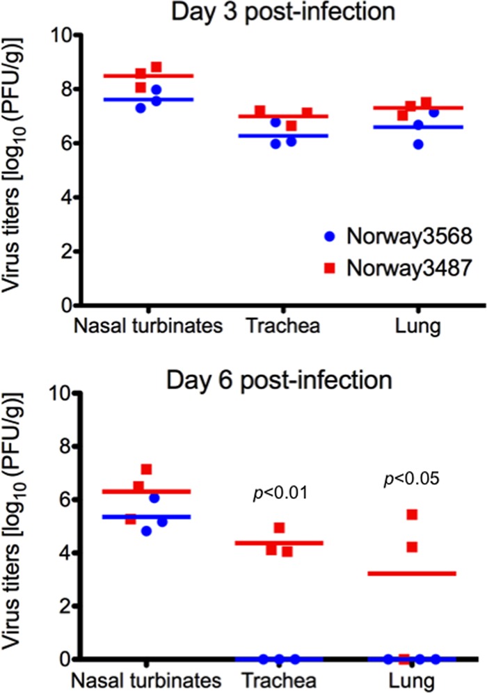
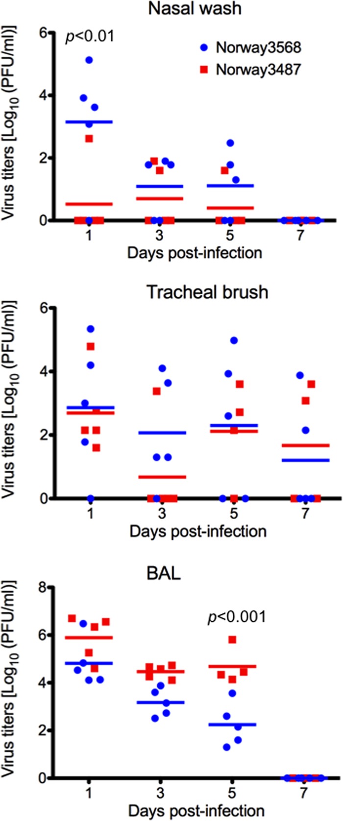
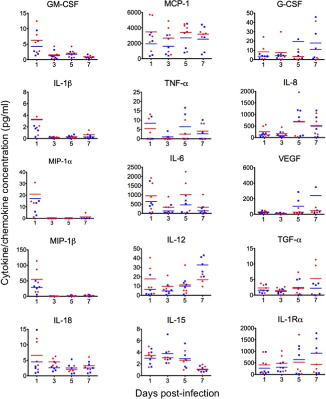
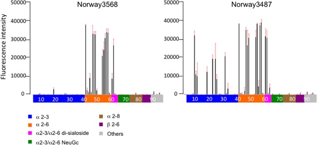
The first influenza pandemic of the 21st century was caused by novel H1N1 viruses that emerged in early 2009. Molecular evolutionary analyses of the 2009 pandemic influenza A H1N1 [A(H1N1)pdm09] virus revealed two major clusters, cluster I and cluster II. Although the pathogenicity of viruses belonging to cluster I, which became extinct by the end of 2009, has been examined in a nonhuman primate model, the pathogenic potential of viruses belonging to cluster II, which has spread more widely in the world, has not been studied in this animal model. Here, we characterized two Norwegian isolates belonging to cluster II, namely, A/Norway/3568/2009 (Norway3568) and A/Norway/3487-2/2009 (Norway3487), which caused distinct clinical symptoms, despite their genetic similarity. We observed more efficient replication in cultured cells and delayed virus clearance from ferret respiratory organs for Norway3487 virus, which was isolated from a severe case, compared with the efficiency of replication and time of clearance of Norway3568 virus, which was isolated from a mild case. Moreover, Norway3487 virus to some extent caused more severe lung damage in nonhuman primates than did Norway3568 virus. Our data suggest that the distinct replicative and pathogenic potentials of these two viruses may result from differences in their biological properties (e.g., the receptor-binding specificity of hemagglutinin and viral polymerase activity).


Figures

Fig 1
Fig 1
Viral growth kinetics in MDCK cells (A) and normal human alveolar epithelial cells (B). Cells were infected with Norway3568 or Norway3487 at a multiplicity of infection of 0.001. At different times postinfection, virus titers in the supernatants of infected cells were determined by use of plaque assays in MDCK cells. The mean from triplicate independent cultures ± standard deviation is shown. The mean titers of Norway3487 virus were significantly higher than those of Norway3568 virus (*, P < 0.001; **, P < 0.01).
Fig 2
Fig 2
Virus replication in respiratory organs of ferrets. Ferrets were intranasally infected with 106 PFU/500 μl of virus. Three animals from each group were euthanized on days 3 and 6 postinfection for virus titration. Red and blue horizontal bars show the mean titers for Norway3487 and Norway3568 viruses, respectively. No statistically significant differences between virus titers in respiratory tissues of ferrets infected with Norway3487 virus and those infected with Norway3568 virus were found at day 3 postinfection. The titers of Norway3487 virus in trachea and lungs were significantly higher than those of Norway3568 virus at day 6 postinfection (P < 0.01 in trachea; P < 0.05 in lungs).
Fig 3
Fig 3
Virus titers in respiratory washes from infected cynomolgus macaques. Cynomolgus macaques were infected with 6.7 × 10⁷ PFU of viruses through multiple routes. Nasal wash, tracheal brush, and BAL fluid samples were collected every other day for virus titration. Red and blue horizontal bars show the mean titers for Norway3487 and Norway3568 viruses, respectively. The titers of Norway3487 virus were significantly higher than those of Norway3568 virus in BAL fluid samples on day 5 postinfection (P < 0.001), whereas those of Nowary3487 in nasal wash samples were lower than those of Norway3568 virus at day 1 postinfection (P < 0.01).
Fig 4
Fig 4
Pathological analyses of the lungs of infected macaques. (A) Infection with Norway3568 virus (panel 1) resulted in moderate to severe bronchointerstitial pneumonia (panel 4) with some minimally affected to unaffected regions (panel 7). Infection with Norway3487 virus (panel 2) resulted in moderate to severe bronchointerstitial pneumonia with severe lung edema and inflammatory changes (panel 5), even in areas with less severe gross changes (panel 8). Lungs derived from a noninfected control animal did not have any gross or histological changes (panels 3 and 6). Arrowheads, gross lesions. Boxes drawn with dotted lines depict the areas shown in the microscopic images. Bars, 200 μm. (B) Pathological severity scores in infected animals on day 7 postinfection. To represent comprehensive histological changes, respiratory tissue slides were evaluated by scoring the pathological changes. The pathological scores were determined for each animal in each group (n = 4 and n = 3 for Norway3487 and Norway3568 viruses, respectively). Error bars denote standard deviations. The mean pathological severity score in the right cranial lung lobe was higher for Norway3487 virus than for Norway3568 virus (Student t test, P = 0.0096).
Fig 5
Fig 5
Cytokine/chemokine production in infected cynomolgus macaques. The concentrations of various cytokines and chemokines in the BAL fluid of infected cynomolgus macaques were measured on days 1, 3, 5, and 7 postinfection by use of protein array analysis with a Milliplex MAP nonhuman primate cytokine/chemokine panel (premixed 23-plex; Millipore, Bedford, MA) with a Bio-Plex 200 system (Bio-Rad Laboratories, Hercules, CA). No statistically significant differences between cytokine/chemokine levels for Norway3487 virus and those for Norway3568 virus were found.
Fig 6
Fig 6
Receptor specificity of Norwegian viruses. Sialylated glycan binding by Norway3568 and Norway3487 viruses; purified whole virions were analyzed by glycan microarray. The microarrays displayed 86 sialylated and 9 asialo glycans printed on coated glass slides. Different types of glycans on the array (x axis) are highlighted in different colors; the identity of each numbered glycan is provided in Table S1 in the supplemental material. Black bars denote the mean fluorescent binding signal intensity (y axis) of 4 spots; the standard error is shown as a red extension.
Fig 7
Fig 7
Polymerase activity of Norwegian viruses measured in a minigenome replicon assay. Four protein expression plasmids for PB2, PB1, PA, and NP derived from Norway3487 or Norway3568, pPolI-WNA-Flu expressing the NA gene encoding the firefly luciferase gene, and the pGL4.74[hRuc/TK] control plasmid were transfected into 293T cells and assayed for luciferase activity after a 24-h incubation at 37°C. Since Norway3487 and Norway3568 viruses do not differ in their PA and NP sequences, only combinations of PB1 and PB2 proteins were tested. 3487 and 3568, Norway3487 and Norway3568 viruses, respectively. The values shown are means ± standard deviations for the results of three independent experiments and are standardized to the activities of the expression plasmids for the Norway3568 RNP complex proteins (100%). Polymerase activities of replication complexes possessing the PB2 protein from Norway3487 virus were higher than those possessing PB2 from Norway3568 virus (P < 0.001).

References

    1. Baskin CR, et al. 2009. Early and sustained innate immune response defines pathology and death in nonhuman primates infected by highly pathogenic influenza virus. Proc. Natl. Acad. Sci. U. S. A. 106:3455–3460 - PMC - PubMed
    1. Bautista E, et al. 2010. Clinical aspects of pandemic 2009 influenza A (H1N1) virus infection. N. Engl. J. Med. 362:1708–1719 - PubMed
    1. Blixt O, et al. 2004. Printed covalent glycan array for ligand profiling of diverse glycan binding proteins. Proc. Natl. Acad. Sci. U. S. A. 101:17033–17038 - PMC - PubMed
    1. Christman MC, Kedwaii A, Xu J, Donis RO, Lu G. 2011. Pandemic (H1N1) 2009 virus revisited: an evolutionary retrospective. Infect. Genet. Evol. 11:803–811 - PMC - PubMed
    1. Chutinimitkul S, et al. 2010. Virulence-associated substitution D222G in the hemagglutinin of 2009 pandemic influenza A(H1N1) virus affects receptor binding. J. Virol. 84:11802–11813 - PMC - PubMed

Publication types

MeSH terms