. 2012 Sep;86(17):8998-9014.
doi: 10.1128/JVI.01209-12. Epub 2012 Jun 20.

A Nuclear localization signal in herpesvirus protein VP1-2 is essential for infection via capsid routing to the nuclear pore

Affiliations

A Nuclear localization signal in herpesvirus protein VP1-2 is essential for infection via capsid routing to the nuclear pore

F Abaitua et al. J Virol. 2012 Sep.

Abstract

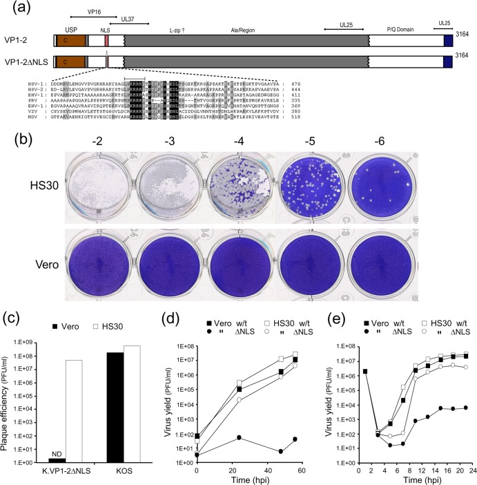
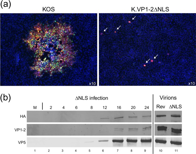
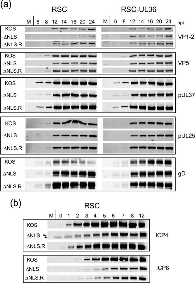
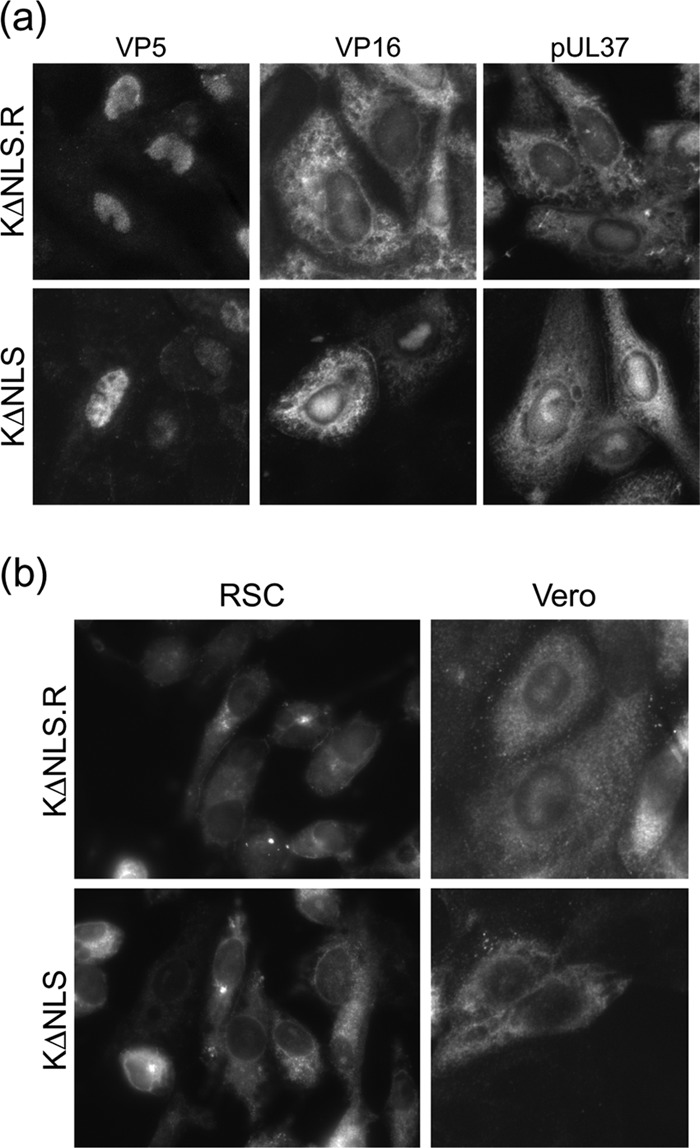
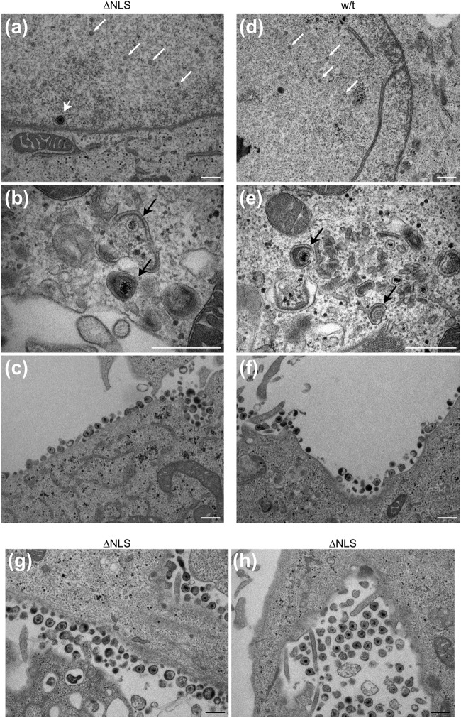
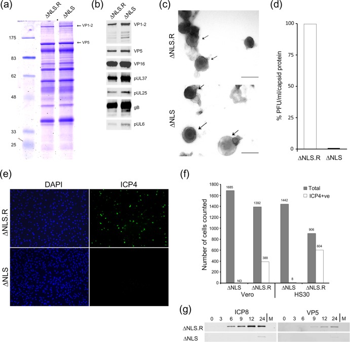
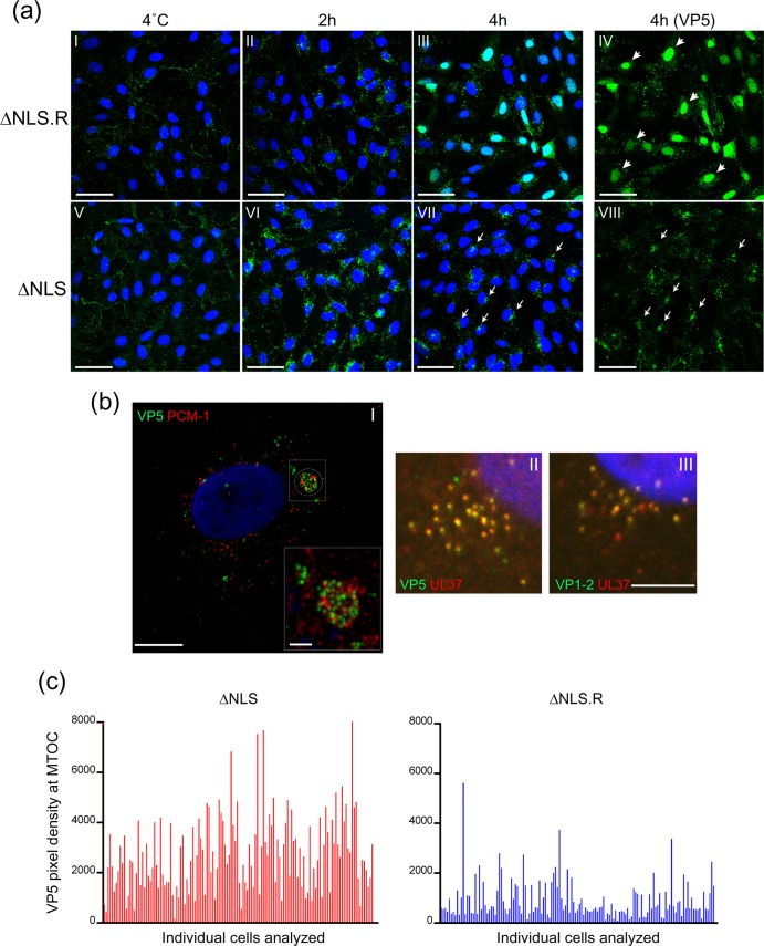
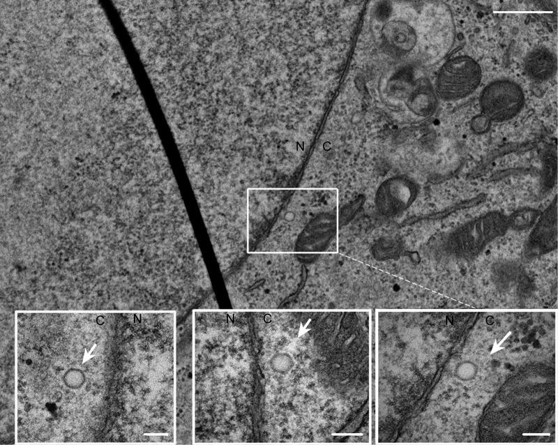
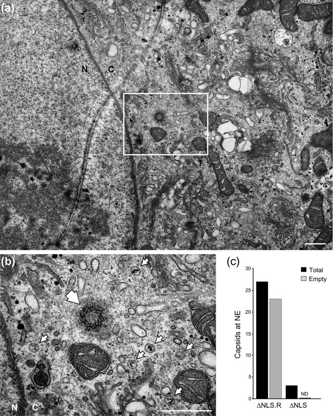
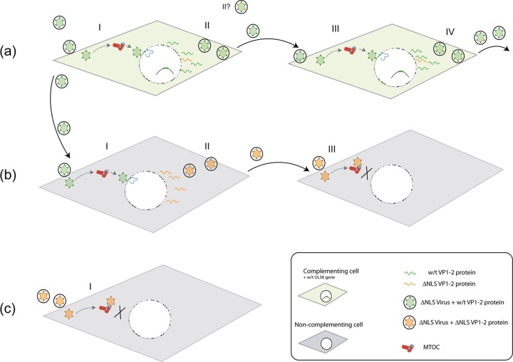
To initiate infection, herpesviruses must navigate to and transport their genomes across the nuclear pore. VP1-2 is a large structural protein of the virion that is conserved in all herpesviruses and plays multiple essential roles in virus replication, including roles in early entry. VP1-2 contains an N-terminal basic motif which functions as an efficient nuclear localization signal (NLS). In this study, we constructed a mutant HSV strain, K.VP1-2ΔNLS, which contains a 7-residue deletion of the core NLS at position 475. This mutant fails to spread in normal cells but can be propagated in complementing cell lines. Electron microscopy (EM) analysis of infection in noncomplementing cells demonstrated capsid assembly, cytoplasmic envelopment, and the formation of extracellular enveloped virions. Furthermore, extracellular virions isolated from noncomplementing cells had similar profiles and abundances of structural proteins. Virions containing VP1-2ΔNLS were able to enter and be transported within cells. However, further progress of infection was prevented, with at least a 500- to 1,000-fold reduction in the efficiency of initiating gene expression compared to that in the revertant. Ultrastructural and immunofluorescence analyses revealed that the K.VP1-2ΔNLS mutant was blocked at the microtubule organizing center or immediately upstream of nuclear pore docking and prior to gene expression. These results indicate that the VP1-2 NLS is not required for the known assembly functions of the protein but is a key requirement for the early routing to the nuclear pore that is necessary for successful infection. Given its conservation, we propose that this motif may also be critical for entry of other classes of herpesviruses.


Figures

Fig 1
Fig 1
Deletion of VP1-2 abolishes plaque formation. (a) Schematic summary of VP1-2, indicating the N-terminal USP domain, the highly basic NLS motif contained within a poorly conserved region, the large central region (gray), and a C-terminal conserved region (blue). Regions relevant to interactions with VP16, pUL37, and pUL25 are also indicated (10, 15, 30, 34, 41, 47, 59). (b) Serial dilutions of K.VP1-2ΔNLS were examined for plaque formation in complementing HS30 cells versus normal Vero cells. (c) Quantitative analysis of plaque-forming efficiency of the mutant versus wt KOS in Vero and HS30 cells. Plaque formation was diminished by at least 7 logs and was essentially undetected in Vero cells compared to HS30 cells. (d) Comparison of the wt and mutant strains by multistep growth curves after low-MOI infection (0.01 PFU/cell, based on titers on complementing cells) of Vero (solid symbols) or HS30 (open symbols) cells. Yields were titrated on HS30 cells. (e) Comparison of wt and mutant strain replication by single-step growth curves after high-MOI infection (5 PFU/cell, based on titers on complementing cells) of Vero or HS30 cells.
Fig 2
Fig 2
Initial infection of single cells by K.VP1-2ΔNLS and failure of the virus to spread. (a) Vero cells were infected with K.VP1-2ΔNLS or wt KOS at an MOI calculated to infect 1/500 cells. Monolayers were examined for virus proteins after 3 days by immunofluorescence. Green, ICP4; red, pUL37; blue, nuclear counterstain. For the wt virus, plaques had obviously grown and contained numerous antigen-positive cells. In contrast, for the mutant, single antigen-positive cells were observed (both antigens were detected, though the red channel obscures the green in this merged example), but infection was never detected and did not progress in surrounding cells. (b) To examine the induction of expression of wt VP1-2 during propagation in complementing cells, RSC-HAUL36 cells were infected with K.VP1-2ΔNLS at an MOI of 5 (based on titers on HS30 cells), and samples were harvested at the indicated times and examined for expression of endogenous wt VP1-2 by virtue of the HA tag, of total VP1-2 by using anti-VP1-2, or of the major capsid protein VP5. Extracellular virus (also see Fig. 5) from K.VP1-2ΔNLS and K.VP1-2ΔNLS.R (Rev), amplified in complementing RSC-HAUL36 cells and purified by Ficoll density gradient centrifugation, was analyzed to assess the incorporation of endogenous VP1-2 into the virion particles produced in these cells in relation to that of VP5 (lanes 10 and 11).
Fig 3
Fig 3
Comparison of protein expression during infection by K.VP1-2ΔNLS and its revertant. (a) RSC or RSC-HAUL36 cells were infected in parallel with the mutant virus K.VP1-2ΔNLS, the revertant virus K.VP1-2ΔNLS.R, or wt KOS virus at an MOI of 5. Samples were harvested at the indicated times, and expression of a series of delayed early or late proteins, i.e., VP5, pUL37, UL25, gD, and VP1-2 itself, was examined by Western blotting using fluorescent antibodies and quantitative detection as described in Materials and Methods. (b) Same as panel a, but examining immediate early (ICP4) and delayed early (ICP8) proteins every hour for 12 h. Some reduction in the abundance of ICP4 (long arrow) was observed, though there was little difference in the amount of ICP8. (A nonspecific band migrating just below ICP4 was occasionally observed, as indicated by the short arrow.) The parental KOS strain exhibited slightly increased expression of both proteins, estimated by quantitative analysis as a 2- to 4-fold increase over expression by the ΔNLS and ΔNLS.R viruses.
Fig 4
Fig 4
Comparison of protein localization during infection by K.VP1-2ΔNLS and wt KOS. (a) Vero cells were infected with KOS or the mutant virus K.VP1-2ΔNLS at an MOI of 5. Cultures were fixed after 16 h and processed for immunofluorescence as described in the text. Patterns of localization discussed in the text were similar for both the wt and mutant viruses. (b) RSC or Vero cells were infected and processed as described for panel a and were stained for VP1-2 localization.
Fig 5
Fig 5
Ultrastructural analysis of capsid and virion formation in K.VP1-2ΔNLS-infected cells. RSC were infected in parallel with KOS or the mutant virus K.VP1-2ΔNLS at an MOI of 5, fixed at 16 h postinfection, and processed for thin-section electron microscopy as described in Materials and Methods. (a to c) K.VP1-2ΔNLS; (d to f) KOS. Capsid formation was readily apparent for both the mutant and wt viruses (a and d; small white arrows indicate nucleocapsids, and the large white arrowhead indicates a particle in primary envelopment). Cytoplasmic envelopment was also observed for the mutant and wt viruses (b and e; black arrows indicate wrapping during secondary envelopment). Finally, panels c and f show the presence of assembled virions for the mutant and the revertant, respectively. (g and h) Further examples of assembled extracellular virions, which were readily observed for the mutant virus. In general, little difference in the main ultrastructural features of infection were observed for the wt versus the mutant strain, and all qualitative features of capsid and virion assembly could be observed. Similar results were obtained with the K.VP1-2ΔNLS revertant virus and with different cell lines (Vero and HaCaT cells). Bars, 500 nm.
Fig 6
Fig 6
Characterization of extracellular K.VP1-2ΔNLSNC virus produced from noncomplementing cells. HaCaT cells were infected with K.VP1-2ΔNLSC or the revertant virus at an MOI of 5 (estimated from titration on complementing cells). We used HaCaT cells because we find that they help to increase overall yields of all herpesvirus strains. The medium was harvested 16 h after infection and clarified, and extracellular virus was pelleted by centrifugation at 19,000 rpm for 90 min. Samples of pelleted virus showed slightly increased amounts of virion proteins for the mutant. Samples were adjusted (based on the major capsid protein VP5) and analyzed by SDS-PAGE. (a) Mutant and revertant viruses exhibited similar qualitative and quantitative profiles of structural proteins, including VP1-2 and VP5. (b) Equalized samples were analyzed by Western blotting for the presence of a number of structural components, as indicated. (c) Samples of extracellular particles from revertant- or mutant-infected cell medium, analyzed by negative staining. Various degrees of stain penetration were observed for both the mutant and the revertant, and examples illustrating partially stained intact particles are shown for both. Bars, 200 nm. (d) Comparison of specific infectivities of K.VP1-2ΔNLSNC and revertant extracellular virus particles, equalized on the basis of the major capsid protein and titrated for plaque formation on complementing HS30 cells. On a capsid-standardized basis, K.VP1-2ΔNLSNC particles exhibited approximately 600-fold lower infectivity. (e) Comparison of infectivities at the single-cell level. Vero or HS30 cells were infected with serial dilutions of the revertant virus designed to give low-MOI infections within the range of 1:3 to 1:100 cells infected. Cells were infected in parallel with K.VP1-2ΔNLSNC particles over an equivalent range based on equalized capsid protein. Cells were fixed 6 h after infection, stained with antibody to detect ICP4, and counterstained with DAPI (4′,6-diamidino-2-phenylindole) to enumerate cells. Numerous cells were counted for each virus at each dilution. Typical fields are shown for Vero cells with the largest amount of input virus to emphasize the difference in infectivity between the revertant and the mutant, as quantified in panel f. (g) Based on the titer of the revertant virus, cells were infected at a high MOI (5 PFU/cell) with the revertant virus or an equivalent input of mutant virus, based on standardization of the capsid protein in extracellular virus particles. Cells were harvested for Western blotting at the indicated times, with analysis in this case of a typical delayed early protein (ICP8) or the late major capsid protein VP5.
Fig 7
Fig 7
Immunofluorescence analysis of K.VP1-2ΔNLSNC virus entry. Cells (RSC) were infected with extracellular virus of either the revertant ΔNLS.R virus or the mutant ΔNLS virus produced from noncomplementing cells. Infection was done at an MOI of 100 for the revertant and with an equivalent amount of the mutant, based on standardization of VP5 in the extracellular purified virus. To help to synchronize infection, cells were washed with cold medium and incubated at 4°C after addition of the inoculum. After 1 h, warmed medium was added, and the cells were further incubated for 2 or 4 h at 37°C prior to fixation and permeabilization. Control samples maintained at 4°C were also examined as controls. Samples were stained for the major capsid protein VP5 and counterstained with DAPI to detect nuclei. The panels show typical examples of many fields examined (using a ×40 objective to capture multiple cells in each field) and present both channels for each virus at the indicated time. The VP5 channel only is shown in panels IV and VIII, highlighting the contrast between the revertant and mutant viruses. Bars, 50 μm. (b) (I) High-resolution confocal section of cells infected with the ΔNLS virus and stained with anti-VP5 (green), anti-PCM1 (red), and DAPI. Bar, 10 μm. The inset (bar, 2 μm) shows the pericentriolar PCM1 accumulations with congregating capsids in the same location, though the capsids do not necessarily directly overlap the PCM1-specific material. (II and III) Higher-magnification images of capsids (VP5) and tegument proteins (pUL37 and VP1-2) colocalizing at the MTOC perinuclear accumulations during ΔNLS virus infection. Bar, 5 μm. (c) Graph showing quantification of MTOC accumulation for revertant and mutant capsids. Defined and unbiased areas of interest were applied around the PCM1 pericentriolar region, and the accumulated density of pixels in the green channel for VP5 in the same areas was calculated. Each vertical bar represents an individual cell. Approximately 130 cells were evaluated for each virus.
Fig 8
Fig 8
EM analysis showing nuclear pore interaction during entry of the ΔNLS revertant virus. Vero cells were infected with the revertant virus K.VP1-2ΔNLS.R at an MOI of 500 PFU/cell. The cells were washed with cold medium and incubated at 4°C after addition of the inoculum. After 1 h, warmed medium was added, and the cells were incubated for 4 h at 37°C and then fixed and processed for thin-section (70 nm) ultrastructural analysis as described in Materials and Methods. N, nucleus; C, cytoplasm. An example section is shown, with the inset illustrating the presence of empty capsids in close proximity to the nuclear pore. Additional insets are shown for additional independent sections illustrating the same features of nuclear pore proximity and empty capsids. Bars, 500 nm in the main image and 250 nm in the insets. Like the case in thin-section analysis, capsids immediately adjacent to the NE represented only a subset of infecting capsids but were readily observed for the wt virus. Total numbers of capsids within 100 nm of the NE are quantitated in Fig. 9c.
Fig 9
Fig 9
EM analysis of the block in K.VP1-2ΔNLSNC virus entry. In parallel with the analysis shown in Fig. 8, cells were infected with the mutant virus K.VP1-2ΔNLSNC, fixed, and processed. N, nucleus; C, cytoplasm. We readily detected cytoplasmic capsids, frequently in close proximity to the centrioles of the MTOC, but capsids close to the NE were rare. (a and b) Representative field (a) indicating the position within the cell of a higher-magnification inset (b) showing full capsids of the infecting mutant. The single large arrowhead indicates the position of one of the centrioles, with small arrows pointing to 6 capsids accumulated around the MTOC area. (c) Quantification of full and empty capsids from K.VP1-2ΔNLS- and revertant-infected cells. The total perimeter of the NE in 100 cells was scanned for capsids in close proximity to the NE (within 100 nm of the NE), which were scored as empty or DNA-containing capsids. We found only rare examples of capsids close to the NE, and these were not adjacent to pores and contained DNA. Bars, 500 nm.
Fig 10
Fig 10
Summary of propagation of K.VP1-2ΔNLS in complementing and normal cell types. The three rows (a, b, and c) summarize the outcomes of K.VP1-2ΔNLS infection. (a) Complementing cells (shaded in green) contain the wt UL36 gene, which is induced after infection with the K.VP1-2ΔNLS mutant. The wt VP1-2 protein is recruited onto virions (green virions with green dots, indicating functional VP1-2). Note that a population of mutant VP1-2 (indicated in orange) could be recruited onto particles, and virions may have mixed populations and different distributions (II?). It may be that not all of the VP1-2 incorporated needs to be wt, though neither the distribution of wt versus mutant VP1-2 nor the amount of wt VP1-2 required for successful infection is known. The mutant can replicate, spread, and form plaques in complementing cells. (b) Upon infection of noncomplementing cells (shaded in gray) with the complemented virus, the virus can infect, undergo gene expression, and produce extracellular virus (orange virions), which now contains mutant VP1-2ΔNLS (orange dots; II). Our results indicate that this virus contains VP1-2ΔNLS, appears structurally similar to wt virus, with a similar cohort of proteins, and can infect cells but is blocked at the MTOC prior to successful nuclear engagement. (c) Producing K.VP1-2ΔNLS virus in noncomplementing cells results in the formation of extracellular noncomplemented virions. These virions have a profound block in infection at the MTOC prior to nuclear engagement.

References

    1. Abaitua F, Daikoku T, Crump CM, Bolstad M, O'Hare P. 2011. A single mutation responsible for temperature sensitive entry and assembly defects in the VP1-2 protein of HSV. J. Virol. 85:2024–2036 - PMC - PubMed
    1. Abaitua F, O'Hare P. 2008. Identification of a highly conserved, functional nuclear localization signal within the N-terminal region of herpes simplex virus type 1 VP1-2 tegument protein. J. Virol. 82:5234–5244 - PMC - PubMed
    1. Abaitua F, Souto RN, Browne H, Daikoku T, O'Hare P. 2009. Characterization of the herpes simplex virus (HSV)-1 tegument protein VP1-2 during infection with the HSV temperature-sensitive mutant tsB7. J. Gen. Virol. 90:2353–2363 - PubMed
    1. Batterson W, Furlong D, Roizman B. 1983. Molecular genetics of herpes simplex virus. VIII. Further characterization of a temperature-sensitive mutant defective in release of viral DNA and in other stages of the viral reproductive cycle. J. Virol. 45:397–407 - PMC - PubMed
    1. Batterson W, Roizman B. 1983. Characterization of the herpes simplex virion-associated factor responsible for the induction of alpha genes. J. Virol. 46:371–377 - PMC - PubMed

Publication types

Substances