. 2012 Aug;23(16):3156-66.
doi: 10.1091/mbc.E12-02-0079. Epub 2012 Jun 20.

Characterization of an apical ceramide-enriched compartment regulating ciliogenesis

Affiliations

Characterization of an apical ceramide-enriched compartment regulating ciliogenesis

Qian He et al. Mol Biol Cell. 2012 Aug.

Abstract

We show that in Madin-Darby canine kidney (MDCK) cells, an apical ceramide-enriched compartment (ACEC) at the base of primary cilia is colocalized with Rab11a. Ceramide and Rab11a vesicles isolated by magnetic sorting contain a highly similar profile of proteins (atypical protein kinase C [aPKC], Cdc42, Sec8, Rab11a, and Rab8) and ceramide species, suggesting the presence of a ciliogenic protein complex associated with ceramide at the ACEC. It is intriguing that C16 and C18 ceramide, although less abundant ceramide species in MDCK cells, are highly enriched in ceramide and Rab11a vesicles. Expression of a ceramide-binding but dominant-negative mutant of aPKC suppresses ciliogenesis, indicating that the association of ceramide with aPKC is critical for the formation of this complex. Our results indicate that ciliogenic ceramide is derived from apical sphingomyelin (SM) that is endocytosed and then converted to the ACEC. Consistently, inhibition of acid sphingomyelinase with imipramine disrupts ACEC formation, association of ciliogenic proteins with Rab11a vesicles, and cilium formation. Ciliogenesis is rescued by the histone deacetylase (HDAC) inhibitor trichostatin A, indicating that ceramide promotes tubulin acetylation in cilia. Taken together, our results suggest that the ACEC is a novel compartment in which SM-derived ceramide induces formation of a ciliogenic lipid-protein complex that sustains primary cilia by preventing deacetylation of microtubules.


Figures

FIGURE 1:
FIGURE 1:
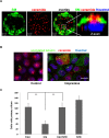
Ceramide is associated with the initiation of the primary cilium at the ACEC. MDCK cells were serum deprived, and ceramide (red) and acetylated tubulin (green) were visualized by immunocytochemistry. (A) At 24 h after serum deprivation. Right, arrow points at ACEC shown at higher magnification. Bar, 5 (left), 1 μm (right). (B) At 72 h after serum deprivation. Right, Z-scan of the ciliated cell. Arrows point at the cilium attachment site at the ACEC. Bar, 2 µm (left), 1 μm (right). (C) Primary cilium with ceramide distribution to the tip and along the cilium shaft (right). Arrows point at ceramide. Bar, 0.5 μm (left), 0.1 μm (right).
FIGURE 2:
FIGURE 2:
The ACEC is derived from apical SM hydrolyzed by ASMase. (A) Colony of MDCK cells serum deprived for 72 h, followed by immunocytochemistry for SM (green) and ceramide (red). Right, the image of a Z-scan. Consistent with the assumption that apical SM was gradually converted to ceramide during ACEC formation, the fluorescence signal for SM was reduced, whereas that of ceramide was increased. (B) Incubation of MDCK cells with the ASMase inhibitor imipramine (40 μM) led to delocalization of ceramide and acetylated tubulin, and prevented formation of primary cilia. Bar, 10 μm. (C) Quantification of data shown in B (number of cilia in 150 cells). Cilium formation was restored by incubation of imipramine (Imi)-treated cells with the novel ceramide analogue S18. n = 5, **p < 0.01.
FIGURE 3:
FIGURE 3:
ACEC formation relies on endolysosomal processing of SM involving Rab11a(+) compartments. (A) Projection view from top onto the apical plane of control cells and cells treated with two inhibitors in the salvage pathway for endolysosomal processing and remodeling of SM and ceramide (imipramine and FB1). Immunocytochemistry for SM (green) and ceramide (red) shows that the two lipids disappear from the apical plane when SM and ceramide processing is inhibited. Bar, 10 μm. (B) Z-scan of serum-deprived MDCK cells shows colocalization of ceramide (red) with Rab11a (blue) at the base of primary cilia (acetylated tubulin, green). Bar, 5 μm. (C) Colocalization of ceramide, SM, and Rab11a at the ACEC. Bar, 5 μm.
FIGURE 4:
FIGURE 4:
Serum deprivation induces codistribution of SM, ceramide, and Rab11a at the ACEC. (A) Immunocytochemistry for ceramide (red), SM (blue), and Rab11a (green) showing colocalization with the ACEC. Right, shows a pixel profile along the axis indicated at the left. Bar, 5 μm. (B) Immunocytochemistry for ceramide (red) and Rab11a (green) before and 15 h after serum deprivation. Bar, 10 μm. (C) Codistribution of ceramide (red) and Rab11a (green) by serum deprivation is prevented in the presence of imipramine. It can be rescued by incubation with a ceramide analogue (S18). Arrow points at primary cilium. Bar, 5 μm.
FIGURE 5:
FIGURE 5:
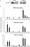
Ceramide and Rab11a vesicles contain almost identical ceramide pools enriched with C16 and C18 ceramide. Analysis of ceramide and Rab11a vesicles isolated from ciliated MDCK cells using anti-ceramide rabbit IgG or anti-Rab11a rabbit IgG immobilized on magnetic beads. Eluted vesicles were analyzed for the presence of Rab11a using immunoblotting (A) and for different ceramide species using sphingolipidomics/mass spectrometry (B). (A) Left, input control (corresponding to 50% of cell homogenate used for the isolation procedure). Right, output. (B) Sphingolipidomics (liquid chromatography–tandem mass spectrometry) analysis of ceramide species from ciliated MDCK cells (72 h after serum deprivation) and ceramide or Rab11a vesicles. Note that the ceramide profile of ceramide and Rab11a vesicles is almost identical and encompasses the entire pool of C16 and C18 ceramide in ciliated MDCK cells (as quantified by the amount of ceramide/lipid-bound phosphate). n = 3.
FIGURE 6:
FIGURE 6:
Ceramide is critical for association of Rab11a vesicles with aPKC and other ciliogenic proteins. (A) Expression of the ceramide-binding, dominant-negative PKCζ mutant C20ζ-GFP in MDCK cells. Left, immunocytochemistry for ceramide shows codistribution with C20ζ-GFP; right, relative number of cilia in GFP-expressing cells. n = 5, p < 0.01. Bar, 5 μm. (B) Magnetic isolation of ceramide and Rab11a vesicles, followed by immunoblotting for various proteins involved in ciliogenesis. Note that the protein profile of ceramide and Rab11a vesicles is almost identical, although acetylated tubulin was only recovered from ceramide vesicles. Input corresponds to 20% of cell homogenate used for the isolation procedure. (C) As in B, however, MDCK cells were serum deprived in the presence of inhibitors for SM and ceramide processing (FB1 and imipramine). GAPDH was used as a negative control not involved in Rab11a vesicle binding or ciliogenesis. (D) Immunocytochemistry for ceramide, Rab11a, and Rab8 shows colocalization at the ACEC. (E) As in D, except using an antibody against Sec8. Arrows point at location of primary cilium as determined by the colocalization of ceramide and acetylated tubulin in the same sample (data not shown). Bars, 5 μm.
FIGURE 7:
FIGURE 7:
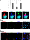
The ACEC inhibits deacetylation of acetylated tubulin: a potential mechanism for the initiation and stabilization of the primary cilium. (A) Incubation of imipramine-treated MDCK cells with the HDAC inhibitor TSA (0.5 μM) rescued ciliogenesis (figure shows number of cilia in 150 cells). n = 5; p < 0.01. (B) Codistribution of BBS1 (blue) with ceramide (red) and acetylated tubulin (green) as determined by immunocytochemistry (figure shows Z-scan). Bar, 5 μm. (C, D) In ciliated control MDCK cells (C), ceramide (red), BBS1 (green), and HDAC6 (blue) are codistributed at the ACEC, which is inhibited by imipramine (D).
FIGURE 8:
FIGURE 8:
Model for the ACEC-mediated stabilization of the primary cilium. SM from the cell membrane or Golgi is converted to ceramide, which binds to an aPKC-Par6-Cdc42 core complex. This core complex is associated with the exocyst (via Cdc42/Par6 binding to Sec10-Sec8-Sec15) and may regulate fusion of ceramide vesicles with the ACEC. The exocyst also mediates association of the ceramide–aPKC core complex with the Rab activation complex (Rab11a-Rabin8-Rab8), which is critical for primary cilium formation. Recruitment of the BBsome to the Rab activation complex (via binding of Rabin8 to BBS1) inhibits HDAC6 and initiates or stabilizes the cilium by promoting acetylation of tubulin at the ACEC. In bold are proteins we have shown to colocalize or copurify with ceramide in the ACEC or lipid vesicles.

Similar articles

Cited by

References

    1. Albouz S, Le Saux F, Wenger D, Hauw JJ, Baumann N. Modifications of sphingomyelin and phosphatidylcholine metabolism by tricyclic antidepressants and phenothiazines. Life Sci. 1986;38:357–363. - PubMed
    1. Babbey CM, Bacallao RL, Dunn KW. Rab10 associates with primary cilia and the exocyst complex in renal epithelial cells. Am J Physiol Renal Physiol. 2010;299:F495–F506. - PMC - PubMed
    1. Bieberich E. Lipid vesicle-mediated affinity chromatography using magnetic activated cell sorting (LIMACS): a novel method to analyze protein-lipid interaction. J Vis Exp. 2011;(50):e2657. doi: 10.3791/2657. - PMC - PubMed
    1. Bieberich E, Hu B, Silva J, MacKinnon S, Yu RK, Fillmore H, Broaddus WC, Ottenbrite RM. Synthesis and characterization of novel ceramide analogs for induction of apoptosis in human cancer cells. Cancer Lett. 2002;181:55–64. - PubMed
    1. Bieberich E, Kawaguchi T, Yu RK. N-acylated serinol is a novel ceramide mimic inducing apoptosis in neuroblastoma cells. J Biol Chem. 2000;275:177–181. - PubMed

Publication types

MeSH terms

LinkOut - more resources