. 2012 Aug;23(16):3041-56.
doi: 10.1091/mbc.E12-03-0194. Epub 2012 Jun 20.

Molecular chaperones and stress-inducible protein-sorting factors coordinate the spatiotemporal distribution of protein aggregates

Affiliations

Molecular chaperones and stress-inducible protein-sorting factors coordinate the spatiotemporal distribution of protein aggregates

Liliana Malinovska et al. Mol Biol Cell. 2012 Aug.

Abstract

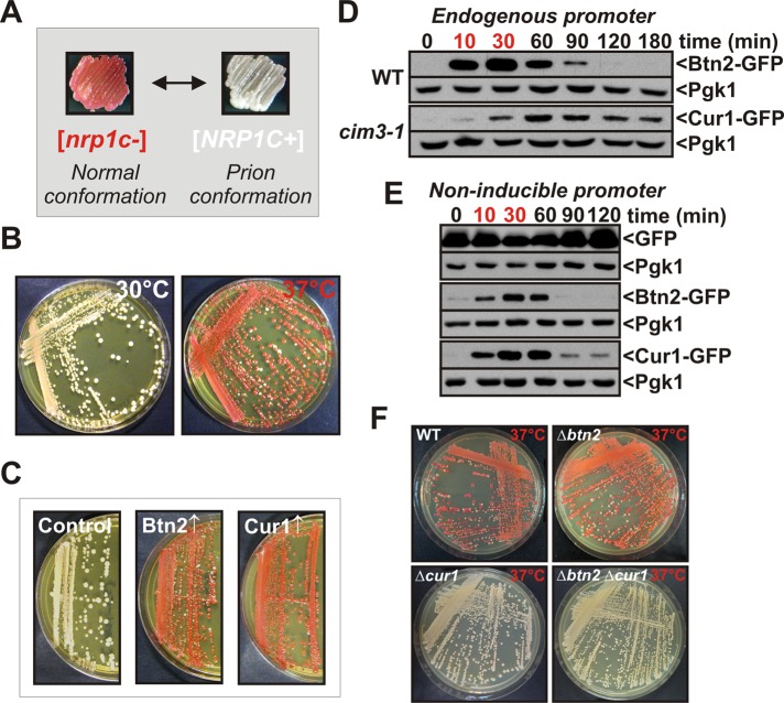
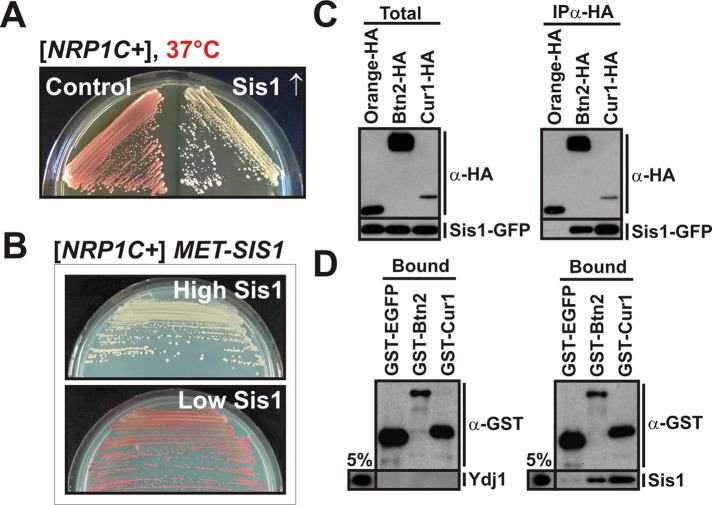
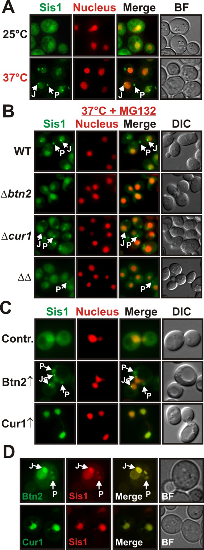
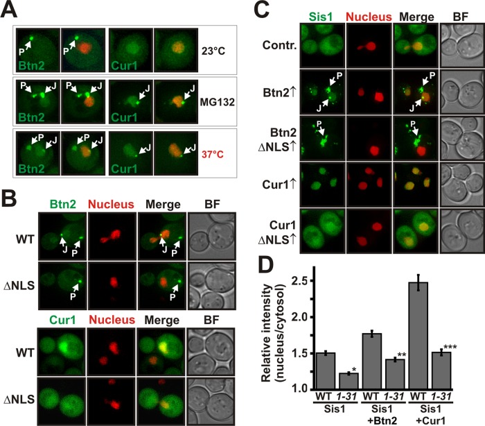
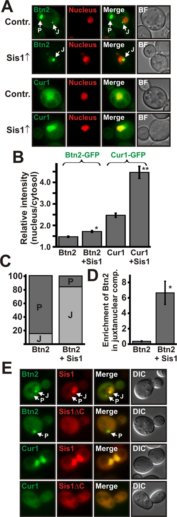
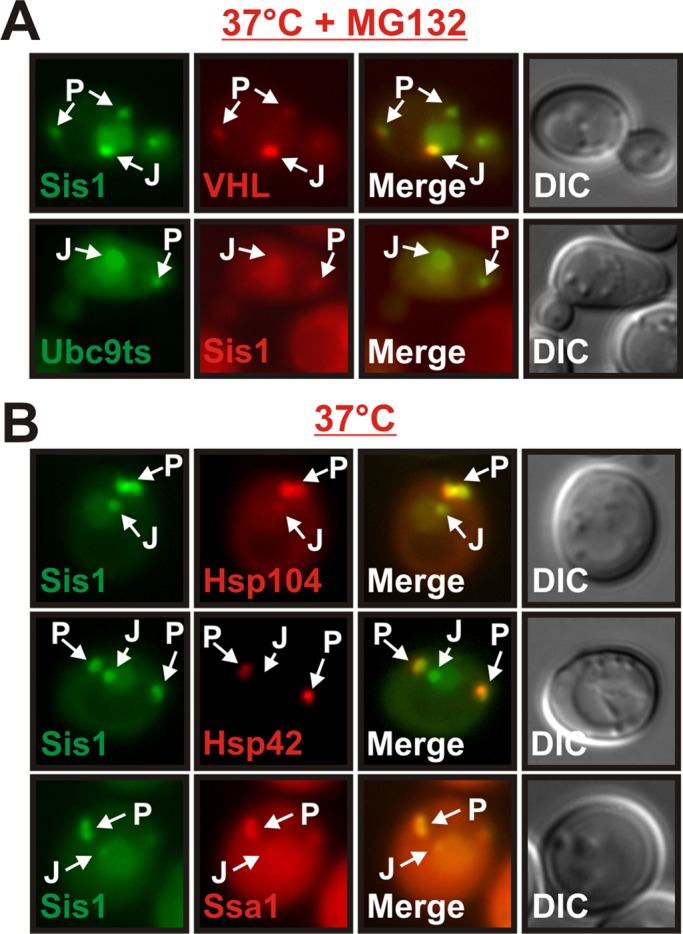
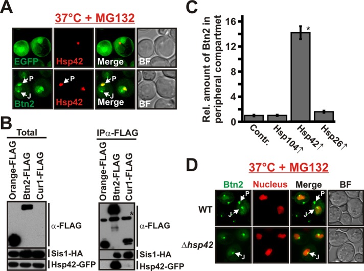
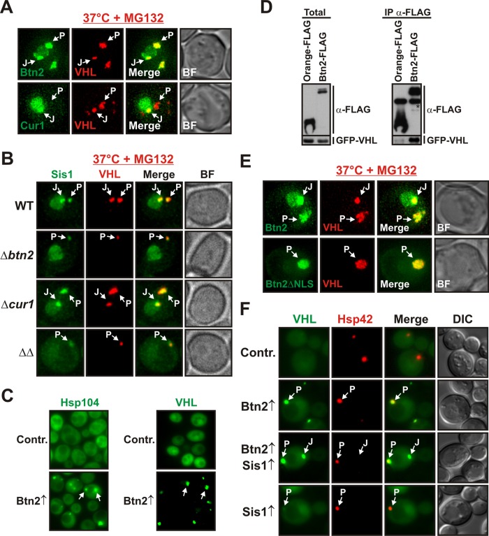
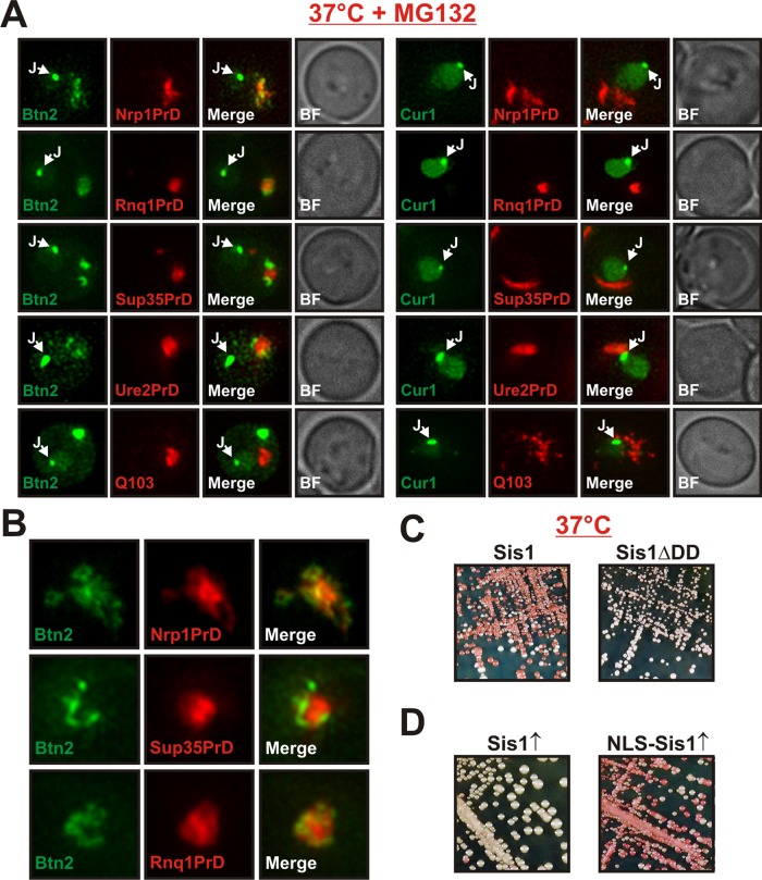
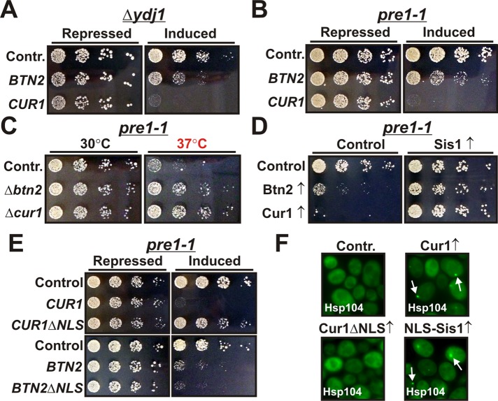
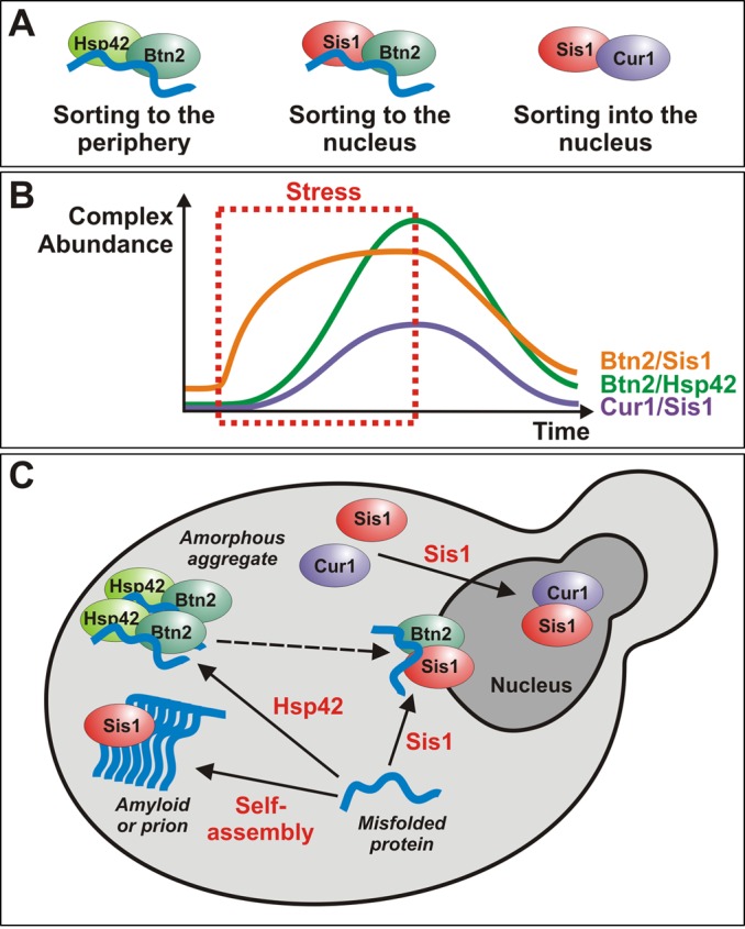
Acute stress causes a rapid redistribution of protein quality control components and aggregation-prone proteins to diverse subcellular compartments. How these remarkable changes come about is not well understood. Using a phenotypic reporter for a synthetic yeast prion, we identified two protein-sorting factors of the Hook family, termed Btn2 and Cur1, as key regulators of spatial protein quality control in Saccharomyces cerevisiae. Btn2 and Cur1 are undetectable under normal growth conditions but accumulate in stressed cells due to increased gene expression and reduced proteasomal turnover. Newly synthesized Btn2 can associate with the small heat shock protein Hsp42 to promote the sorting of misfolded proteins to a peripheral protein deposition site. Alternatively, Btn2 can bind to the chaperone Sis1 to facilitate the targeting of misfolded proteins to a juxtanuclear compartment. Protein redistribution by Btn2 is accompanied by a gradual depletion of Sis1 from the cytosol, which is mediated by the sorting factor Cur1. On the basis of these findings, we propose a dynamic model that explains the subcellular distribution of misfolded proteins as a function of the cytosolic concentrations of molecular chaperones and protein-sorting factors. Our model suggests that protein aggregation is not a haphazard process but rather an orchestrated cellular response that adjusts the flux of misfolded proteins to the capacities of the protein quality control system.


Figures

FIGURE 1:
FIGURE 1:
Stress-inducible Btn2 and Cur1 interfere with prion inheritance. (A) A prion reporter based on the translation termination activity of the C-terminal domain of Sup35 was used to monitor [NRP1C+] in yeast (see the text for details). (B) [NRP1C+] yeast cells were incubated on YPD plates at 30°C or 37°C for 3 d. (C) Galactose-regulatable expression plasmids coding for BTN2 or CUR1 were introduced into [NRP1C+] yeast. The transformants were transferred onto galactose-containing plates and incubated for 3 d at 30°C. Subsequently, the strains were transferred onto fresh YPD plates to allow for colony color development. (D) Chromosomal BTN2 was tagged with GFP in the BY4741 strain background. CUR1 was modified with GFP in a strain carrying a temperature-sensitive mutation in the proteasome subunit CIM3. The strains were grown at 25°C, shifted to 39°C for 30 min (time points in red), and then again exposed to 25°C. Cell lysates were prepared at the indicated time points and analyzed by immunoblotting with an anti-GFP antibody. The extended presence of Cur1 is probably an artifact of the reduced proteasomal activity of the cim3-1 strain. (E) Cells were treated as in D except that Btn2-GFP and Cur1-GFP were expressed from a low-copy plasmid that carried a GPD promoter. (F) [NRP1C+] yeast carrying deletions of BTN2 and/or CUR1 were incubated at 37°C for 3 d.
FIGURE 2:
FIGURE 2:
Btn2 and Cur1 functionally and physically interact with Sis1 to modify prion inheritance. (A) [NRP1C+] cells were transformed with a control plasmid or a low-copy expression plasmid coding for SIS1 and incubated at 37°C for 3 d. (B) Endogenous SIS1 was deleted in [NRP1C+] yeast, and the deletion was covered with a plasmid carrying SIS1 behind a methionine-regulatable promoter. The cells were grown in the presence (low Sis1) or absence (high Sis1) of methionine. (C) BY4741 yeast cells carrying a GFP-tagged chromosomal copy of SIS1 were transformed with low-copy expression plasmid for HA-tagged Orange (control), BTN2, or CUR1. HA-tagged proteins were immunoprecipitated from cell lysates with a HA-specific antibody. (D) Protein-binding assay with bacterially purified GST-EGFP (control), GST-Btn2, GST-Cur1, His6-Ydj1 (control), or His6-Sis1. Proteins were detected by immunoblotting with an anti-GST or anti-His antibody. The pull-down efficiency was ∼20% for GST-tagged proteins. Five percent of the input is shown for comparison.
FIGURE 3:
FIGURE 3:
Btn2 and Cur1 promote the sorting of Sis1 to the nucleus and to stress-inducible cytosolic compartments. (A) BY4741 yeast cells carrying a GFP-tagged chromosomal copy of SIS1 were grown at 25 or 37°C and subjected to fluorescence microscopy. J and P denote juxtanuclear and peripheral compartments, respectively. See Supplemental Information for details on image interpretation, as well as on control experiments. (B) Wild-type, Δbtn2, Δcur1, or Δbtn2 Δcur1 BY4741 cells carrying a GFP-tagged chromosomal copy of SIS1 were grown at 37°C in the presence of MG132 (MG132 was used because compartment formation was more pronounced) and were subjected to fluorescence microscopy. (C) Low-copy expression plasmids for BTN2 and CUR1 were introduced into a BY4741 strain expressing Sis1-GFP and an mCherry-tagged nuclear marker. Fluorescence microscopy was performed at 25°C. (D) Plasmid-expressed Btn2-GFP or Cur1-GFP was coexpressed with Sis1-mCherry in BY4741 yeast for colocalization analysis at 25°C.
FIGURE 4:
FIGURE 4:
Nuclear targeting of Sis1 is dependent on nuclear localization sequences in Btn2 and Cur1 and requires the α-importin Srp1. (A) W303 yeast expressing GFP-tagged Btn2 or Cur1 from a low-copy plasmid were grown at 23 or 37°C or in the presence of the proteasome inhibitor MG132. Left, the GFP channel. Right, an overlay with an mCherry-tagged nuclear marker. (B) W303 yeast cells expressing GFP-tagged Btn2, Cur1, Btn2ΔNLS, or Cur1ΔNLS were subjected to fluorescence microscopy at 30°C. (C) BY4741 yeast cells carrying a GFP-tagged chromosomal copy of SIS1 were transformed with low-copy plasmids for Btn2, Cur1, Btn2ΔNLS, or Cur1ΔNLS. The resulting transformants were grown at 25°C and subjected to fluorescence microscopy. (D) Wild-type yeast (WT) or yeast carrying a temperature-sensitive mutation in SRP1 (srp1-31) were cotransformed with expression plasmids for Sis1-GFP and Orange (control), Btn2, or Cur1. The cells were subjected to fluorescence microscopy after a shift to the nonpermissive temperature for 1 h. The relative nuclear:cytosolic GFP pixel intensity of the strains was determined. *p = 2.7 × 10⁻⁸; **p = 0.0027; ***p = 2.9 × 10⁻⁸.
FIGURE 5:
FIGURE 5:
Complex formation between Sis1 and Btn2 or Sis1 and Cur1 is required for targeting to the nucleus. (A) Low-copy expression plasmids for GFP-tagged Btn2 and Cur1 were introduced into a BY4741 strain that contained a control plasmid or a low-copy expression plasmid for Sis1. Fluorescence microscopy was performed at 25°C. (B) Quantification of the relative nuclear:cytosolic GFP pixel intensity of the strains shown in A. *p = 3.3 × 10⁻⁵; **p = 2.6 × 10⁻¹⁰. (C) Quantification of the fraction of cells containing Btn2-positive juxtanuclear (J) and/or peripheral (P) foci. On the basis of the distribution of Bnt2-GFP, we arbitrarily divided cells into the two categories J and P (see Materials and Methods for details). (D) Juxtanuclear and peripheral signals were quantified (total integrated pixel intensity) in 30 cells that simultaneously contained one juxtanuclear and one peripheral compartment; p = 0.000094. (E) Chromosomal SIS1 was deleted in BY4741 yeast, and the deletion was covered with expression plasmids for mCherry-tagged SIS1 or SISΔC. Expression plasmids for GFP-tagged BTN2 and CUR1 were introduced, and the cells were observed by fluorescence microscopy.
FIGURE 6:
FIGURE 6:
Sis1 localizes to stress-inducible compartments that contain misfolded proteins and molecular chaperones. (A) Fluorescence microscopy of BY4741 yeast cells expressing Sis1-GFP and mCherry-VHL or GFP-Ubc9ts and Sis1-mCherry. The cells were incubated at 37°C for 1 h in the presence of MG132. (B) BY4741 yeast cells carrying a GFP-tagged chromosomal copy of SIS1 and an mCherry-tagged chromosomal copy of HSP104, HSP42, or SSA1 were observed by fluorescence microscopy after growth at 37°C for 1 h.
FIGURE 7:
FIGURE 7:
Btn2 localization to a peripheral compartment is dependent on Hsp42. (A) BY4741 yeast carrying an mCherry-tagged chromosomal copy of HSP42 were transformed with low-copy expression plasmids for GFP (control) or GFP-tagged Btn2. (B) BY4741 yeast carrying a GFP-tagged chromosomal copy of HSP42 were transformed with low-copy expression plasmids for HA-tagged Sis1 and FLAG-tagged Orange (control), Btn2, or Cur1. FLAG-tagged proteins were immunoprecipitated with a specific antibody. The anti-FLAG immunoblot on the top right received only 1/10 of the control (Orange-FLAG) sample. Due to its low expression level, Cur1 was only detected in the total after longer exposure times (data not shown). The asterisk marks the heavy chain of the antibody that was used for immunoprecipitation. (C) The amount of Btn2-GFP in the peripheral compartment was quantified (total integrated pixel intensity) in BY4741 cells that expressed Hsp26, Hsp104, or Hsp42 from a low-copy plasmid; *p = 9.4 × 10⁻²³. (D) Wild-type or Hsp42-deficient BY4741 yeast were transformed with a low-copy expression plasmid for GFP-tagged Btn2. The cells were incubated at 37°C for 1 h in the presence of MG132.
FIGURE 8:
FIGURE 8:
Btn2 promotes the sorting of misfolded proteins to cytosolic protein deposition sites. (A) Low-copy expression plasmids for Btn2-GFP or Cur1-GFP were introduced into a W303 strain that expressed mCherry-VHL. Fluorescence microscopy was performed at 37°C in the presence of MG132. (B) Wild-type, Δbtn2, Δcur1, or Δbtn2 Δcur1 BY4741 cells carrying a GFP-tagged chromosomal copy of SIS1 were transformed with a plasmid for expression of mCherry-VHL. Cells were treated as in A. (C) Low-copy expression plasmids coding for Btn2 were introduced into a BY4741 [pin-] strain expressing Hsp104-GFP from its chromosomal locus (left) or a BY4741 strain that contained an expression plasmid for GFP-VHL (right). The cells were processed for fluorescence microscopy at 25°C. (D) BY4741 yeast cells were transformed with low-copy expression plasmids for GFP-VHL and FLAG-tagged Orange (control) or Btn2. FLAG-tagged proteins were immunoprecipitated with a specific antibody. The asterisk denotes the heavy chain of the antibody that was used for immunoprecipitation. (E) Low-copy expression plasmids for Btn2-GFP or Btn2ΔNLS-GFP were introduced into W303 cells that expressed mCherry-VHL. Fluorescence microscopy was performed at 37°C in the presence of MG132. (F) Low-copy expression plasmids for Btn2 and Cur1 were introduced into a W303 strain that expressed GFP-VHL from a plasmid and Hsp42-mCherry from the endogenous locus. Strains that overexpressed Sis1 contained an additional copy of SIS1 that was integrated into the genome. Fluorescence microscopy was performed at 25°C.
FIGURE 9:
FIGURE 9:
Btn2 and Cur1 influence prion propagation indirectly through changes in the availability of Sis1. (A) Low-copy expression plasmids for the expression of mCherry-tagged Nrp1PrD, Rnq1PrD, Sup35PrD, Ure2PrD, or glutamine-expanded huntingtin exon 1 (Q103) were introduced into BY4741 yeast cells that expressed GFP-tagged Btn2 or Cur1 from a plasmid. Fluorescence microscopy was performed at 25°C. (B) Magnification of peripheral aggregates in cells that coexpressed Btn2-GFP and mCherry-tagged Nrp1PrD, Sup35PrD, or Rnq1PrD. (C) Wild-type [NRP1C+] cells or cells in which chromosomal Sis1 was replaced with Sis1ΔDD were incubated at 37°C for 3 d. (D) [NRP1C+] cells were transformed with galactose-regulatable expression plasmids for Sis1 or NLS-Sis1. The transformants were streaked onto galactose plates, incubated for 3 d, transferred onto YPD plates for color development, and photographed.
FIGURE 10:
FIGURE 10:
Cur1 regulates the partitioning of substrate proteins between the juxtanuclear and peripheral compartments. (A) BY4741 yeast cells carrying a chromosomal deletion of YDJ1 were transformed with galactose-regulatable expression plasmids for BTN2 or CUR1. Cells were grown overnight in glucose-containing media. Fivefold serial dilutions were prepared and spotted onto either glucose-containing (repressing) or galactose-containing (inducing) plates. (B) Yeast cells carrying a temperature-dependent mutation in PRE1 (pre1-1) were transformed with galactose-regulatable expression plasmids for BTN2 and CUR1. Cells were treated as in A. (C) Deletions of BTN2 or CUR1 were introduced into the pre1-1 strain and tested for growth on rich medium at the indicated temperatures. (D) Pre1-1 yeast cells were transformed with a plasmid for constitutive Sis1 expression and galactose-regulatable expression plasmids for Btn2 and Cur1. The cells were spotted onto either glucose- or galactose-containing plates. The plates were incubated at 30°C. (E) Cells were treated as in D using plasmids for the indicated proteins. (F) BY4741 yeast expressing Hsp104-GFP from the endogenous locus were transformed with low-copy expression plasmids for Cur1, Cur1ΔNLS, or NLS-Sis1. The transformants were grown at 25°C and analyzed by fluorescence microscopy.
FIGURE 11:
FIGURE 11:
A model for the coordinated action of sorting factors and molecular chaperones during acute heat stress. (A) Schematic representation of the different sorting complexes that were identified in this work. A bound misfolded protein is depicted in blue. Note that only the complexes containing Btn2 are involved in aggregate sorting, whereas the Cur1 complex mediates sorting of Sis1 to the nucleus. (B) Schematic representation of the changes in complex abundance that happen before during and after a stress stimulus. (C) A model that summarizes the role of Btn2, Cur1, Sis1, and Hsp42 in the sorting of misfolded proteins to deposition sites. The events that induce sorting (association with a factor or assembly into an amyloid structure) are indicated in red. Arrows indicate the direction of sorting. The dashed arrow indicates that it is unclear whether misfolded proteins can directly be sorted from the peripheral to the juxtanuclear compartment.

Similar articles

Cited by

References

    1. Alberti S, Gitler AD, Lindquist S. A suite of Gateway cloning vectors for high-throughput genetic analysis in Saccharomyces cerevisiae. Yeast. 2007;24:913–919. - PMC - PubMed
    1. Alberti S, Halfmann R, King O, Kapila A, Lindquist S. A systematic survey identifies prions and illuminates sequence features of prionogenic proteins. Cell. 2009;137:146–158. - PMC - PubMed
    1. Alberti S, Halfmann R, Lindquist S. Biochemical, cell biological, and genetic assays to analyze amyloid and prion aggregation in yeast. Methods Enzymol. 2010;470:709–734. - PubMed
    1. Allen KD, Wegrzyn RD, Chernova TA, Muller S, Newnam GP, Winslett PA, Wittich KB, Wilkinson KD, Chernoff YO. Hsp70 chaperones as modulators of prion life cycle: novel effects of Ssa and Ssb on the Saccharomyces cerevisiae prion [PSI+] Genetics. 2005;169:1227–1242. - PMC - PubMed
    1. Aron R, Higurashi T, Sahi C, Craig EA. J-protein co-chaperone Sis1 required for generation of [RNQ+] seeds necessary for prion propagation. EMBO J. 2007;26:3794–3803. - PMC - PubMed

Publication types

MeSH terms