

Trafficking defects in WASH-knockout fibroblasts originate from collapsed endosomal and lysosomal networks
- PMID: 22718907
- PMCID: PMC3418315
- DOI: 10.1091/mbc.E12-02-0101
Trafficking defects in WASH-knockout fibroblasts originate from collapsed endosomal and lysosomal networks
Abstract
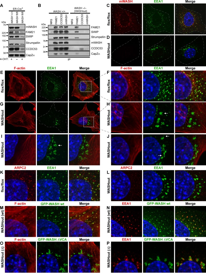
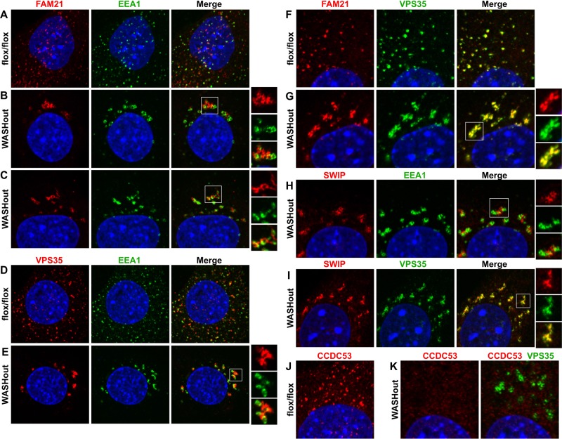
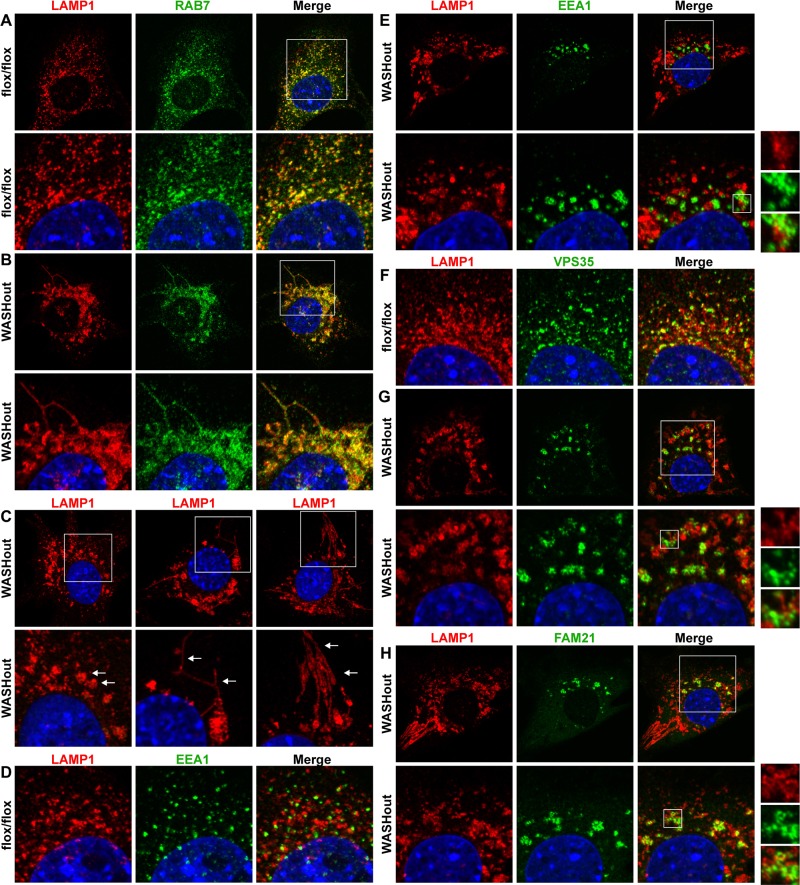
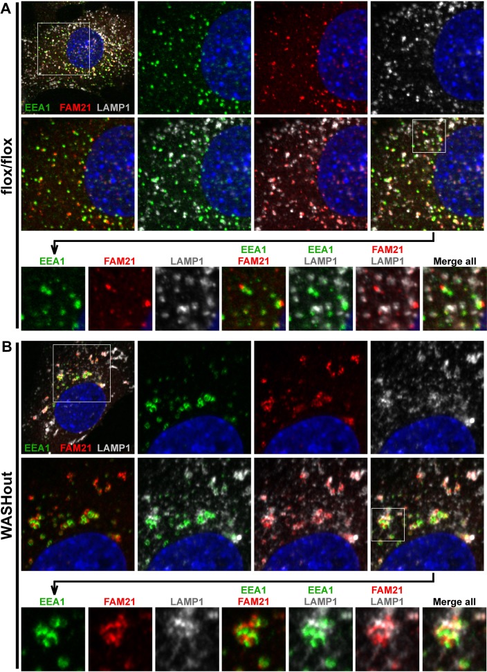
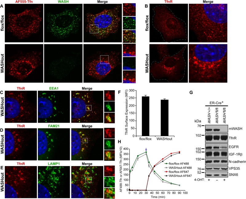
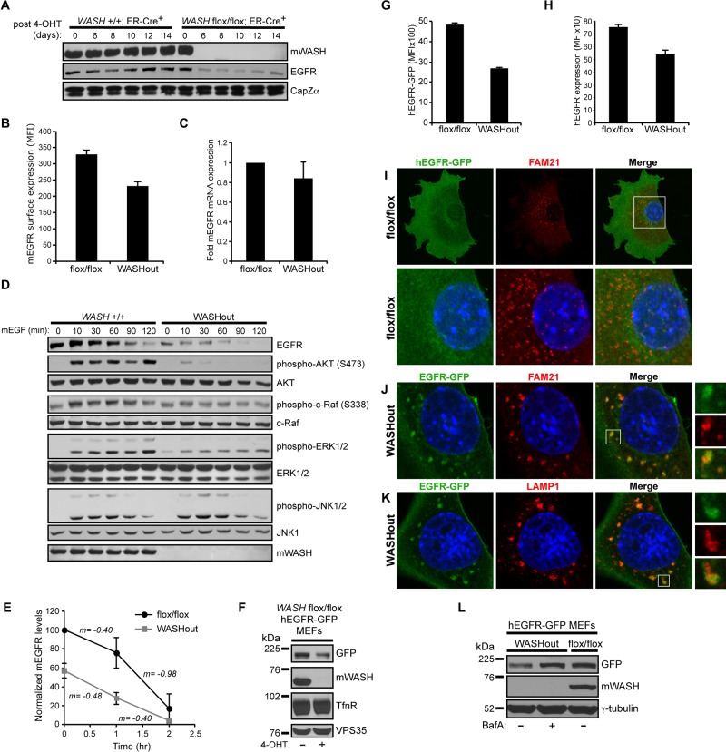
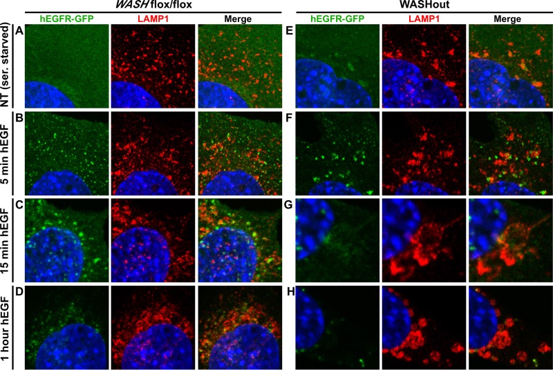
The Arp2/3-activator Wiskott-Aldrich syndrome protein and Scar homologue (WASH) is suggested to regulate actin-dependent membrane scission during endosomal sorting, but its cellular roles have not been fully elucidated. To investigate WASH function, we generated tamoxifen-inducible WASH-knockout mouse embryonic fibroblasts (WASHout MEFs). Of interest, although EEA1(+) endosomes were enlarged, collapsed, and devoid of filamentous-actin and Arp2/3 in WASHout MEFs, we did not observe elongated membrane tubules emanating from these disorganized endomembranes. However, collapsed WASHout endosomes harbored segregated subdomains, containing either retromer cargo recognition complex-associated proteins or EEA1. In addition, we observed global collapse of LAMP1(+) lysosomes, with some lysosomal membrane domains associated with endosomes. Both epidermal growth factor receptor (EGFR) and transferrin receptor (TfnR) exhibited changes in steady-state cellular localization. EGFR was directed to the lysosomal compartment and exhibited reduced basal levels in WASHout MEFs. However, although TfnR was accumulated with collapsed endosomes, it recycled normally. Moreover, EGF stimulation led to efficient EGFR degradation within enlarged lysosomal structures. These results are consistent with the idea that discrete receptors differentially traffic via WASH-dependent and WASH-independent mechanisms and demonstrate that WASH-mediated F-actin is requisite for the integrity of both endosomal and lysosomal networks in mammalian cells.
Figures
Similar articles
-
WASH is required for lysosomal recycling and efficient autophagic and phagocytic digestion.Mol Biol Cell. 2013 Sep;24(17):2714-26. doi: 10.1091/mbc.E13-02-0092. Epub 2013 Jul 24. Mol Biol Cell. 2013. PMID: 23885127 Free PMC article.
-
Endosomal recruitment of the WASH complex: active sequences and mutations impairing interaction with the retromer.Biol Cell. 2013 May;105(5):191-207. doi: 10.1111/boc.201200038. Epub 2013 Mar 7. Biol Cell. 2013. PMID: 23331060
-
Actin polymerization in the endosomal pathway, but not on the Coxiella-containing vacuole, is essential for pathogen growth.PLoS Pathog. 2018 Apr 18;14(4):e1007005. doi: 10.1371/journal.ppat.1007005. eCollection 2018 Apr. PLoS Pathog. 2018. PMID: 29668757 Free PMC article.
-
Actin-dependent endosomal receptor recycling.Curr Opin Cell Biol. 2019 Feb;56:22-33. doi: 10.1016/j.ceb.2018.08.006. Epub 2018 Sep 15. Curr Opin Cell Biol. 2019. PMID: 30227382 Review.
-
Retromer-mediated endosomal protein sorting: all WASHed up!Trends Cell Biol. 2013 Nov;23(11):522-8. doi: 10.1016/j.tcb.2013.04.010. Epub 2013 May 28. Trends Cell Biol. 2013. PMID: 23721880 Free PMC article. Review.
Cited by
-
WAVE/SCAR promotes endocytosis and early endosome morphology in polarized C. elegans epithelia.Dev Biol. 2013 May 15;377(2):319-32. doi: 10.1016/j.ydbio.2013.03.012. Epub 2013 Mar 17. Dev Biol. 2013. PMID: 23510716 Free PMC article.
-
Genetic disruption of WASHC4 drives endo-lysosomal dysfunction and cognitive-movement impairments in mice and humans.Elife. 2021 Mar 22;10:e61590. doi: 10.7554/eLife.61590. Elife. 2021. PMID: 33749590 Free PMC article.
-
Loss of ARPC1B impairs cytotoxic T lymphocyte maintenance and cytolytic activity.J Clin Invest. 2019 Dec 2;129(12):5600-5614. doi: 10.1172/JCI129388. J Clin Invest. 2019. PMID: 31710310 Free PMC article.
-
Toll-Dorsal signaling regulates the spatiotemporal dynamics of yolk granule tubulation during Drosophila cleavage.Dev Biol. 2022 Jan;481:64-74. doi: 10.1016/j.ydbio.2021.09.009. Epub 2021 Oct 7. Dev Biol. 2022. PMID: 34627795 Free PMC article.
-
The Proteome of BLOC-1 Genetic Defects Identifies the Arp2/3 Actin Polymerization Complex to Function Downstream of the Schizophrenia Susceptibility Factor Dysbindin at the Synapse.J Neurosci. 2016 Dec 7;36(49):12393-12411. doi: 10.1523/JNEUROSCI.1321-16.2016. J Neurosci. 2016. PMID: 27927957 Free PMC article.
References
Publication types
MeSH terms
Substances
Grants and funding
LinkOut - more resources
Full Text Sources
Molecular Biology Databases
Research Materials
Miscellaneous
