. 2012 Aug;23(16):3122-32.
doi: 10.1091/mbc.E12-03-0235. Epub 2012 Jun 20.

Control of the mitotic exit network during meiosis

Affiliations

Control of the mitotic exit network during meiosis

Michelle A Attner et al. Mol Biol Cell. 2012 Aug.

Abstract

The mitotic exit network (MEN) is an essential GTPase signaling pathway that triggers exit from mitosis in budding yeast. We show here that during meiosis, the MEN is dispensable for exit from meiosis I but contributes to the timely exit from meiosis II. Consistent with a role for the MEN during meiosis II, we find that the signaling pathway is active only during meiosis II. Our analysis further shows that MEN signaling is modulated during meiosis in several key ways. Whereas binding of MEN components to spindle pole bodies (SPBs) is necessary for MEN signaling during mitosis, during meiosis MEN signaling occurs off SPBs and does not require the SPB recruitment factor Nud1. Furthermore, unlike during mitosis, MEN signaling is controlled through the regulated interaction between the MEN kinase Dbf20 and its activating subunit Mob1. Our data lead to the conclusion that a pathway essential for vegetative growth is largely dispensable for the specialized meiotic divisions and provide insights into how cell cycle regulatory pathways are modulated to accommodate different modes of cell division.


Figures
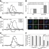
FIGURE 1:
FIGURE 1:
The MEN is dispensable for exit from meiosis I but is required for the timely exit from anaphase II. (A–D) Wild-type (A, A14201) or cdc15-as1 (B, A19440) cells harboring a GAL-NDT80 fusion as the sole source of NDT80 were induced to sporulate. β-Estradiol, 1 μM, and 1-NA-PP1, 10 μM, were added to cultures 6 h after transfer into sporulation medium. The percentage of cells with metaphase I (closed squares), anaphase I (open squares), metaphase II (closed circles), anaphase II (open circles) was determined at the indicated times. Two hundred cells were analyzed at each time point. (C) Comparison of the percentage of anaphase II cells in wild type (closed squares) and cdc15-as1 mutants (closed circles). (D) Quantification of the accumulation of anaphase I and anaphase II spindles in wild type and cdc15-as1 mutants (n = 6 experiments). The area under the lines for anaphase I and anaphase II spindles was determined and expressed as a fold change of wild type (anaphase I: mean, 1.083, SD = 0.122, p = 0.598 [two-tailed, paired t test]; anaphase II: mean, 1.757, SD = 0.362, p = 0.005 [two-tailed, paired t test]). (E, F) Examples of anaphase II cells with Cdc14 sequestered in the nucleolus (top; E) or released into the nucleus and cytoplasm (bottom; E). Cdc14-HA is shown in red, DNA in blue, and microtubules in green. (F) Quantification of Cdc14 release in anaphase II cells. WT (A22130; white bars) or cdc15-as1 (A22129; gray bars) cells carrying CDC14-HA fusions were induced to sporulate as in A. At the time points indicated, the percentage of anaphase I or anaphase II cells with Cdc14 released was determined (n = 100 for each time point except for wild type 8.25 h, n = 35, and wild type 9 h, n = 36).
FIGURE 2:
FIGURE 2:
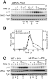
Dbf20 kinase activity peaks in anaphase II and depends on Cdc15 kinase activity. (A) Cells containing a DBF20-ProA fusion (A23162) were sporulated as described in Figure 1A, except that 1-NA-PP1 was not added. Samples were taken at the indicated times to determine Dbf20-associated kinase activity and Dbf20-ProA protein levels. Dbf20-associated kinase activity was assessed by phosphorylation of the substrate histone H1 (p-H1). Pgk1 was used as a loading control. The peak of each stage of meiosis is indicated below the blot (MI, metaphase I; AI, anaphase I; MII, metaphase II; AII, anaphase II). (B) Quantification of Dbf20-associated kinase activity (closed circles) and anaphase II spindles (closed squares; n = 200 cells counted). (C) Wild-type (A23162) or cdc15-as1 (A23733) cells containing a DBF20-ProA fusion were induced to sporulate as described in Figure 1A. Dbf20-associated kinase activity and Dbf20-ProA protein levels were determined at the indicated times.
FIGURE 3:
FIGURE 3:
MEN signaling occurs in a NUD1-independent manner during meiosis. (A–C) The temperature 34°C is restrictive for nud1-44. Wild-type (A8499) and nud1-44 (A28757) cells containing a 3MYC-DBF2 fusion were arrested in G1 with α-factor pheromone (5 μg/ml) in YEPD medium for 2 h at room temperature and then shifted to 34°C for 30 min. Cells were released into pheromone-free YEPD medium at 34°C thereafter. Dbf2 protein and kinase activity (A) and the percentage of cells in metaphase and anaphase (B, C) were analyzed at the indicated times (n = 100 cells per time point). (D–F) Wild-type (A23162) or nud1-44 (A27697) cells containing a DBF20-ProA fusion were induced to sporulate via the Ndt80-block release protocol. Cells were incubated in sporulation medium at room temperature and shifted to 34°C after 5 h. At 6 h, cultures were induced with 1 μM β-estradiol. Cells were maintained at 34°C throughout the rest of the experiment. Samples were taken at the indicated times to determine Dbf20 protein levels and Dbf20 kinase activity (D) and meiotic progression (E, F; n = 100 cells per time point).
FIGURE 4:
FIGURE 4:
Differential regulation of Dbf2 and Dbf20 activity. (A–D) pCUP1-DBF2-ProA (A25020) or pCUP1-DBF20-ProA (A25193) cells were arrested in G1 with α-factor pheromone (5 μg/ml) in YEPD medium. CuSO4, 50 μM, was added to the medium 2 h into the arrest. When the arrest was complete (2 h, 30 min), cells were released into pheromone-free YEPD medium containing 50 μM CuSO4. Dbf2 and Dbf20 kinase activity, Dbf2 and Dbf20 protein (A, B), and mitotic progression (C, D; n = 100 cells per time point) were examined at the indicated times. Quantifications of Dbf2 and Dbf20 kinase activity are shown by the graph in B. (E–H) pCUP1-DBF2-ProA (A25028) or pCUP1-DBF20-ProA (A25195) cells were sporulated via the Ndt80 block-release protocol. β-Estradiol, 1 μM, and CuSO4, 50 μM, were added to the medium 6 h after transfer into sporulation medium. Dbf2 and Dbf20 kinase activity, Dbf2 and Dbf20 protein (E, F), and meiotic progression (G, H; n = 100 cells per time point) were examined at the indicated times. Quantifications of Dbf2 and Dbf20 kinase activity are shown by the graph in F.
FIGURE 5:
FIGURE 5:
The Dbf20–Mob1 interaction is under cell cycle control in meiosis but not mitosis. (A, B) Cells containing pCUP1-DBF2-ProA and a MOB1-V5 fusion (A27687) or MOB1 (A25020, no tag control) were arrested in G1 with α-factor pheromone (5 μg/ml) in YEPD medium. CuSO4, 50 μM, was added to the medium 2 h into the arrest. When the arrest was complete (2 h, 30 min), cells were released into pheromone-free YEPD medium containing 50 μM CuSO4. Western blots in A show total Dbf2-ProA protein (input), immunoprecipitated Mob1-V5, and coimmunoprecipitated Dbf2-ProA at the indicated time points. (B) Quantification of coimmunoprecipitation expressed as the amount of Dbf2 coimmunoprecipitated over the amount of Mob1-V5 immunoprecipitated. Values were normalized so that the maximum value was set to 1.0 (closed triangles). For comparison, the percentage of cells in metaphase (open squares) and anaphase (open circles) is shown. (C, D) Cells containing pCUP1-DBF20-ProA and a MOB1-V5 fusion (A27367) or MOB1 (A25191, no tag control) were grown and analyzed as described in A and B. (E, F) Cells containing pCUP1-DBF20-ProA and a MOB1-V5 fusion (A27370) or MOB1 (A25195, no tag control) were sporulated via the Ndt80 block-release protocol. Cells were induced with 1 μM β-estradiol and 50 μM CuSO4 at 6 h after transfer to sporulation medium. Western blots show immunoprecipitated Mob1-V5, coimmunoprecipitated Dbf20-ProA, and total Dbf20 protein (input) at the indicated time points (E). Quantification of the amount of Dbf20 coimmunoprecipitated with Mob1-V5 is shown as a ratio of the two values. Values were normalized so that the maximum value was set to 1.0. For comparison, meiotic progression was also analyzed.
FIGURE 6:
FIGURE 6:
The Dbf20-Mob1 interaction depends on CDC15 in meiosis. (A–C) Cells containing pCUP1-DBF20-ProA, a MOB1-V5 fusion, and CDC15 (A27371) or cdc15-as1 (A29149) were induced to sporulate via the Ndt80 block-release protocol as in Figure 1A with the addition of 10 μM 1-NA-PP1 and 50 μM CuSO4 at 6 h after transfer to sporulation medium. Western blots in A show total Dbf20-ProA protein (input), immunoprecipitated Mob1-V5, and coimmuno­precipitated Dbf20-ProA at the indicated time points. The genotype of no tag A is pCUP1-DBF20-ProA (A25195), and the genotype of no tag B is pCUP1-DBF20-ProA cdc15-as1 (A29150). (B, C) Meiotic progression in the two strains. (D–F) Cells containing pCUP1-DBF20-ProA, a MOB1-V5 fusion, and CDC15 (A27367) or cdc15-as1 (A29125) were arrested in G1 with α-factor pheromone (5 μg/ml) in YEPD medium as in Figure 4A. CuSO4, 50 μM, was added to the medium 2 h into the arrest. When the arrest was complete (2 h, 30 min), cells were released into YEPD medium containing 10 μM 1-NA-PP1 and 50 μM CuSO4. (D) Total Dbf20-ProA protein (input), coimmunoprecipitated Dbf20-ProA, and immunoprecipitated Mob1-V5 for the indicated time points. (E, F) Mitotic progression of the two strains. The genotype of no tag A is pCUP1-DBF20-ProA (A25193), and the genotype of no tag B is pCUP1-DBF20-ProA cdc15-as1 (A29124).

Similar articles

Cited by

References

    1. Adams IR, Kilmartin JV. Localization of core spindle pole body (SPB) components during SPB duplication in Saccharomyces cerevisiae. J Cell Biol. 1999;145:809–823. - PMC - PubMed
    1. Asakawa K, Yoshida S, Otake F, Toh-e A. A novel functional domain of Cdc15 kinase is required for its interaction with Tem1 GTPase in Saccharomyces cerevisiae. Genetics. 2001;157:1437–1450. - PMC - PubMed
    1. Bardin AJ, Visintin R, Amon A. A mechanism for coupling exit from mitosis to partitioning of the nucleus. Cell. 2000;102:21–31. - PubMed
    1. Bertazzi DT, Kurtulmus B, Pereira G. The cortical protein Lte1 promotes mitotic exit by inhibiting the spindle position checkpoint kinase Kin4. J Cell Biol. 2011;193:1033–1048. - PMC - PubMed
    1. Bishop AC, et al. A chemical switch for inhibitor-sensitive alleles of any protein kinase. Nature. 2000;407:395–401. - PubMed

Publication types

MeSH terms