. 2012 Sep;40(16):8144-54.
doi: 10.1093/nar/gks548. Epub 2012 Jun 19.

How an exonuclease decides where to stop in trimming of nucleic acids: crystal structures of RNase T-product complexes

Affiliations

How an exonuclease decides where to stop in trimming of nucleic acids: crystal structures of RNase T-product complexes

Yu-Yuan Hsiao et al. Nucleic Acids Res. 2012 Sep.

Abstract

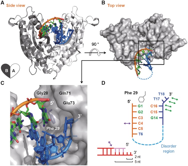
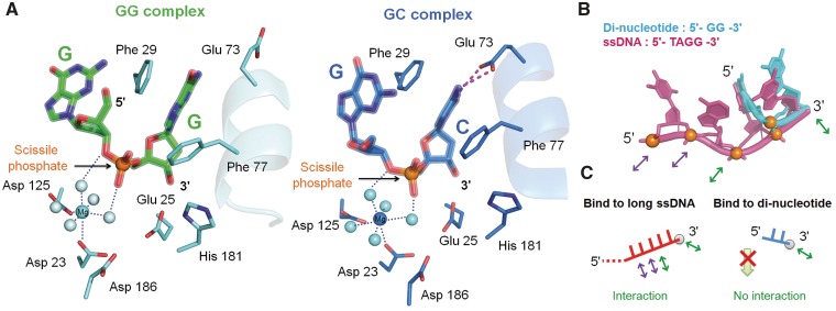
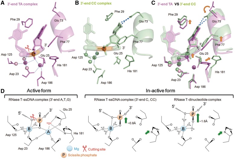
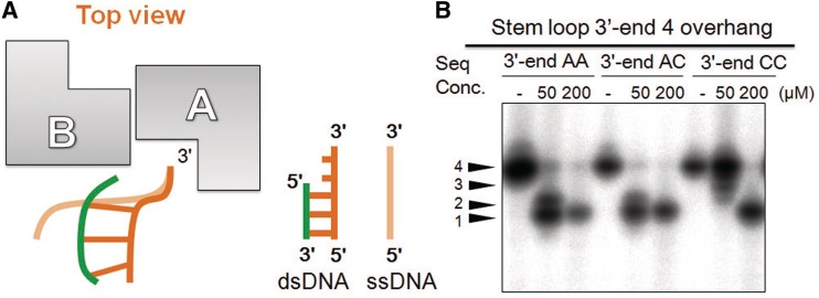
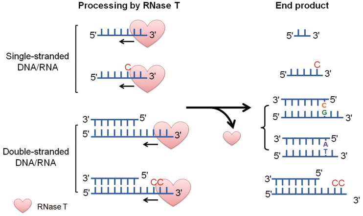
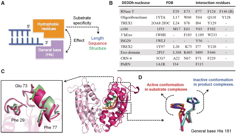
Exonucleases are key enzymes in the maintenance of genome stability, processing of immature RNA precursors and degradation of unnecessary nucleic acids. However, it remains unclear how exonucleases digest nucleic acids to generate correct end products for next-step processing. Here we show how the exonuclease RNase T stops its trimming precisely. The crystal structures of RNase T in complex with a stem-loop DNA, a GG dinucleotide and single-stranded DNA with different 3'-end sequences demonstrate why a duplex with a short 3'-overhang, a dinucleotide and a ssDNA with a 3'-end C cannot be further digested by RNase T. Several hydrophobic residues in RNase T change their conformation upon substrate binding and induce an active or inactive conformation in the active site that construct a precise machine to determine which substrate should be digested based on its sequence, length and structure. These studies thus provide mechanistic insights into how RNase T prevents over digestion of its various substrates, and the results can be extrapolated to the thousands of members of the DEDDh family of exonucleases.


Figures

Figure 1.
Figure 1.
The crystal structure of RNase T bound to a stem-loop DNA product with a 2-nt 3′-overhang. (A) The ribbon model of RNase T bound with a stem-loop DNA displayed in a side view. The schematic heart-shaped diagram at the bottom left represents that RNase T is a dimeric protein with two protomers A and B. Nucleotides 1–6 near the 5′-end are shown in orange and nucleotides 14–18 near the 3′-end are shown in blue. (B) Molecular surface of RNase T bound to the stem-loop DNA displayed in a top view. Disordered region of the stem-loop DNA is shown by a blue dotted line. (C) Phe29 stacking with the 5′-end nucleotide G1 and the ‘wall’ made of Gly28, Gln71 and Glu73 stop further digestion at the 3′-end of the duplex by RNase T. (D) Schematic diagram of the interactions between the stem-loop DNA and RNase T. The interactions between RNase T and DNA are displayed by green arrows (protomer A) and purple arrows (protomer B). The schematic diagram in the bottom shows the rationale for the preference of RNase T in digesting a duplex substrate with a 3′-overhang of 2 to 5 nt.
Figure 2.
Figure 2.
The active site of RNase T-dinucleotide complexes in an inactive conformation. (A) Inactive conformation of the active site of RNase T bound to the dinucleotide GG and GC. The two active sites share a similar conformation with the only difference in Glu73, which rotates its side chain to hydrogen bonds (red dotted lines) with the 3′-C in the GC complex. Water molecules are displayed as light blue balls. (B) The superposition of the dinucleotide GG onto a longer ssDNA substrate (7 nt) bound at RNase T shows that the dinucleotide shifts its position. The GG dinucleotide (PDB entry: 3VA0) is colored in blue, and the long single-stranded DNA (PDB entry: 3NH1) is colored in red with phosphates colored in orange. The interactions between DNA and RNase T are marked by green (protomer A) and purple (protomer B) arrows. (C) The schematic diagram of the interactions between RNase T and a single-stranded DNA, 7 nt versus 2 nt. The dinucleotide interacts with RNase T less extensively, and therefore it shifts its position and cannot be further cleaved into mononucleotides. See Supplementary Figure S2 for a detailed comparison and interactions.
Figure 3.
Figure 3.
Crystal structures of RNase T bound to single-stranded substrates (3′-end AA, TA or AT) and products (3′-end AC and CC). (A) Active site of RNase T bound with a single-stranded DNA with a 3′-end TA reveals an active conformation with two Mg2+ ions and a nearby general base His181. The ssDNA with a 3′-end AT and GG bind to RNase T in a similar way as shown in Supplementary Figure S3. The red scissor marks the cutting site. (B) Active site of RNase T bound with a single-stranded DNA product with a 3′-end CC reveals an inactive conformation with only one Mg2+ ion and a shift-away His181. Hydrogen bonds between Gln73 and the 3′-end C are indicated by blue dotted lines. (C) Superposition of the active site of RNase T bound with a single-stranded substrate (3′-end TA) and product (3′-end CC) shows the shifts in the product complex as marked by the orange arrows. (D) Schematic diagrams of the three types of active sites in RNase T: substrate-bound active form and product-bound inactive forms (3′-end C and dinucleotide). The nucleophilic water molecule (colored in red) between metal B and the general base His181 was not present in the structures. Green arrows show the shifts induced upon product binding.
Figure 4.
Figure 4.
The different binding and cleavage modes of RNase T for single- and double-stranded substrates. (A) In binding a single-stranded substrate, both protomers A and B of RNase T interact with the same strand of the substrate, whereas in binding a double-stranded substrate, protomer A and B interact with different strands of the substrate. (B) Stem-loop DNA with a 4-nt 3′-overhang of different sequences were digested by RNase T. The stem-loop DNA with a 3′-end CC was most resistant to digestion as compared to the ones with a 3′-end AA or AC. The length of the 3′-overhang of the stem-loop DNA is indicated by arrows to the left of the figure.
Figure 5.
Figure 5.
General rules for RNase T in trimming of nucleic acids. RNase T digests single-stranded nucleic acids to generate either a dinucleotide (without C in the middle of the chain) or an end product with a 3′-end C (with a C in the middle of the chain). Double-stranded nucleic acids with a 3′-overhang of 2–5 nt are processed by RNase T to generate an end product with a 3′-overhang of 1–4 nt, depending on the sequence of the substrate in double-stranded and overhang regions. Without a CC sequence in the 3′-overhang region, a 1-nt overhang is generated if the last base pair is AT, or a 2-nt overhang is generated if the last base pair is GC, whereas with a CC sequence in the 3′-overhang region, the trimming stops at CC.
Figure 6.
Figure 6.
Hydrophobic residues play key roles in regulating the substrate specificity and enzyme activity of the DEDDh family of exonucleases. (A) A number of hydrophobic residues of RNase T interact with nucleic acid substrates and regulate the activity of RNase T in digestion of nucleic acids of different lengths, sequences and structures. (B) A table lists the putative substrate-binding residues in the DEDDh exonucleases that correlate with Phe29, Glu73, Phe77, Phe124 and Phe146 in RNase T by structural alignment. (C) Crystal structures of RNase T–DNA complexes show that the hydrophobic residues, including Phe29 and Phe77, located in a flexible helix, shift their positions upon binding different types of substrates/products. Red and green molecules are the apo- and substrate-bound form of RNase T, respectively (ssDNA with a 3′-end C). (D) The general base H181 is located in a flexible loop region with two conformations: active conformation in apo-form (orange) and all the substrate complexes (red: 3′-end TA), and inactive conformation in all the product complexes (light green: 3′-end CC; light blue: dinucleotide; gray: duplex DNA with 2 nt 3′-overhang).

Similar articles

Cited by

References

    1. Shevelev IV, Hubscher U. The 3′-5′ exonucleases. Nat. Rev. Mol. Cell. Biol. 2002;3:364–376. - PubMed
    1. Zuo Y, Deutscher MP. Exoribonuclease superfamilies: structural analysis and phylogenetic distribution. Nucleic Acids Res. 2001;29:1017–1026. - PMC - PubMed
    1. Uhrhammer NA, Lafarge L, Dos Santos L, Domaszewska A, Lange M, Yang Y, Aractingi S, Bessis D, Bignon YJ. Werner syndrome and mutations of the WRN and LMNA genes in France. Hum. Mutat. 2006;27:718–719. - PubMed
    1. Kavanagh D, Spitzer D, Kothari PH, Shaikh A, Liszewski MK, Richards A, Atkinson JP. New roles for the major human 3′-5′ exonuclease TREX1 in human disease. Cell Cycle. 2008;7:1718–1725. - PMC - PubMed
    1. Crow YJ, Hayward BE, Parmar R, Robins P, Leitch A, Ali M, Black DN, van Bokhoven H, Brunner HG, Hamel BC, et al. Mutations in the gene encoding the 3′-5′ DNA exonuclease TREX1 cause Aicardi-Goutieres syndrome at the AGS1 locus. Nat. Genet. 2006;38:917–920. - PubMed

Publication types