

MRI and ultrasonography in Morton's neuroma: Diagnostic accuracy and correlation
- PMID: 22719120
- PMCID: PMC3377144
- DOI: 10.4103/0019-5413.96390
MRI and ultrasonography in Morton's neuroma: Diagnostic accuracy and correlation
Abstract
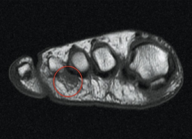
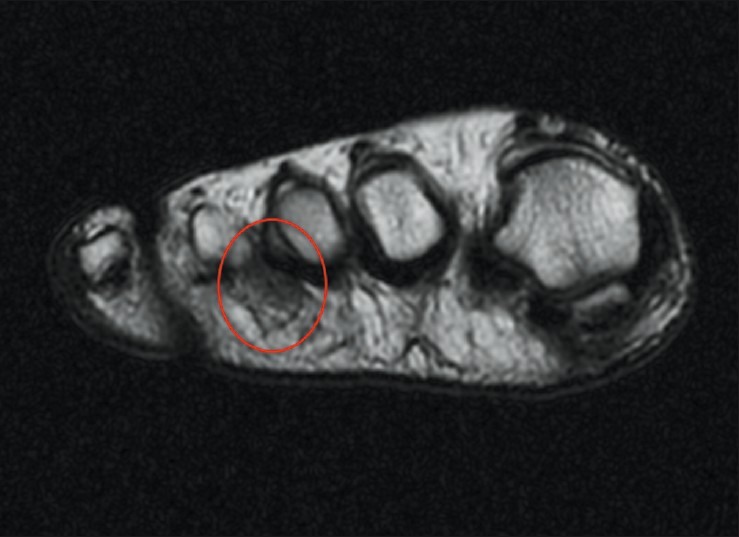
Background: The diagnosis of Morton's neuroma is based primarily on clinical findings. Ultrasonography (US) and magnetic resonance image (MRI) studies are considered complementary diagnostic techniques. The aim of this study was to establish the correlation and sensitivity of both techniques used to diagnose Morton's neuroma.
Materials and methods: Thirty seven patients (43 intermetatarsal spaces) with Morton's neuroma operated were retrospectively reviewed. In all cases MRI or ultrasound was performed to complement clinical diagnosis of Morton's neuroma. In all cases, a histopathological examination confirmed the diagnosis. Estimates of sensitivity were made and correlation (kappa statistics) was assessed for both techniques.
Results: Twenty seven women and 10 men participated with a mean age of 60 years. Double lesions presented in six patients. The second intermetatarsal space was affected in 10 patients and the third in 33 patients. An MRI was performed in 41 cases and a US in 23 cases. In 21 patients, both an MRI and a US were performed. With regard to the 41 MRIs performed, 34 were positive for Morton's neuroma and 7 were negative. MRI sensitivity was 82.9% [95% confidence interval (CI): 0.679-0.929]. Thirteen out of 23 US performed were positive and 10 US were negative. US sensitivity was 56.5% (95% CI: 0.345-0.768). Relative to the 21 patients on whom both techniques were carried out, the agreement between both techniques was poor (kappa statistics 0.31).
Conclusion: Although ancillary studies may be required to confirm the clinical diagnosis in some cases, they are probably not necessary for the diagnosis of Morton's neuroma. MRI had a higher sensitivity than US and should be considered the technique of choice in those cases. However, a negative result does not exclude the diagnosis (false negative 17%).
Keywords: Correlation; MRI; Morton; neuroma; ultrasonography.
Conflict of interest statement
Figures
References
-
- Civinni F. Su di un gangliare rigonfiamento della piñata del piede. Mem Chir Archiespedale Pistoia. 1835:4–17.
-
- Durlacher L. A treeatise on corns, bunions, the disease of nails, and the generalmanagement of the feet. London: Simpkin, Marshall; 1845. p. 52.
-
- Morton TG. A peculiar and painful affection of the fourth metarsophalangeal articulation. Am J Med Sci. 1876;71:37–9.
-
- Levine SE, Myerson MS, Shapiro PP, Shapiro SL. Ultrasonographic diagnosis ofrecurrence after excision of an interdigital neuorma. Foot Ankle Int. 1998;19:79–84. - PubMed
-
- Klenerman L, MacClellan GE, Gulloff RJ, Scadding JW. Mortons's metatarsalgia: A retrospective and prospective study. In: Proceedings of the British OrthopaedicAssociation. J Bone Joint Surg Br. 1983;28:78–82.
LinkOut - more resources
Full Text Sources