Review
. 2012:2012:764017.
doi: 10.1155/2012/764017. Epub 2012 May 8.

The use of functional MRI to study appetite control in the CNS

Affiliations
Review

The use of functional MRI to study appetite control in the CNS

Akila De Silva et al. Exp Diabetes Res. 2012.

Abstract

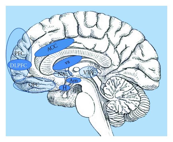
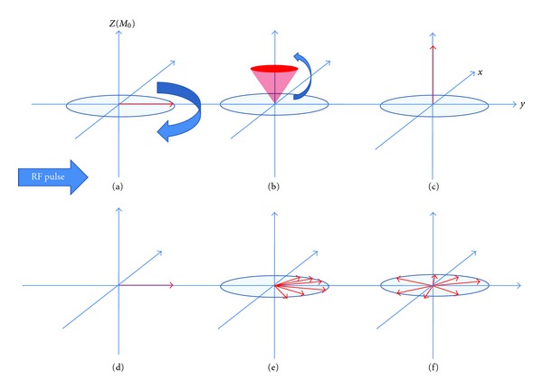
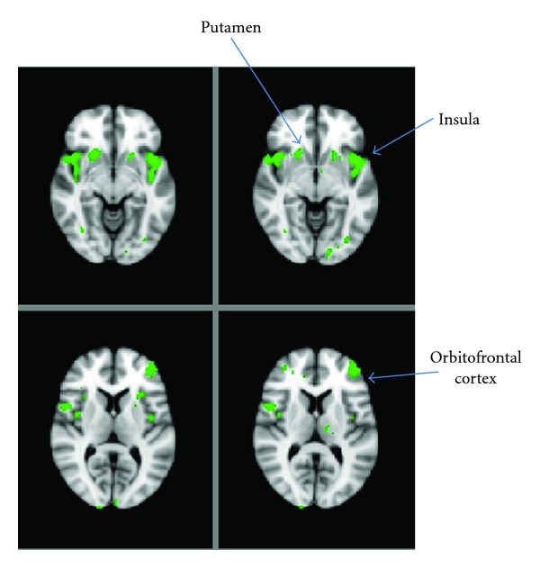
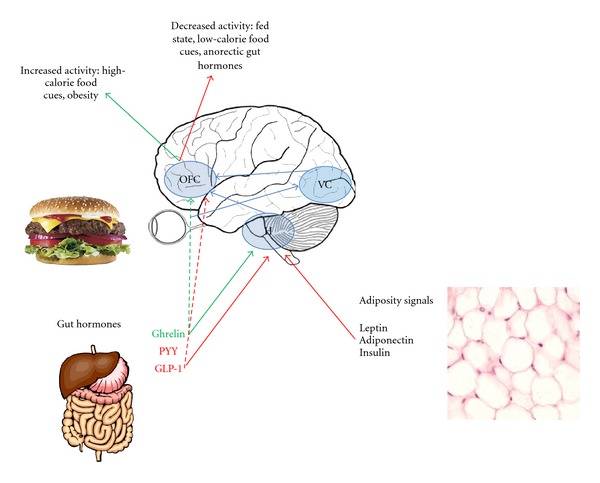
Functional magnetic resonance imaging (fMRI) has provided the opportunity to safely investigate the workings of the human brain. This paper focuses on its use in the field of human appetitive behaviour and its impact in obesity research. In the present absence of any safe or effective centrally acting appetite suppressants, a better understanding of how appetite is controlled is vital for the development of new antiobesity pharmacotherapies. Early functional imaging techniques revealed an attenuation of brain reward area activity in response to visual food stimuli when humans are fed-in other words, the physiological state of hunger somehow increases the appeal value of food. Later studies have investigated the action of appetite modulating hormones on the fMRI signal, showing how the attenuation of brain reward region activity that follows feeding can be recreated in the fasted state by the administration of anorectic gut hormones. Furthermore, differences in brain activity between obese and lean individuals have provided clues about the possible aetiology of overeating. The hypothalamus acts as a central gateway modulating homeostatic and nonhomeostatic drives to eat. As fMRI techniques constantly improve, functional data regarding the role of this small but hugely important structure in appetite control is emerging.


Figures

Figure 1
Figure 1
Brain reward centres: the hypothalamus (H), as a homeostatic gatekeeper, has numerous connections with higher brain centres which process salience and reward. The hypothalamus transmits to these higher centres information received from the periphery, such as nutritional status signalled via the postprandial release of gut hormones, and in turn modulates metabolic rate via the sympathetic nervous system. This sagittal section of the brain reveals the important areas involved in the hedonic control of eating behaviour; amygdale (Am): emotional and aversive processing; nucleus accumbens (Nac): anticipatory reward processing; ventral tegmental area (VTA): numerous dopaminergic projections to other limbic areas; ventral striatum (VS): motivation reward; expectancy and novelty processing; anterior cingulate cortex (ACC): decision making; orbitofrontal cortex (OFC): reward encoding; prefrontal cortex (PFC): translation of external and internal cues into behavioural responses; dorsolateral prefrontal cortex (DLPFC): self-control. Not shown is the insular cortex (a more lateral structure), which is also important in gustatory processing.
Figure 2
Figure 2
Schematic of T 1 and T 2 relaxation. MRI utilises the behaviour of protons within varying magnetic fields to produce signals which can be converted into images. Each hydrogen nucleus in the brain can be thought of as a vector (in the z and x-y planes) representing the strength and direction of its magnetic field as it spins on its axis (its magnetic dipole moment, MDM). The MDMs of the imaged protons try to align with the main external magnetic field of the scanner (referred to here as B 0 and conventionally shown along the z axis in 3D coordinates). A second magnetic field (in the form of a short radiofrequency RF pulse) is applied, which flips all of the MDMs from alignment in the z direction into the x-y plane (a). Before application of the RF pulse the, amplitude in the z-axis is maximal while the amplitude in the x-y plane is zero. Just after application of the RF pulse the, amplitude in the z-axis is zero (a) while the amplitude in the x-y plane is maximal (d). During relaxation, the amplitude in the z-axis will slowly increase ((b) and (d)) while the amplitude in the x-y plane slowly decreases ((e) and (f)).  T 1 relaxation is the time taken for the z vector to regain in strength, whereas T 2 relaxation is the time taken for the x-y vector to decay. These changing magnetic vectors invoke their own RF signals, which are picked up by the receiver coils and interpreted into information about the proton density of the subject being scanned.
Figure 3
Figure 3
Modulation of neuronal activity in the fed versus fasted state. Representative whole-brain fMRI sections showing regions where the difference in BOLD signal between viewing food images and nonfood images is blunted in the fed state compared with the fasted state. Unpublished image from [37].
Figure 4
Figure 4
Modulation of the rewarding aspects of food. In this highly simplified schematic, the orbitofrontal cortex (OFC) is highlighted as the most important hub in the reward encoding network. External food cues, via the visual cortex (VC), modulate the OFC response, with increased activity seen in fasted and obese patients and in response to high- versus low-calorie foods. OFC activity is also thought to be modulated by inputs from the hypothalamus, which senses internal information about nutritional status in the form of adiposity signals (such as leptin, which gives information about longer-term energy stores) and gut hormones (which are meal dependent and therefore give information about shorter-term nutrient availability). Anorectic (postprandial) gut hormones, such as PYY and GLP-1, attenuate OFC activity and, in fasted individuals induce, an OFC response to visual food cues more similar to that measured when fed. Conversely, the orexigenic hormone ghrelin upregulates reward centre activity.

References

    1. von Ruesten A, Steffen A, Floegel A, et al. Trend in obesity prevalence in European adult cohort populations during follow-up since 1996 and their predictions to 2015. PLoS ONE. 2011;6(11) Article ID e27455. - PMC - PubMed
    1. Valentino MA, Colon-Gonzalez F, Lin JE, Waldman SA. Current trends in targeting the hormonal regulation of appetite and energy balance to treat obesity. Expert Review of Endocrinology and Metabolism. 2010;5(5):765–783. - PMC - PubMed
    1. Sam AH, Troke RC, Tan TM, Bewick GA. The role of the gut/brain axis in modulating food intake. Neuropharmacology. 2012;63(1):46–56. - PubMed
    1. Ritter RC. Gastrointestinal mechanisms of satiation for food. Physiology and Behavior. 2004;81(2):249–273. - PubMed
    1. Gautron L, Elmquist JK. Sixteen years and counting: an update on leptin in energy balance. Journal of Clinical Investigation. 2011;121(6):2087–2093. - PMC - PubMed

Publication types