

A Pilot Study for the Neuroprotective Effect of Gongjin-dan on Transient Middle Cerebral Artery Occlusion-Induced Ischemic Rat Brain
- PMID: 22719787
- PMCID: PMC3375177
- DOI: 10.1155/2012/682720
A Pilot Study for the Neuroprotective Effect of Gongjin-dan on Transient Middle Cerebral Artery Occlusion-Induced Ischemic Rat Brain
Abstract
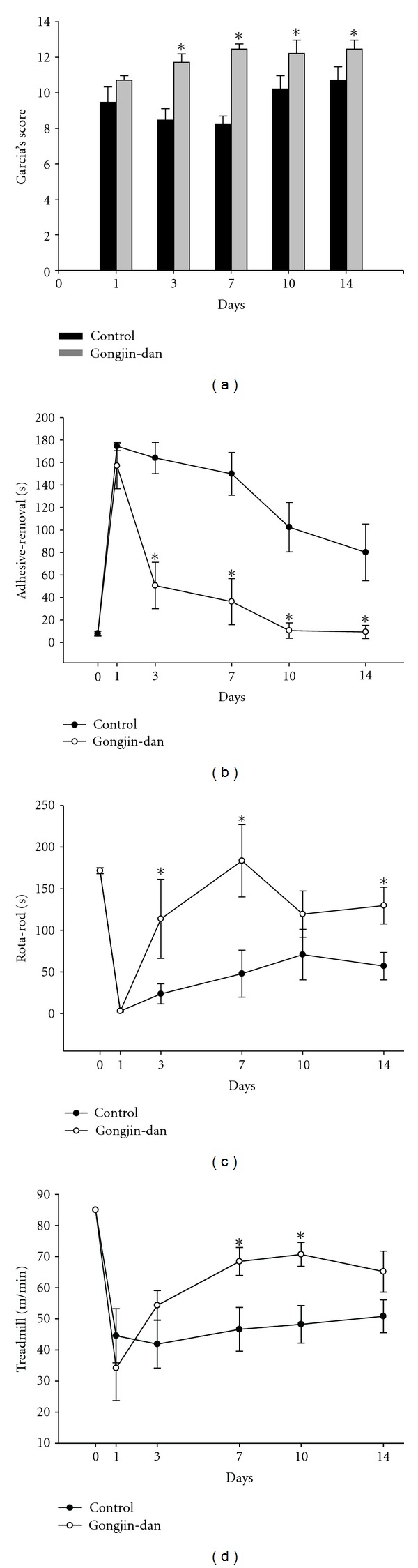
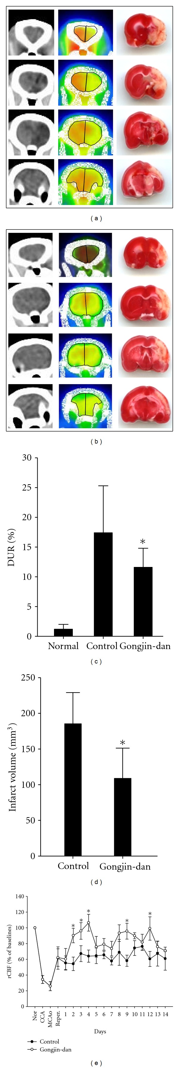
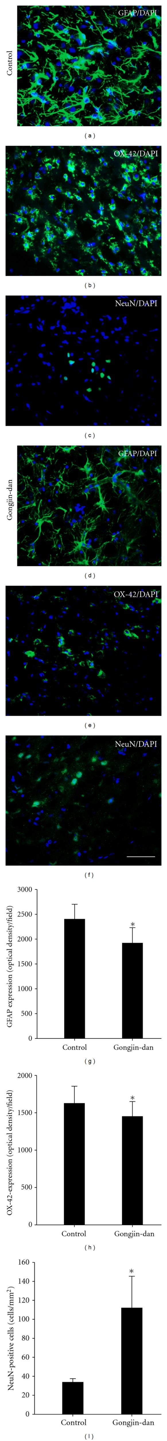
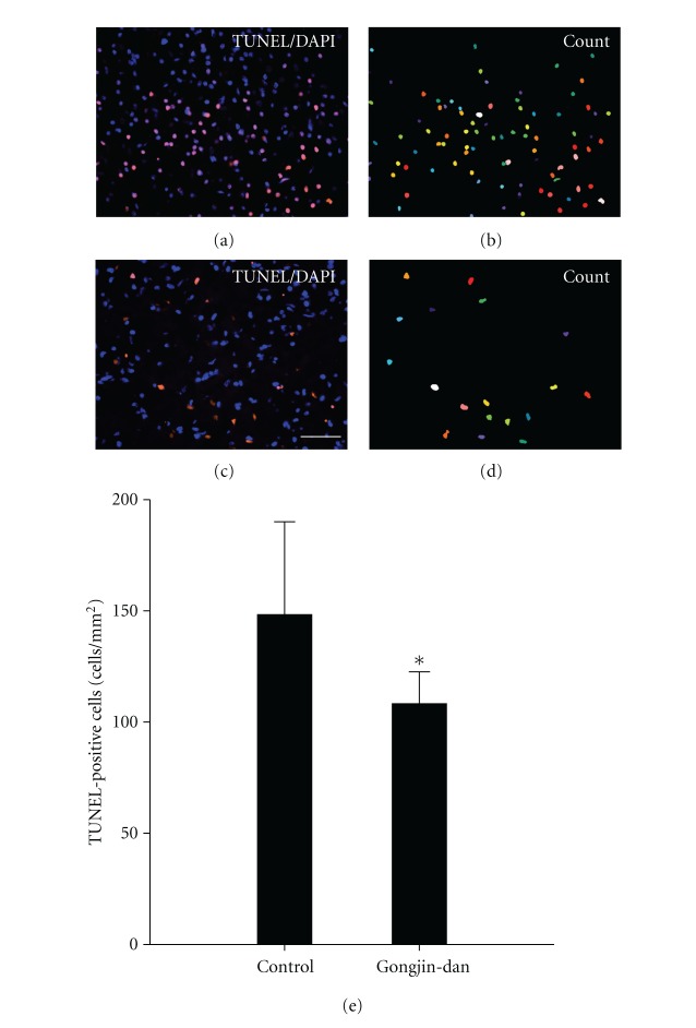
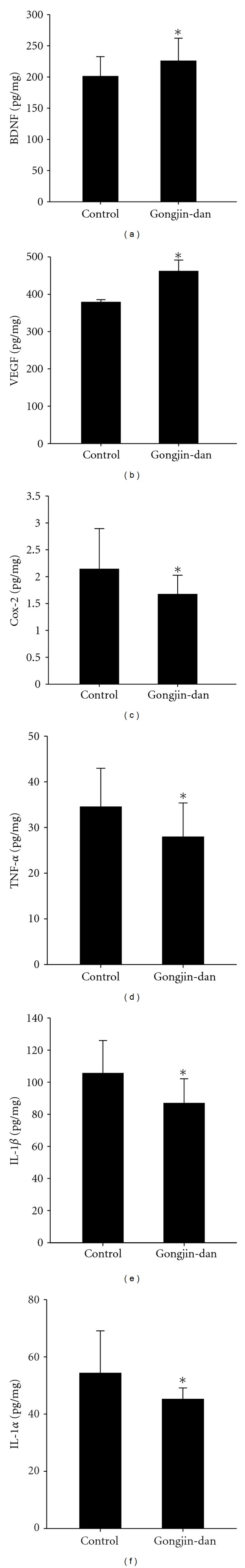
In this study, we investigated whether gongjin-dan improves functional recovery and has neuroprotective effects on reducing the infarct volume after transient middle cerebral artery occlusion (MCAo). Infarct volume was measured using TTC staining and glucose utilization by F-18 FDG PET. Functional improvement was evaluated with the Rota-rod, treadmill, Garcia score test, and adhesive removal test. At 14 days after MCAo, neuronal cell survival, astrocytes expansion, and apoptosis were assessed by immunohistofluorescence staining in the peri-infarct region. Also, the expression of neurotrophic factors and inflammatory cytokines such as VEGF, BDNF, Cox-2, TNF-α, IL-1β, and IL-1α was measured in ischemic hemisphere regions. The gongjin-dan-treated group showed both reduced infarct volume and increased glucose utilization. Behavior tests demonstrated a significant improvement compared to the control. Also in the gongjin-dan treated group, NeuN-positive cells were increased and number of astrocytes, microglia, and apoptotic cells was significantly decreased compared with the control group in the ischemic peri-infarct area. Furthermore, the expression of VEGF and BDNF was increased and level of Cox-2, TNF-α, IL-1β, and IL-1α was decreased. These results suggest that gongjin-dan may improve functional outcome through the rapid restoration of metabolism and can be considered as a potential neuroprotective agent.
Figures
Similar articles
-
Effect of combination therapy with sodium ozagrel and panax ginseng on transient cerebral ischemia model in rats.J Biomed Biotechnol. 2010;2010:893401. doi: 10.1155/2010/893401. Epub 2011 Jan 3. J Biomed Biotechnol. 2010. PMID: 21274269 Free PMC article.
-
The traditional drug Gongjin-Dan ameliorates chronic fatigue in a forced-stress mouse exercise model.J Ethnopharmacol. 2015 Jun 20;168:268-78. doi: 10.1016/j.jep.2015.04.001. Epub 2015 Apr 10. J Ethnopharmacol. 2015. PMID: 25865680
-
Preischemic neuroprotective effect of minocycline and sodium ozagrel on transient cerebral ischemic rat model.Brain Res. 2015 Mar 2;1599:85-92. doi: 10.1016/j.brainres.2014.12.051. Epub 2014 Dec 31. Brain Res. 2015. PMID: 25555371
-
Yangyin Tongnao granules enhance neurogenesis in the peri-infarct area and upregulate brain-derived neurotrophic factor and vascular endothelial growth factor after focal cerebral ischemic infarction in rats.Mol Biol Rep. 2019 Aug;46(4):3817-3826. doi: 10.1007/s11033-019-04824-5. Epub 2019 Apr 20. Mol Biol Rep. 2019. PMID: 31006100
-
The novel exercise-induced hormone irisin protects against neuronal injury via activation of the Akt and ERK1/2 signaling pathways and contributes to the neuroprotection of physical exercise in cerebral ischemia.Metabolism. 2017 Mar;68:31-42. doi: 10.1016/j.metabol.2016.12.003. Epub 2016 Dec 11. Metabolism. 2017. PMID: 28183451
Cited by
-
Delayed treatment of α5 GABAA receptor inverse agonist improves functional recovery by enhancing neurogenesis after cerebral ischemia-reperfusion injury in rat MCAO model.Sci Rep. 2019 Feb 19;9(1):2287. doi: 10.1038/s41598-019-38750-0. Sci Rep. 2019. PMID: 30783142 Free PMC article.
-
Gongjin-Dan Enhances Neurite Outgrowth of Cortical Neuron by Ameliorating H2O2-Induced Oxidative Damage via Sirtuin1 Signaling Pathway.Nutrients. 2021 Nov 27;13(12):4290. doi: 10.3390/nu13124290. Nutrients. 2021. PMID: 34959841 Free PMC article.
-
Neuronal Regeneration after Electroacupuncture Treatment in Ischemia-Reperfusion-Injured Cerebral Infarction Rats.Biomed Res Int. 2017;2017:3178014. doi: 10.1155/2017/3178014. Epub 2017 Aug 24. Biomed Res Int. 2017. PMID: 28913350 Free PMC article.
-
Immune-boosting effect of Yookgong-dan against cyclophosphamide-induced immunosuppression in mice.Heliyon. 2024 Jan 4;10(2):e24033. doi: 10.1016/j.heliyon.2024.e24033. eCollection 2024 Jan 30. Heliyon. 2024. PMID: 38293434 Free PMC article.
-
Oldenlandia diffusa Promotes Antiproliferative and Apoptotic Effects in a Rat Hepatocellular Carcinoma with Liver Cirrhosis.Evid Based Complement Alternat Med. 2015;2015:501508. doi: 10.1155/2015/501508. Epub 2015 Mar 10. Evid Based Complement Alternat Med. 2015. PMID: 25852766 Free PMC article.
References
-
- Connolly WE. Suffering, justice, and the politics of becoming. Culture, Medicine and Psychiatry. 1996;20(3):251–277. - PubMed
-
- Zhou XQ, Zeng XN, Kong H, Sun XL. Neuroprotective effects of berberine on stroke models in vitro and in vivo . Neuroscience Letters. 2008;447(1):31–36. - PubMed
-
- Loh KP, Huang SH, Tan BKH, Zhu YZ. Cerebral protection of purified Herba Leonuri extract on middle cerebral artery occluded rats. Journal of Ethnopharmacology. 2009;125(2):337–343. - PubMed
-
- Zemke D, Smith JL, Reeves MJ, Majid A. Ischemia and ischemic tolerance in the brain: an overview. NeuroToxicology. 2004;25(6):895–904. - PubMed
-
- Yoshikawa T, Akiyoshi Y, Susumu T, et al. Ginsenoside Rb1 reduces neurodegeneration in the peri-infarct area of a thromboembolic stroke model in non-human primates. Journal of Pharmacological Sciences. 2008;107(1):32–40. - PubMed
LinkOut - more resources
Full Text Sources
Medical
Research Materials
Miscellaneous
