. 2012;7(6):e38672.
doi: 10.1371/journal.pone.0038672. Epub 2012 Jun 14.

Respiratory insufficiency correlated strongly with mortality of rodents infected with West Nile virus

Affiliations

Respiratory insufficiency correlated strongly with mortality of rodents infected with West Nile virus

John D Morrey et al. PLoS One. 2012.

Abstract

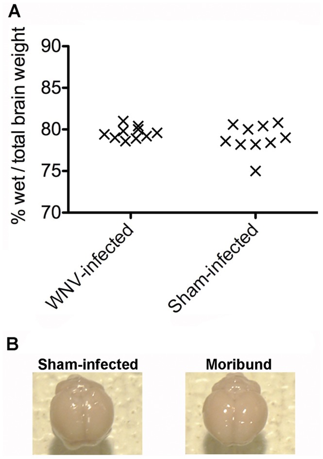
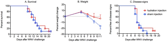
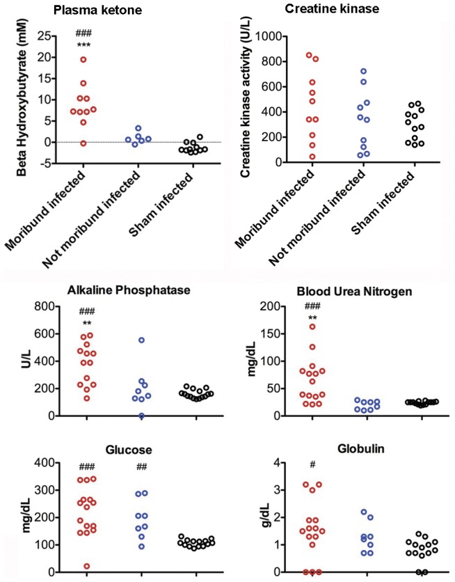
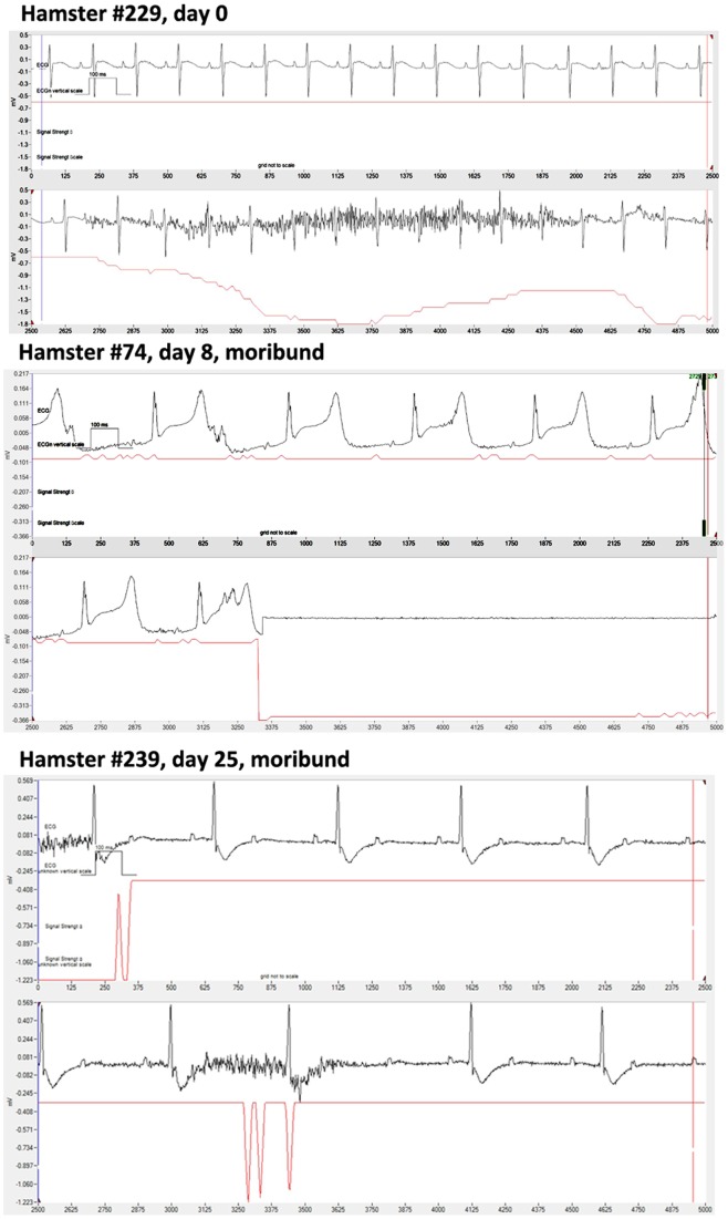
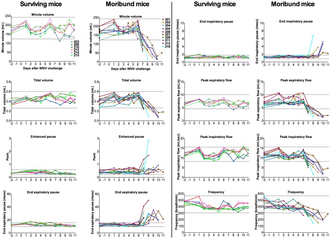
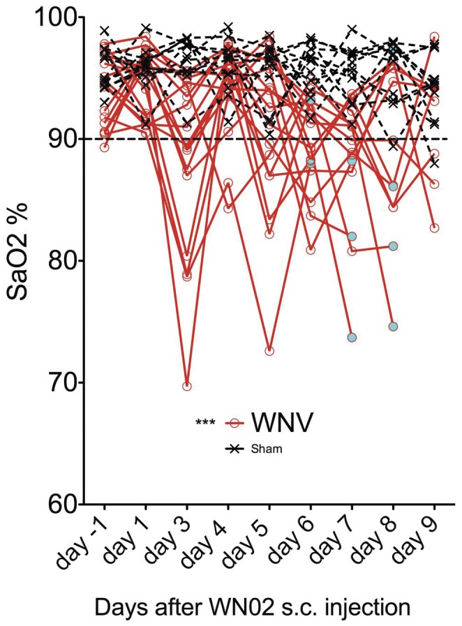
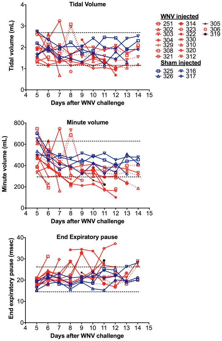
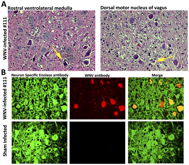
West Nile virus (WNV) disease can be fatal for high-risk patients. Since WNV or its antigens have been identified in multiple anatomical locations of the central nervous system of persons or rodent models, one cannot know where to investigate the actual mechanism of mortality without careful studies in animal models. In this study, depressed respiratory functions measured by plethysmography correlated strongly with mortality. This respiratory distress, as well as reduced oxygen saturation, occurred beginning as early as 4 days before mortality. Affected medullary respiratory control cells may have contributed to the animals' respiratory insufficiency, because WNV antigen staining was present in neurons located in the ventrolateral medulla. Starvation or dehydration would be irrelevant in people, but could cause death in rodents due to lethargy or loss of appetite. Animal experiments were performed to exclude this possibility. Plasma ketones were increased in moribund infected hamsters, but late-stage starvation markers were not apparent. Moreover, daily subcutaneous administration of 5% dextrose in physiological saline solution did not improve survival or other disease signs. Therefore, infected hamsters did not die from starvation or dehydration. No cerebral edema was apparent in WNV- or sham-infected hamsters as determined by comparing wet-to-total weight ratios of brains, or by evaluating blood-brain-barrier permeability using Evans blue dye penetration into brains. Limited vasculitis was present in the right atrium of the heart of infected hamsters, but abnormal electrocardiograms for several days leading up to mortality did not occur. Since respiratory insufficiency was strongly correlated with mortality more than any other pathological parameter, it is the likely cause of death in rodents. These animal data and a poor prognosis for persons with respiratory insufficiency support the hypothesis that neurological lesions affecting respiratory function may be the primary cause of human WNV-induced death.


Conflict of interest statement

Competing Interests: The authors have declared that no competing interests exist.

Figures

Figure 1
Figure 1. Lack of evidence for brain edema in WNV-infected moribund hamsters.
A) Hamsters were injected s.c. with WNV or sham. Brains were collected from WNV-infected hamsters when they were moribund. A corresponding sham-injected hamster was necropsied with every WNV-moribund hamster. Weights of brains before and after drying at 110°C for 24 hr were used to calculate the ratio of wet/total weight of each brain as an indication of edema. B) Fifteen hamsters were injected with WNV and five were injected with sham. When one WNV-injected hamster became moribund, a corresponding non-moribund WNV-injected hamster was selected for i.v. injection of Evans blue dye. Four hours later, brains were examined for any blue color due to breakdown of the blood-brain-barrier. Examples of brains from WNV- and sham-injected hamsters are shown. No blue color was evident in all of the WNV- or sham-injected hamsters.
Figure 2
Figure 2. Hydration supportive therapy for WNV-infected hamsters.
Six days after viral challenge just before the earliest disease signs appeared and up to 14 days after viral challenge, 10 hamsters were treated s.c. with D5 dextrose solution (5% dextrose, 0.9% physiological saline); and 10 hamsters were sham-injected. The animals were monitored through day 21 for A) mortality, B) weight change, and C) disease signs such as hind limb paralysis, front leg tremors, eye lacrimation, and diarrhea. Mortality was considered a disease sign too when constructing the graph. ***P≤0.001 using log rank test.
Figure 3
Figure 3. Plasma ketone, creatine kinase, and blood chemistries in WNV-infected moribund hamsters.
Twenty-three hamsters were injected with WNV and fifteen were injected with sham. When one WNV-injected hamster became moribund, the plasma was obtained from a corresponding non-moribund WNV-injected hamster. Numbers were unequal between the assays, because of insufficient sample volume to perform all assays. ###P≤0.001 compared to sham-infected, ***P≤0.001 compared to not-moribund infected using t-test.
Figure 4
Figure 4. Telemetric 2-lead electrocardiograms from WNV-infected hamsters taken over the course of disease.
Example of normal ECG from a hamster (#229) at day 0 where the erroneous signal was identified by a drop in the signal strength. Example of abnormal ECGs of two infected hamsters at time of death (#74) and within 6 hr of death (#239).
Figure 5
Figure 5. Plethysmography of C57BL/7 mice infected with WNV.
Sixteen mice were injected s.c. with WNV. Plethysmograph readings were obtained 2 days before viral challenge and from days 1 to 11 after viral challenge. Data were divided between the surviving or moribund mice. The dotted lines are two standard deviations of the mean of readings on days −2, 1, and 2 for all animals.
Figure 6
Figure 6. Pulse oximetry of BALB/c mice infected with WNV or sham.
White female BALB/c mice (>7 weeks old) were infected s.c. with WNV and monitored over time for Sa02 using pulse oximetry. Seventeen animals were included in each group. The cyan-colored circles identified animals that had become moribund. ***P≤0.001 using one-way analysis of variance with Newman-Keuls comparison.
Figure 7
Figure 7. Respiratory function as measured by plethysmography of hamsters infected with WNV.
All but three animals (#251, #303, #304) eventually became moribund. The dotted lines are two standard deviations of the means (n = 46) of sham-infected animals.
Figure 8
Figure 8. Histopathology of moribund WNV-infected hamster (#111) with respiratory distress.
A) H&E staining of rostral ventrolateral medulla containing respiratory control cells and dorsal motor nucleus of vagus. Yellow arrows indicate necrotic neurons. B) Immunohistochemical staining for neuron specific enolase (green) and WNV (red).

Similar articles

Cited by

References

    1. Lindsey NP, Staples JE, Lehman JA, Fischer M. Surveillance for human West Nile virus disease - United States, 1999–2008. MMWR Surveill Summ. 2010;59:1–17. - PubMed
    1. Morrey JD, Day CW, Julander JG, Olsen AL, Sidwell RW, et al. Modeling hamsters for evaluating West Nile virus therapies. Antiviral Res. 2004;63:41–50. - PubMed
    1. Xiao SY, Guzman H, Zhang H, Travassos da Rosa AP, Tesh RB. West Nile virus infection in the golden hamster (Mesocricetus auratus): a model for West Nile encephalitis. Emerg Infect Dis. 2001;7:714–721. - PMC - PubMed
    1. Oliphant T, Engle M, Nybakken GE, Doane C, Johnson S, et al. Development of a humanized monoclonal antibody with therapeutic potential against West Nile virus. Nat Med. 2005;11:522–530. - PMC - PubMed
    1. Leis AA, Stokic DS. Neuromuscular Manifestations of Human West Nile Virus Infection. Curr Treat Options Neurol. 2005;7:15–22. - PubMed

Publication types