. 2012;7(6):e38896.
doi: 10.1371/journal.pone.0038896. Epub 2012 Jun 12.

Evaluation of a multiparametric immunofluorescence assay for standardization of neuromyelitis optica serology

Affiliations

Evaluation of a multiparametric immunofluorescence assay for standardization of neuromyelitis optica serology

Letizia Granieri et al. PLoS One. 2012.

Abstract

Background: Neuromyelitis optica (NMO) is a severely disabling autoimmune disorder of the central nervous system, which predominantly affects the optic nerves and spinal cord. In a majority of cases, NMO is associated with antibodies to aquaporin-4 (AQP4) (termed NMO-IgG).

Aims: In this study, we evaluated a new multiparametric indirect immunofluorescence (IIF) assay for NMO serology.

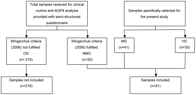
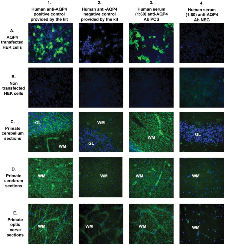
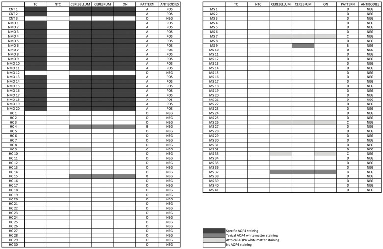
Methods: Sera from 20 patients with NMO, 41 patients with multiple sclerosis (MS), 30 healthy subjects, and a commercial anti-AQP4 IgG antibody were tested in a commercial composite immunofluorescence assay ("Neurology Mosaic 17"; Euroimmun, Germany), consisting of five different diagnostic substrates (HEK cells transfected with AQP4, non-transfected HEK cells, primate cerebellum, cerebrum, and optic nerve tissue sections).

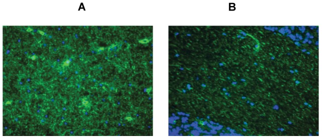
Results: We identified AQP4 specific and non-specific fluorescence staining patterns and established positivity criteria. Based on these criteria, this kit yielded a high sensitivity (95%) and specificity (100%) for NMO and had a significant positive and negative likelihood ratio (LR+ = ∞, LR- = 0.05). Moreover, a 100% inter- and intra-laboratory reproducibility was found.

Conclusions: The biochip mosaic assay tested in this study is a powerful tool for NMO serology, fast to perform, highly sensitive and specific for NMO, reproducible, and suitable for inter-laboratory standardization as required for multi-centre clinical trials.


Conflict of interest statement

Competing Interests: LG and PV received a contribution for attending a scientific meeting from Euroimmun, Luebeck, Germany. PN received a contribution for a fellowship in Oxford by Euroimmun, Luebeck, Germany. LG, FM and PV received consultation fees from Biogen Idec and Merck Serono. MC received consultation fees from Biogen Idec, Merck Serono and Novartis. AB has been on steering committees in clinical trials sponsored by Biogen Idec, Roche, and has received speaker honoraria from Biogen Idec, Merck Serono, TEVA, Bayer Schering, Sanofi-aventis, and Novartis; and his institution has received grant support from Biogen Idec, Bayer Schering, Merck Serono and Sanofi-Aventis. There are no patents, products in development or marketed products to declare. This does not alter the authors' adherence to all the PLoS ONE policies on sharing data and materials, as detailed online in the guide for authors. The other authors have no financial links with Euroimmun, and they have no competing financial interests.

Figures

Figure 1
Figure 1. Recruitment of NMO patients, MS patients and HC.
OD: Other diseases, NMO: neuromyelitis optica, MS: multiple sclerosis, HC: healthy controls.
Figure 2
Figure 2. Fluorescence staining patterns as observed with positive and negative controls provided by the manufacturer (column 1 and 2, respectively), an anti-AQP4 antibody positive human serum sample (column 3), and an anti-AQP4 antibody negative human control sample (column 4).
The biochip mosaic consists of 5 substrates: HEK cells transfected with full length recombinant human AQP4 (row A), non-transfected HEK cells (row B) and cryosections of primate cerebellum (row C), cerebrum (row D) and optic nerve (row E). Bound IgG was visualized using secondary antibodies labeled with FITC (green). Cell nuclei, stained with DAPI, are shown in blue. Magnification 40×. GL: granular layer; WM: white matter; DAPI: 4,6-diamidino-2-phenylindole; FITC: fluorescein isothiocyanate.
Figure 3
Figure 3. “Typical” (A) and “atypical” (B) white matter fluorescence staining as observed with anti-AQP4 positive NMO (A) and anti-AQP4 negative MS (B) human serum samples, respectively.
Bound IgG was visualized using secondary antibodies labeled with FITC (green). Cell nuclei, stained with DAPI, are shown in blue. Magnification 40×. NMO: neuromyelitis optica; MS: multiple sclerosis; DAPI: 4,6-diamidino-2-phenylindole; FITC: Fluorescein isothiocyanate.
Figure 4
Figure 4. Differential distribution of IIF staining patterns in five diagnostic substrates following incubation with serum samples from patients with NMO or controls.
The corresponding staining patterns (A, B, C, D; typical and atypical white matter staining), as defined in the results section, are indicated, together with the final evaluation of positivity or negativity for anti-AQP4 antibodies, and the healthy volunteers' clinical status. NMO: neuromyelitis optica, MS: multiple sclerosis, HC: healthy controls, CNT 1: commercial goat polyclonal anti-human AQP4 IgG (H19, Santa Cruz Biotechnology), CNT 2: human anti-AQP4 positive serum provided by the manufacturer, CNT 3: human anti-AQP4 negative serum provided by the manufacturer, TC = transfected cells, NTC = non-transfected cells, ON = optic nerve.

Similar articles

Cited by

References

    1. Wingerchuk DM, Hogancamp WF, O'Brien PC, Weinshenker BG. The clinical course of neuromyelitis optica (Devic's syndrome). Neurology. 1999;53(5):1107–1114. - PubMed
    1. Wingerchuk DM, Lennon VA, Pittock SJ, Lucchinetti CF, Weinshenker BG. Revised diagnostic criteria for neuromyelitis optica. Neurology 23: 66(10),1485–1489. 2006. - PubMed
    1. Lennon VA, Wingerchuk DM, Kryzer TJ, Pittock SJ, Lucchinetti CF, et al. A serum autoantibody marker of neuromyelitis optica: distinction from multiple sclerosis. Lancet. 2004;364:2106–2112. - PubMed
    1. Lennon VA, Kryzer TJ, Pittock SJ, Verkman AS, Hinson SR. IgG marker of optic-spinal multiple sclerosis binds to the aquaporin-4 water channel. J Exp Med. 2005;202:473–477. - PMC - PubMed
    1. Weinshenker BG, Wingerchuk DM, Vukusic S, Linbo L, Pittock SJ, et al. Neuromyelitis optica IgG predicts relapse after longitudinally extensive transverse myelitis. Ann Neurol. 2006;59:566–569. - PubMed

Publication types

Substances