. 2012;7(6):e38912.
doi: 10.1371/journal.pone.0038912. Epub 2012 Jun 14.

Local oxidative and nitrosative stress increases in the microcirculation during leukocytes-endothelial cell interactions

Affiliations

Local oxidative and nitrosative stress increases in the microcirculation during leukocytes-endothelial cell interactions

Saptarshi Kar et al. PLoS One. 2012.

Abstract

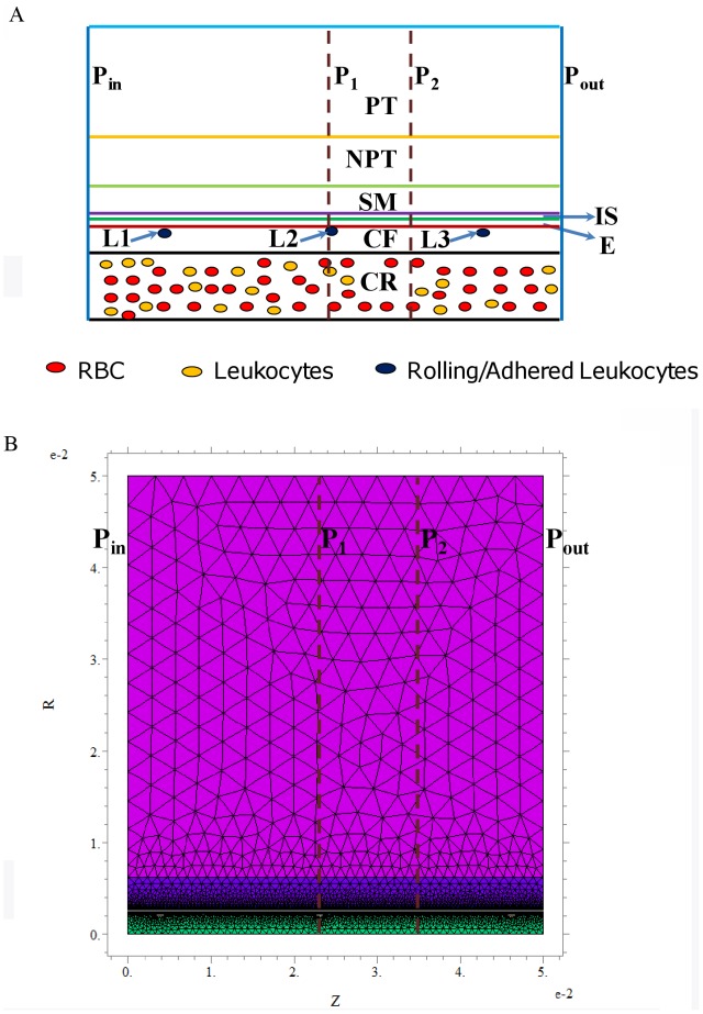
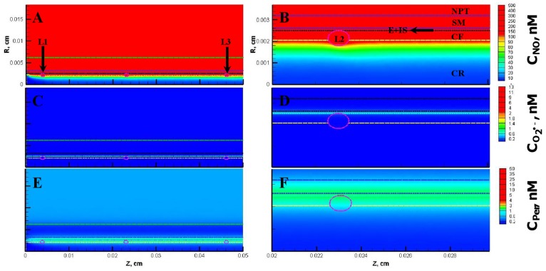
Leukocyte-endothelial cell interactions and leukocyte activation are important factors for vascular diseases including nephropathy, retinopathy and angiopathy. In addition, endothelial cell dysfunction is reported in vascular disease condition. Endothelial dysfunction is characterized by increased superoxide (O(2) (•-)) production from endothelium and reduction in NO bioavailability. Experimental studies have suggested a possible role for leukocyte-endothelial cell interaction in the vessel NO and peroxynitrite levels and their role in vascular disorders in the arterial side of microcirculation. However, anti-adhesion therapies for preventing leukocyte-endothelial cell interaction related vascular disorders showed limited success. The endothelial dysfunction related changes in vessel NO and peroxynitrite levels, leukocyte-endothelial cell interaction and leukocyte activation are not completely understood in vascular disorders. The objective of this study was to investigate the role of endothelial dysfunction extent, leukocyte-endothelial interaction, leukocyte activation and superoxide dismutase therapy on the transport and interactions of NO, O(2)(•-) and peroxynitrite in the microcirculation. We developed a biotransport model of NO, O(2)(•-) and peroxynitrite in the arteriolar microcirculation and incorporated leukocytes-endothelial cell interactions. The concentration profiles of NO, O(2)(•-) and peroxynitrite within blood vessel and leukocytes are presented at multiple levels of endothelial oxidative stress with leukocyte activation and increased superoxide dismutase accounted for in certain cases. The results showed that the maximum concentrations of NO decreased ~0.6 fold, O(2)(•-) increased ~27 fold and peroxynitrite increased ~30 fold in the endothelial and smooth muscle region in severe oxidative stress condition as compared to that of normal physiologic conditions. The results show that the onset of endothelial oxidative stress can cause an increase in O(2)(•-) and peroxynitrite concentration in the lumen. The increased O(2) (•-) and peroxynitrite can cause leukocytes priming through peroxynitrite and leukocytes activation through secondary stimuli of O(2)(•-) in bloodstream without endothelial interaction. This finding supports that leukocyte rolling/adhesion and activation are independent events.


Conflict of interest statement

Competing Interests: The authors have declared that no competing interests exist.

Figures

Figure 1
Figure 1. Schematic representation of the role of endothelial dysfunction on leukocyte related events through interactions between free radical species (NO, ROS and peroxynitrite).
The free radical species are represented by the orange ovals, the leukocyte related events and endothelial dysfunction are represented by the yellow compartments and the chemical species expressed as a result of the interactions of free radicals (cytokines, adhesion molecules and inflammatory agents) are represented by the light green compartments. Endothelial dysfunction leads to increased ROS production from endothelium and a possible reduction in NO availability (indicated by the dashed lines). The ROS and NO combine to form peroynitrite (Per). ROS and peroxynitrite increase expression of adhesion molecules and cytokines leading to leukocyte recruitment and priming. Peroxynitrite and ROS can also prime and activate primed leukocytes, respectively. The dashed lines connecting the leukocyte related events shows the uncertaintly associated with their sequential nature.
Figure 2
Figure 2. Geometrical description of the problem.
Panel A shows the schematic of the arteriolar geometry. The geometry consists of concentric cylinders representing the different regions of the arteriole. The different regions fall under the category of either luminal or abluminal region. The luminal and abluminal regions are separated by the endothelial region (E). The luminal region consists of the RBC rich core (CR) and RBC free plasma region (CF). The abluminal region consists of the interstitial region (IS), smooth muscle region (SM), non-perfused (NPT) and capillary perfused (PT) parenchymal regions. L1, L2 and L3 represent the leukocytes interacting with the endothelium. Pin and Pout represent the inlet and outlet of the arteriolar/vessel segment, respectively. P1 and P2 represent the locations where the radial concentration profiles of NO, O2 •− and peroxynitrite were obtained and are located at distances of 230 and 345 µm, respectively from Pin. Panel B shows the schematic of finite element mesh grid with relative accuracy set to 0.001.
Figure 3
Figure 3. Concentration distribution under normal physiological conditions (Case 1).
Panel A, C and E shows the NO, O2 •− and peroxynitrite (referred as CPer) concentration distribution, respectively across the entire arteriolar geometry. Panel B, D and F shows the NO, O2 •− and peroxynitrite concentration distribution, respectively across a segment of the arteriolar geometry between 200 and 300 µm encompassing the luminal, E, SM and NPT regions. The endothelial and capillary based O2 •− production rates in this case were 5% of their respective NO production rates and the leukocytes were considered inactive.
Figure 4
Figure 4. Radial concentration profiles at locations P1 and P2 for the Case 1.
Panel A and B shows the radial concentration profiles of NO, O2 •− and peroxynitrite at the location P1 and P2, respectively.
Figure 5
Figure 5. Concentration distribution under conditions of endothelial oxidative stress (Case 2).
The NO, O2 •− and peroxynitrite concentration distribution are shown for the entire arteriolar geometry in Panels A, C, and E, respectively and across the 200–300 µm region in Panels B, D and F, respectively. The endothelial and capillary based O2 •− production rates in this case were 20% of their respective NO production rates and the leukocytes were considered inactive.
Figure 6
Figure 6. Radial concentration profiles at locations P1 and P2 for the Case 2.
Panel A and B shows the radial concentration profiles of NO, O2 •− and peroxynitrite at the location P1 and P2, respectively.
Figure 7
Figure 7. Concentration distribution under combination of endothelial oxidative stress and activation of leukocytes (Case 3).
The NO, O2 •− and peroxynitrite concentration distribution are shown for the entire arteriolar geometry in Panels A, C, and E, respectively and across the 200–300 µm region in Panels B, D and F, respectively. The O2 •− production in the endothelium and capillary in this case were 20% of their respective NO production and the leukocytes were in activated state producing NO and O2 •−.
Figure 8
Figure 8. Radial concentration profiles at locations P1 and P2 for the Case 3.
Panel A and B shows the radial concentration profiles of NO, O2 •− and peroxynitrite at the location P1 and P2, respectively.
Figure 9
Figure 9. Concentration distribution under endothelial oxidative stress, activated leukocytes and increased SOD concentration (Case 4).
The NO, O2 •− and peroxynitrite concentration distribution are shown for the entire arteriolar geometry in Panels A, C, and E, respectively and across the 200–300 µm region in Panels B, D and F, respectively. The O2 •− production in the endothelium and capillary in this case were 20% of their respective NO production and the leukocytes were in activated state producing NO and O2 •−. The SOD concentration across all the regions of the arteriole and within the leukocytes was set at 10 µM.
Figure 10
Figure 10. Radial concentration profiles at locations P1 and P2 for the Case 4.
Panel A and B shows the radial concentration profiles of NO, O2 •− and peroxynitrite at the location P1 and P2, respectively.
Figure 11
Figure 11. Concentration distribution under severe oxidative stress conditions (Case 5).
The NO, O2 •− and peroxynitrite concentration distribution are shown for the entire arteriolar geometry in Panels A, C, and E, respectively and across the 200–300 µm region in Panels B, D and F, respectively. The O2 •− production in the endothelium and capillary in this case were equal to their respective NO production and the leukocytes were in activated state producing NO and O2 •−.
Figure 12
Figure 12. Radial concentration profiles at locations P1 and P2 for the Case 5.
Panel A and B shows the radial concentration profiles of NO, O2 •− and peroxynitrite at the location P1 and P2, respectively.

Similar articles

Cited by

References

    1. Chow FY, Nikolic-Paterson DJ, Ozols E, Atkins RC, Tesch GH. Intercellular adhesion molecule-1 deficiency is protective against nephropathy in type 2 diabetic db/db mice. J Am Soc Nephrol. 2005;16:1711–1722. - PubMed
    1. Geraldes P, King GL. Activation of protein kinase C isoforms and its impact on diabetic complications. Circ Res. 2010;106:1319–1331. - PMC - PubMed
    1. Al-Shabrawey M, Rojas M, Sanders T, Behzadian A, El-Remessy A, et al. Role of NADPH oxidase in retinal vascular inflammation. Invest Ophthalmol Vis Sci. 2008;49:3239–3244. - PMC - PubMed
    1. Julius U, Drel VR, Grassler J, Obrosova IG. Nitrosylated proteins in monocytes as a new marker of oxidative-nitrosative stress in diabetic subjects with macroangiopathy. Exp Clin Endocrinol Diabetes. 2009;117:72–77. - PMC - PubMed
    1. Sumagin R, Lomakina E, Sarelius IH. Leukocyte-endothelial cell interactions are linked to vascular permeability via ICAM-1-mediated signaling. Am J Physiol Heart Circ Physiol. 2008;295:H969–H977. - PMC - PubMed

Publication types