. 2012;7(6):e39080.
doi: 10.1371/journal.pone.0039080. Epub 2012 Jun 14.

Activation of type III interferon genes by pathogenic bacteria in infected epithelial cells and mouse placenta

Affiliations

Activation of type III interferon genes by pathogenic bacteria in infected epithelial cells and mouse placenta

Hélène Bierne et al. PLoS One. 2012.

Abstract

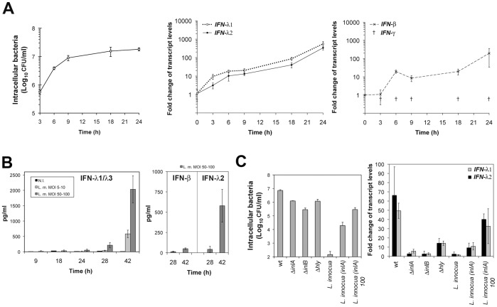
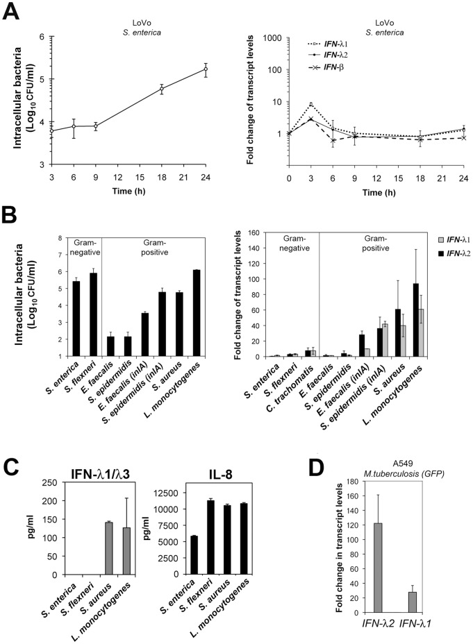
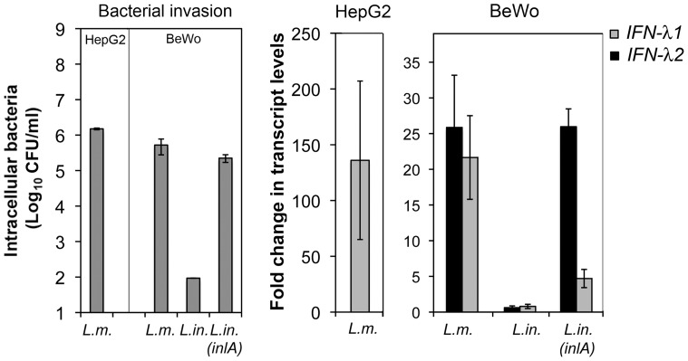
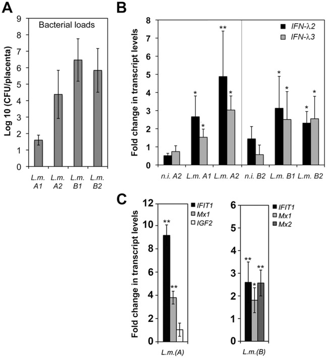
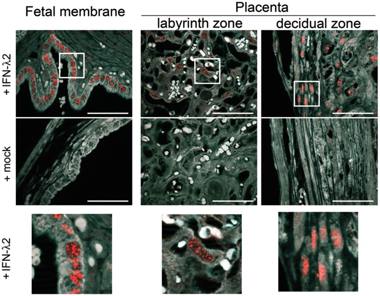
Bacterial infections trigger the expression of type I and II interferon genes but little is known about their effect on type III interferon (IFN-λ) genes, whose products play important roles in epithelial innate immunity against viruses. Here, we studied the expression of IFN-λ genes in cultured human epithelial cells infected with different pathogenic bacteria and in the mouse placenta infected with Listeria monocytogenes. We first showed that in intestinal LoVo cells, induction of IFN-λ genes by L. monocytogenes required bacterial entry and increased further during the bacterial intracellular phase of infection. Other Gram-positive bacteria, Staphylococcus aureus, Staphylococcus epidermidis and Enterococcus faecalis, also induced IFN-λ genes when internalized by LoVo cells. In contrast, Gram-negative bacteria Salmonella enterica serovar Typhimurium, Shigella flexneri and Chlamydia trachomatis did not substantially induce IFN-λ. We also found that IFN-λ genes were up-regulated in A549 lung epithelial cells infected with Mycobacterium tuberculosis and in HepG2 hepatocytes and BeWo trophoblastic cells infected with L. monocytogenes. In a humanized mouse line permissive to fetoplacental listeriosis, IFN-λ2/λ3 mRNA levels were enhanced in placentas infected with L. monocytogenes. In addition, the feto-placental tissue was responsive to IFN-λ2. Together, these results suggest that IFN-λ may be an important modulator of the immune response to Gram-positive intracellular bacteria in epithelial tissues.


Conflict of interest statement

Competing Interests: The authors have declared that no competing interests exist.

Figures

Figure 1
Figure 1. Characterization of L. monocytogenes-mediated induction of IFN-III genes in LoVo intestinal cells. A.
LoVo intestinal epithelial cells infected with L. monocytogenes at multiplicity of infection (MOI) of 20 for the indicated time were lysed and processed for quantification of intracellular bacteria by counting colony-forming units (CFU) on agar plates, or for total cellular RNA extraction. Quantitative RT-PCR was performed to determine relative IFN-λ1, IFN-λ2, IFN-β and IFN-γ mRNA levels. The expression values were normalized to GAPDH transcript levels. Fold inductions were calculated from ΔΔCT values, using uninfected control cell values at the beginning of the experiment as a calibrator ( = 1). Values from three independent experiments are expressed as mean ± S.D. of the fold change. IFN-γ mRNA levels were below the limits of detection (†). At 24 h post-infection, fold change values in uninfected cells were 1.12±0.84 for IFN-λ1; 0.62±0.39 for IFN-λ2; 1.43±0.76 for IFN-β. B. Culture supernatants from LoVo cells infected with L. monocytogenes (L.m.), at the indicated time and MOI, were analyzed by ELISA for IFN production. Experiments were done in triplicate and reproduced once. C. LoVo cells were infected for 18 h with wild type (wt) or isogenic ΔinlA, ΔinlB or Δhly L. monocytogenes strains (MOI = 25), or with L. innocua or L. innocua expressing inlA (MOI = 25) or L. innocua expressing inlA (MOI = 100, indicated by “100”). Cells were processed as described in (A). Values are expressed as mean ± S.D of three independent experiments.
Figure 2
Figure 2. Induction of IFN-III genes by different bacterial species in epithelial cells.
A. LoVo intestinal cells infected with S. enterica at multiplicity of infection (MOI) of 20 for the indicated time, were lysed and processed for quantification of intracellular bacterial by counting colony-forming units (CFU) on agar plates, or for total cellular RNA extraction and quantification of IFN gene expression, as described for L. monocytogenes in Figure 1A. Values from three independent experiments are expressed as mean ± S.D. of the fold change relative to uninfected control cell values at the beginning of the experiment. B. LoVo cells, uninfected or infected for 18 h with different bacterial species, were processed for quantification of bacterial load and mRNA. Quantification of internalized bacteria: data are means ± S.D. of CFU for 105 cells (n = 3) at the indicated MOI, except for C. trachomatis, for which the percentage of infected cells (n = 4) were determined by flow cytometry after antibody labelling (see text). IFN-λ1 and IFN-λ2 transcript levels were determined by qRT-PCR and normalized to GAPDH transcript levels. Values are expressed as mean ± S.D. of the fold change relative to that in uninfected cells (n = 3 to 5). C. Quantification of IFN-λ1/λ3 and IL-8 production were done by ELISA, using culture supernatants of LoVo cells infected for 28 h with the indicated bacterium at a MOI of 50. Experiments were done in triplicates and reproduced once. D. Quantification of IFN-λ mRNAs in A549 lung epithelial cells infected with GFP-expressing M. tuberculosis (n = 4). The percentage of infected cells were determined by flow cytometry (see text).
Figure 3
Figure 3. Induction of IFN-III genes by L. monocytogenes in HepG2 hepatocytes and BeWo trophoblastic cells.
Quantification of bacterial loads (CFU) and IFN-λ mRNAs levels in Listeria-infected HepG2 or BeWo cells. IFN-λ1 and IFN-λ2 transcript levels were determined by qRT-PCR and normalized to GAPDH transcript levels. Values are expressed as mean ± S.D. of the fold change relative to that in uninfected cells (n = 3). IFN-λ2 levels in uninfected HepG2 cells were below the detection threshold, preventing measures of fold change. L. monocytogenes (L. m.), L. innocua (L. in.), L. innocua expressing inlA (L. in. (inlA)).
Figure 4
Figure 4. Induction of IFN-III and IFN-stimulated genes in mouse placenta.
Pregnant E16P+/+ mice were inoculated intravenously with 4×10⁴ CFUs L. monocytogenes in PBS (L.m.) or with PBS only (n.i.) in two independent experiments (exp. A and B). Bacterial numbers were determined in livers and placentas 72 h post-infection. Placentas (n = 7 in exp.A; n = 8 in exp.B) from the two mice that displayed the highest bacterial loads in liver lysates, and placentas (n = 3) from two uninfected mice (n.i.), were processed for RNA extraction and bacterial quantifications. A. Quantification of L. monocytogenes loads and IFN-λ transcripts in placenta homogenates. B. Quantification of IFN-λ2 and IFN-λ3 transcript levels in placentas by qRT-PCR, with normalization to GAPDH transcripts. Values are expressed as mean ± S.D. of the fold change relative to that in placenta of uninfected mouse A1 or mouse B1 ( = 1). There is no significant change in uninfected mouse A2 or mouse B2 (n.i. A2, n.i. B2). C. Quantification of transcript levels of IFN-responsive genes (IFIT1, Mx1, Mx2) and a control gene (IGF2) in placentas were determined by qRT-PCR and normalized to YWHAZ housekeeping gene. Values are expressed as mean ± S.D. of the fold change relative to that in placenta of all uninfected mice of exp.A or exp.B. (*p<0.05; **p<0.005, Student t test).
Figure 5
Figure 5. Response to IFN-III in mouse placenta and fetal membrane.
Pregnant B6.A2G-Mx1-IFNAR10/0 mice were treated subcutaneously with 5 µg of mouse IFN-λ2 or PBS (mock) at 24 and 12 h prior to sacrifice. Embryos in utero were extracted and fixed with formaldehyde. Sections of paraffin-embedded tissue were immuno-stained for IFN-inducible Mx1 protein. IFN-λ response was monitored by nuclear Mx1 staining (red) in epithelial cells of the fetal membrane and various regions of the placenta. Counterstaining: background auto-fluorescence (white). Zooms of squared regions are shown below. Scale bar: 50 µm.

References

    1. Mordstein M, Michiels T, Staeheli P. What have we learned from the IL28 receptor knockout mouse? Journal of interferon & cytokine research : the official journal of the International Society for Interferon and Cytokine Research. 2010;30:579–584. - PubMed
    1. Witte K, Witte E, Sabat R, Wolk K. IL-28A, IL-28B, and IL-29: promising cytokines with type I interferon-like properties. Cytokine & growth factor reviews. 2010;21:237–251. - PubMed
    1. Kotenko SV. IFN-lambdas. Current opinion in immunology. 2011;23:583–590. - PMC - PubMed
    1. Sommereyns C, Paul S, Staeheli P, Michiels T. IFN-lambda (IFN-lambda) is expressed in a tissue-dependent fashion and primarily acts on epithelial cells in vivo. PLoS Pathog. 2008;4:e1000017. - PMC - PubMed
    1. Mordstein M, Neugebauer E, Ditt V, Jessen B, Rieger T, et al. Lambda interferon renders epithelial cells of the respiratory and gastrointestinal tracts resistant to viral infections. Journal of virology. 2010;84:5670–5677. - PMC - PubMed

Publication types