. 2012;7(6):e39216.
doi: 10.1371/journal.pone.0039216. Epub 2012 Jun 14.

Microglial activation correlates with disease progression and upper motor neuron clinical symptoms in amyotrophic lateral sclerosis

Affiliations

Microglial activation correlates with disease progression and upper motor neuron clinical symptoms in amyotrophic lateral sclerosis

Johannes Brettschneider et al. PLoS One. 2012.

Abstract

Background/aims: We evaluated clinicopathological correlates of upper motor neuron (UMN) damage in amyotrophic lateral sclerosis (ALS), and analyzed if the presence of the C9ORF72 repeat expansion was associated with alterations in microglial inflammatory activity.

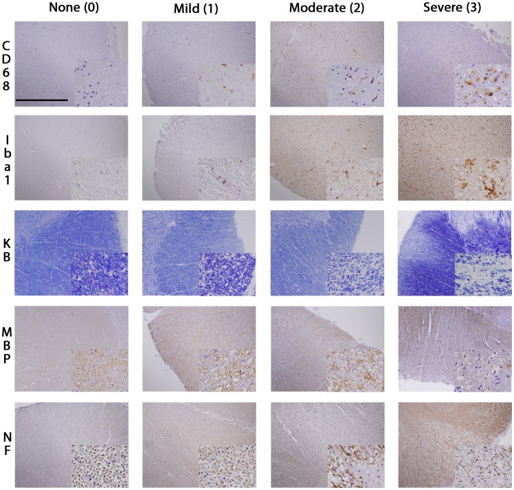
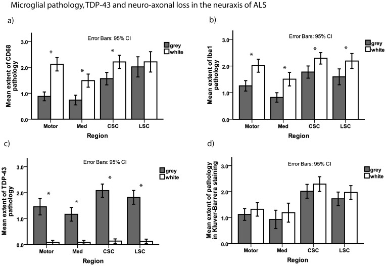
Methods: Microglial pathology was assessed by IHC with 2 different antibodies (CD68, Iba1), myelin loss by Kluver-Barrera staining and myelin basic protein (MBP) IHC, and axonal loss by neurofilament protein (TA51) IHC, performed on 59 autopsy cases of ALS including 9 cases with C9ORF72 repeat expansion.

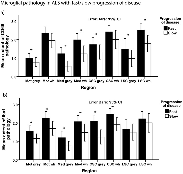
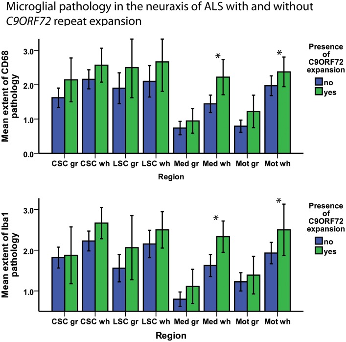
Results: Microglial pathology as depicted by CD68 and Iba1 was significantly more extensive in the corticospinal tract (CST) of ALS cases with a rapid progression of disease. Cases with C9ORF72 repeat expansion showed more extensive microglial pathology in the medulla and motor cortex which persisted after adjusting for disease duration in a logistic regression model. Higher scores on the clinical UMN scale correlated with increasing microglial pathology in the cervical CST. TDP-43 pathology was more extensive in the motor cortex of cases with rapid progression of disease.

Conclusions: This study demonstrates that microglial pathology in the CST of ALS correlates with disease progression and is linked to severity of UMN deficits.


Conflict of interest statement

Competing Interests: The authors have declared that no competing interests exist.

Figures

Figure 1
Figure 1. Staging of CST degeneration in ALS.
The figure illustrates the IHC staging used to grade the extent of microglial activation (CD68, Iba1) and axonal loss (MBP, KB, NF) in the CST of ALS patients. Images are taken from the lateral portion of the cervical CST. KB  =  Kluver-Barrera, MBP =  myelin basic protein, NF  =  neurofilaments (TA51). Large images were taken with 4× objective, Scale bar is 1.0 mm. Small insert images were taken with 60× objective. Small insert image for CD68 stage “0” shows prominent neuronal nuclei, but no activated microglia.
Figure 2
Figure 2. Microglial pathology, TDP-43, and neuro-axonal loss in the neuraxis of ALS.
Bar plots show extent of microglial pathology as detected by staining for CD68 (a) and Iba1 (b) as well as TDP-43 pathology (c) and myelin loss as an indirect measure of axonal loss (d) in different regions of the neuraxis of ALS. For the spinal cord sections, the grey matter examined was the anterior horn and the white matter examined was the anterior and lateral portion of the CST. Whiskers in bar plot indicate 95% confidence interval of mean. CSC  =  cervical spinal cord, grey  =  grey matter, LSC  =  lumbar spinal cord, Med  =  medulla oblongata, motor  =  motor cortex (gyrus praecentralis), white  =  white matter.
Figure 3
Figure 3. Microglial pathology and progression of disease.
Bar plot shows microglial pathology as detected by staining for CD68 (a) and Iba1 (b) in ALS patients with a fast and a slow progression of disease. For the spinal cord sections, the grey matter examined was the anterior horn and the white matter examined was the anterior and lateral portion of the CST. Whiskers in bar plot indicate 95% confidence interval of mean. CSC  =  cervical spinal cord, grey  =  grey matter, LSC  =  lumbar spinal cord, Mot  =  motor cortex (gyrus praecentralis), Med  =  medulla oblongata, wh  =  white matter.
Figure 4
Figure 4. Microglial pathology in ALS with/without C9ORF72 repeat expansion.
Bar plot shows microglial pathology as detected by staining for CD68 (above) and Iba1 (below) in ALS patients with and without presence of a C9ORF72 repeat expansion. For the spinal cord sections, the grey matter examined was the anterior horn and the white matter examined was the anterior and lateral portion of the CST. Whiskers in bar plot indicate 95% confidence interval of mean. CSC  =  cervical spinal cord, gr  =  grey matter, LSC  =  lumbar spinal cord, Mot  =  motor cortex (gyrus praecentralis), Med  =  medulla oblongata, wh  =  white.

References

    1. Kiernan MC, Vucic S, Cheah BC, Turner MR, Eisen A, et al. Amyotrophic lateral sclerosis. Lancet. 2011;377:942–955. - PubMed
    1. Neumann M, Sampathu DM, Kwong LK, Truax AC, Micsenyi MC, et al. Ubiquitinated TDP-43 in frontotemporal lobar degeneration and amyotrophic lateral sclerosis. Science. 2006;314:130–133. - PubMed
    1. Deng HX, Hentati A, Tainer JA, Iqbal Z, Cayabyab A, et al. Amyotrophic lateral sclerosis and structural defects in Cu,Zn superoxide dismutase. Science. 1993;261:1047–1051. - PubMed
    1. Pehar M, Cassina P, Vargas MR, Castellanos R, Viera L, et al. Astrocytic production of nerve growth factor in motor neuron apoptosis: implications for amyotrophic lateral sclerosis. J Neurochem. 2004;89:464–473. - PubMed
    1. Nagai M, Re DB, Nagata T, Chalazonitis A, Jessell TM, et al. Astrocytes expressing ALS-linked mutated SOD1 release factors selectively toxic to motor neurons. Nat Neurosci. 2007;10:615–622. - PMC - PubMed

Publication types