Review
. 2012:2012:203734.
doi: 10.1155/2012/203734. Epub 2012 Jun 6.

Multiple approaches to investigate the transport and activity-dependent release of BDNF and their application in neurogenetic disorders

Affiliations
Review

Multiple approaches to investigate the transport and activity-dependent release of BDNF and their application in neurogenetic disorders

David Hartmann et al. Neural Plast. 2012.

Abstract

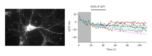
Studies utilizing genetic and pharmacological manipulations in rodent models and neuronal cultures have revealed myriad roles of brain-derived neurotrophic factor (BDNF). Currently, this knowledge of BDNF function is being translated into improvement strategies for several debilitating neurological disorders in which BDNF abnormalities play a prominent role. Common among the BDNF-related disorders are irregular trafficking and release of mature BDNF (mBDNF) and/or its prodomain predecessor, proBDNF. Thus, investigating the conditions required for proper trafficking and release of BDNF is an essential step toward understanding and potentially improving these neurological disorders. This paper will provide examples of disorders related to BDNF release and serve as a review of the techniques being used to study the trafficking and release of BDNF.


Figures

Figure 1
Figure 1
A pyramidal neuron expressing BDNF-eGFP. Insert shows BDNF-eGFP puncta in dendrites, which colocalize with the secretory granule marker SG2 (red).
Figure 2
Figure 2
Schematic illustration of how BDNF-pHluorin fluoresces, both inside the vesicle and upon axonal and dendritic vesicular fusion. Before stimulation, BDNF-pHluorin protein shows little fluorescence. If intracellular calcium increase occurs upon electrical stimulation, then BDNF-pHluorin vesicle may fuse, opening up to the pH 7.4 extracellular space, causing a transient spike in fluorescence that can be detected by TIRF microscopy. Different styles of fusion between axon and dendrite are shown, as explained in [10] and in the text. After sustained vesicular fusion as occurs in dendrites, fluorescence will decrease as a result of BDNF-pHluorin diffusion out of vesicles. Kiss-and-run fusion as occurs in stimulated axons will rather show an increase in fluorescence because minimal pHluorin diffuses out of the vesicle. The sticks represent synaptotagmin-4; see [85] for details. Modified from Figure 4(e) in [85].
Figure 3
Figure 3
Left panel shows a BDNF-pH expressing hippocampal neuron in the presence of 50 mM NH4Cl. Right panel shows the same cell 80 sec after standard ACSF solution was applied. The graph shows the time course of the fractional change of BDNF-pH intensity (background-subtracted delta F/F0) from pixels within the color-coded regions of interest (ROIs) shown in the panels above. The change from NH4Cl-containing ACSF to control buffer quenches BDNF-pH within acidic secretory granules. Time-lapse was performed in an inverted microscope with a Hg-lamp and a cooled CCD camera. Neurons were imaged with a 60x 1.45 NA oil-immersion objective, exposure times ranged from 50–100 ms, and images were taken at 1 frame per second (fps).
Figure 4
Figure 4
To confirm the viability of neurons transfected with BDNF-pH and their responsiveness to extracellular field stimulation, Ca2+ imaging was performed in neurons labeled with fura-2 AM. Upon electrical stimulation through field Pt wires, Ca2+ transiently increased in several dendrites. The graph shows the time course of background-subtracted delta F/F0 of 380 nm-excited fura-2 fluorescence intensity (510 nm emission) from the colored ROIs shown in the image. Images taken at 4 fps.
Figure 5
Figure 5
Neuron expressing BDNF-pHluorin and loaded with the Ca2+ indicator fura-2 AM (image is of 488 nm-excited BDNF-pHluorin). This cell was sensitive to pH (as in Figure 4) and responded to electrical stimulation with Ca2+ transients (using 380 nm excitation, as in Figure 5). Consistent with activity-dependent BDNF release, dendritic BDNF-pHluorin puncta show decreases in intensity (~20% deltaF/F) as a result of BDNF-pHluorin discharge from vesicles following full fusion.

Similar articles

Cited by

References

    1. Reichardt LF. Neurotrophin-regulated signalling pathways. Philosophical Transactions of the Royal Society B. 2006;361(1473):1545–1564. - PMC - PubMed
    1. Lu B, Pang PT, Woo NH. The yin and yang of neurotrophin action. Nature Reviews Neuroscience. 2005;6(8):603–614. - PubMed
    1. Cunha C, Brambilla R, Thomas KL. A simple role for BDNF in learning and memory? Frontiers in Molecular Neuroscience. 2010;3(1):1–15. - PMC - PubMed
    1. Lessmann V, Gottmann K, Malcangio M. Neurotrophin secretion: current facts and future prospects. Progress in Neurobiology. 2003;69(5):341–374. - PubMed
    1. Fahnestock M, Garzon D, Holsinger RMD, Michalski B. Neurotrophic factors and Alzheimer’s disease: are we focusing on the wrong molecule? Journal of Neural Transmission. Supplementum. 2002;(62):241–252. - PubMed

MeSH terms

Substances