. 2012:2012:250421.
doi: 10.1155/2012/250421. Epub 2012 Jun 6.

IGF-1 restores visual cortex plasticity in adult life by reducing local GABA levels

Affiliations

IGF-1 restores visual cortex plasticity in adult life by reducing local GABA levels

José Fernando Maya-Vetencourt et al. Neural Plast. 2012.

Abstract

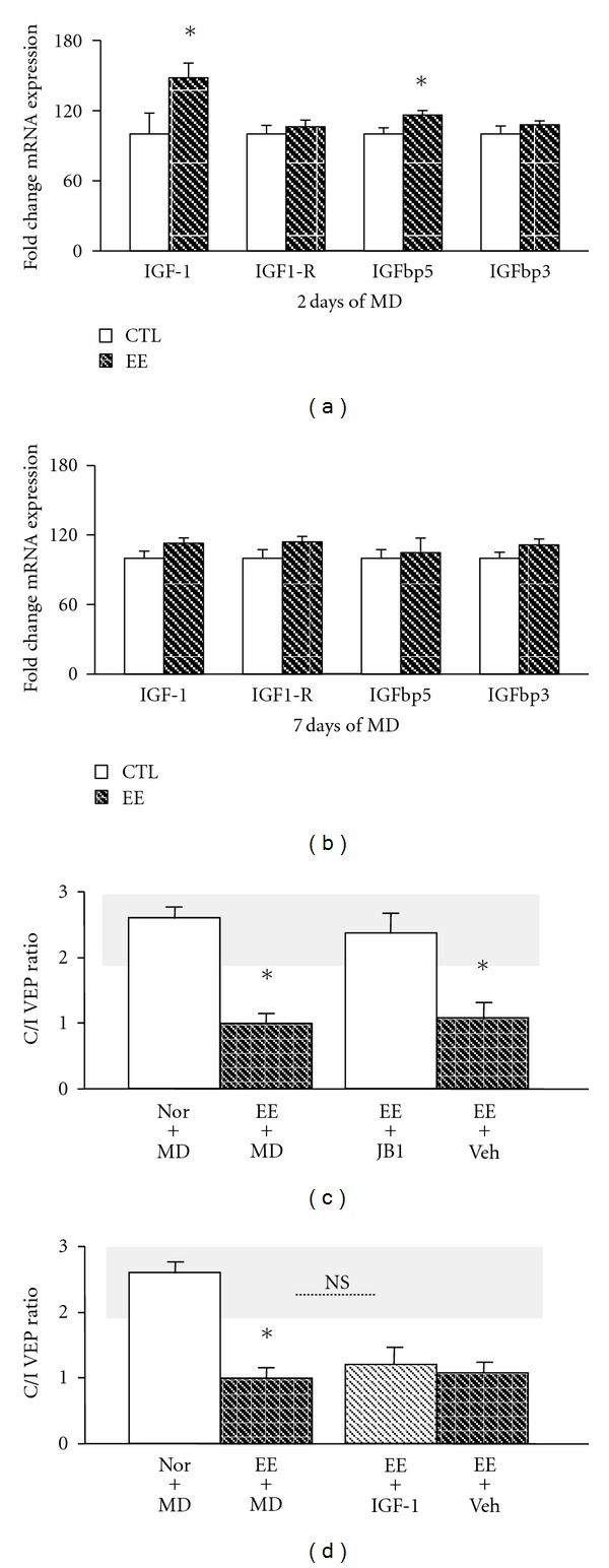
The central nervous system architecture is markedly modified by sensory experience during early life, but a decline of plasticity occurs with age. Recent studies have challenged this dogma providing evidence that both pharmacological treatments and paradigms based on the manipulation of environmental stimulation levels can be successfully employed as strategies for enhancing plasticity in the adult nervous system. Insulin-like growth factor 1 (IGF-1) is a peptide implicated in prenatal and postnatal phases of brain development such as neurogenesis, neuronal differentiation, synaptogenesis, and experience-dependent plasticity. Here, using the visual system as a paradigmatic model, we report that IGF-1 reactivates neural plasticity in the adult brain. Exogenous administration of IGF-1 in the adult visual cortex, indeed, restores the susceptibility of cortical neurons to monocular deprivation and promotes the recovery of normal visual functions in adult amblyopic animals. These effects were accompanied by a marked reduction of intracortical GABA levels. Moreover, we show that a transitory increase of IGF-1 expression is associated to the plasticity reinstatement induced by environmental enrichment (EE) and that blocking IGF-1 action by means of the IGF-1 receptor antagonist JB1 prevents EE effects on plasticity processes.


Figures

Figure 1
Figure 1
IGF-1 treatment reactivates plasticity in the adult visual cortex and decreases intracortical GABA levels. (a) An OD shift in favor of the open eye was evident in IGF-1-infused rats (IGF-1+MD; n = 5, C/I VEP ratio = 1.2 ± 0.17) with respect to adult animals with binocular vision (Nor+Bin; n = 5, C/I VEP ratio = 2.42 ± 0.16, one-way ANOVA P = 0.0002, post hoc Holm Sidak method, P < 0.05). No modification of the C/I VEP ratio was detected in either saline-treated animals (SAL+MD; n = 5, C/I VEP ratio = 2.67 ± 0.15, One-way ANOVA, post hoc Holm Sidak method) or normal adult rats (Nor+MD; n = 7, C/I VEP ratio = 2.53 ± 0.25, One-way ANOVA, post hoc Holm Sidak method). IGF-1 treated rats with binocular vision (IGF-1+Bin; n = 3) exhibited a C/I VEP ratio (2.37 ± 0.12) completely comparable to that of untreated Nor+Bin animals (One-way ANOVA, post hoc Holm Sidak method). Insert: the change of OD in IGF-1-treated rats was due to a decrease of the deprived eye (contra) strength. The amplitude of VEPs recorded in response to the stimulation of the occluded eye in IGF-1+MD rats (0.35 ± 0.05) was significantly lower with respect to that obtained in saline-treated animals following MD (0.68 ± 0.12; t-test; P = 0.047). No difference was detected in VEP amplitudes recorded after the stimulation of the open (Ipsi) eye (IGF-1+MD: 0.38 ± 0.11; SAL+MD: 0.32 ± 0.05; t-test; P = 0.627). VEP amplitudes at the recording site in the VC contralateral to the occlusion were normalized to the sum of the response to stimulation of the contralateral and ipsilateral eye, as described previously [–12]. (b) Full rescue of VA was evident in reverse-sutured IGF-1 treated rats (IGF-1+RS, n = 5): VA of the long-term deprived eye (0.93 ± 0.06 cycles per degree, c/deg) was not different from that of the fellow eye (0.94 ± 0.04 c/deg; paired t-test, P = 0.837). No sign of recovery was detected either in reverse-sutured animals infused with saline (SAL+RS, n = 5; VA for the long-term deprived eye = 0.72 ± 0.03 c/deg; VA for the open eye 0.96 ± 0.01 c/deg; paired t-test, P < 0.001) or in reverse-sutured normal rats (Nor+RS, n = 5; VA for the long-term deprived eye = 0.67 ± 0.01 c/deg; VA for the open eye 1.03 ± 0.03 c/deg; paired t-test; P < 0.001) Insert, while in Nor-RS rats (C/I VEP ratio = 1.02 ± 0.08) there was no recovery of binocularity (One-way ANOVA, post hoc Holm Sidak method); in IGF1+RS the C/I VEP ratio is in the range of normal adult values (C/I VEP ratio 2.12 ± 0.13, One-way ANOVA, F (3-16) = 40.24, P < 0.0001, post hoc Holm Sidak method). No recovery of OD was detected in SAL+RS animals (n = 5; C/I VEP ratio = 0.86 ± 0.11; One-way ANOVA, post hoc Holm Sidak method). (c) Extracellular GABA levels were significantly lower in the VC of IGF-1-treated rats (IGF-1; n = 6; 0.87 ± 0.17 μM) with respect to saline-treated (SAL; n = 5; 4.46 ± 0.17 μM) and untreated animals (Nor; n = 8,4.62 ± 0.97 μM; One-way ANOVA, F (2–16) = 7.445, P = 0.0052, post hoc Tukey test, P < 0.05). (d) No change in glutamate (GLU) levels between IGF-1-treated (3.88 ± 0.4 μM) and control groups (SAL: 4.48 ± 0.6 μM; Nor: 3.74 ± 0.3 μM) was detected (One-way ANOVA, F (2–15) = 0.744, P = 0.491). The grey box denotes the C/I VEP ratio range in adult normal animals. *Statistical significance. Error bars indicate SEM.
Figure 2
Figure 2
The upregulation of IGF-1 expression in EE animals mediates the effects of enriched experience on adult VC plasticity. (a) Analysis of gene expression by RT-PCR revealed that IGF-1 and IGFbp5 expression increased in the VC of EE animals after 2 days of MD with respect to SC animals similarly treated (n = 8 for both experimental groups; t-test; respectively, P = 0.034 and P = 0.030). In contrast, no modifications of either IGF1-R (t-test; P = 0.523) or IGFbp3 (t-test; P = 0.324) expression were detected. (b) The expression of IGF-1, IGF-1R, IGFbp5, and IGFbp3 in the VC was not different between EE animals monocularly deprived for 7 days and SC rats similarly treated. (c) JB1 infusion prevented the OD shift induced by MD in EE animals: no difference in C/I VEP ratio between normal animals (Nor+MD; n = 5, 2.67 ± 0.15) and EE rats treated with JB1 subjected to MD detected (EE+JB1; n = 6; 2.37 ± 0.09; One-way ANOVA, post hoc Holm Sidak method), while monocularly deprived EE animals (EE+MD; n = 6; 0.99 ± 0.07) and EE rats treated with vehicle (EE+Veh; n = 5; 1.08 ± 0.05) showed an OD shift in favor of the open eye (One-way ANOVA, F (5–27) = 46.48, P < 0.0001, post hoc Holm Sidak method). (d) Coupling enriched experience with IGF-1 treatment (EE+IGF1 rats) did not further enhance the OD shift induced by MD in EE animals: the C/I VEP ratio measured in EE+IGF1 rats (n = 5; 1.08 ± 0.07) was completely comparable to that reported for EE animals (EE+MD), while it differed from that recorded in Nor+MD animals (One-way ANOVA F (5–27) = 46.48, P < 0.0001, post hoc Holm Sidak method). The grey box denotes the C/I VEP ratio range in adult normal animals. *Statistical significance. NS: Not significant. Error bars indicate SEM.
Figure 3
Figure 3
The process of plasticity reactivation induced by EE is associated with signal transduction pathways that involve the activation of long-distance neuromodulatory systems and IGF-1 signaling. We propose a model in which the interplay between 5-HT and IGF-1 transmission, in parallel or in series, shifts the inhibitory/excitatory balance in favour of excitation thus activating intracellular mechanisms that eventually promote epigenetic modifications of chromatin structure that, in turn, allow for the expression of plasticity genes in adult life. A pharmacological reduction of inhibitory transmission could promote Bdnf expression and activate physiological mechanisms that may drive the degradation of extracellular matrix (ECM) components that are inhibitory for plasticity. 5-HT and IGF-1 signaling, respectively, may also directly activate Bdnf expression or enhance the ECM remodeling. Bdnf-trkB signaling might upregulate additional gene expression patterns associated with functional modifications in the VC. This could also alter the balance of intracortical inhibition and excitation. Degradation of ECM components may modify the inhibition/excitation ratio in the visual system. The interaction between BDNF-trkB signaling and ECM reorganization has yet to be explored. Continuous arrows represent established interactions between molecular and cellular processes mentioned (boxes). Dashed lines represent interactions that remain to be ascertained.

Similar articles

Cited by

References

    1. Katz LC, Shatz CJ. Synaptic activity and the construction of cortical circuits. Science. 1996;274(5290):1133–1138. - PubMed
    1. Berardi N, Pizzorusso T, Maffei L. Critical periods during sensory development. Current Opinion in Neurobiology. 2000;10(1):138–145. - PubMed
    1. Hensch TK. Critical period plasticity in local cortical circuits. Nature Reviews Neuroscience. 2005;6(11):877–888. - PubMed
    1. Wiesel T, Hubel DH. Single-cell responses in striate cortex of kittens deprived of vision. Journal of Neurophysiology. 1963;26:1003–1017. - PubMed
    1. Hubel DH, Wiesel T. The period of susceptibility to the physiological effects of unilateral eye closure in kittens. Journal of Physiology. 1970;206(2):419–436. - PMC - PubMed

Publication types

Substances