. 2012 Jan 1;1(1):18-27.
doi: 10.4161/onci.1.1.18400.

Lymphoblastoid cell line with B1 cell characteristics established from a chronic lymphocytic leukemia clone by in vitro EBV infection

Affiliations

Lymphoblastoid cell line with B1 cell characteristics established from a chronic lymphocytic leukemia clone by in vitro EBV infection

Anders Rosén et al. Oncoimmunology. .

Abstract

Chronic lymphocytic leukemia (CLL) cells express the receptor for Epstein-Barr virus (EBV) and can be infected in vitro. Infected cells do not express the growth-promoting set of EBV-encoded genes and therefore they do not yield LCLs, in most experiments. With exceptional clones, lines were obtained however. We describe a new line, HG3, established by in vitro EBV-infection from an IGHV1-2 unmutated CLL patient clone. All cells expressed EBNA-2 and LMP-1, the EBV-encoded genes pivotal for transformation. The karyotype, FISH cytogenetics and SNP-array profile of the line and the patient's ex vivo clone showed biallelic 13q14 deletions with genomic loss of DLEU7, miR15a/miR16-1, the two micro-RNAs that are deleted in 50% of CLL cases. Further features of CLL cells were: expression of CD5/CD20/CD27/CD43 and release of IgM natural antibodies reacting with oxLDL-like epitopes on apoptotic cells (cf. stereotyped subset-1). Comparison with two LCLs established from normal B cells showed 32 genes expressed at higher levels (> 2-fold). Among these were LHX2 and LILRA. These genes may play a role in the development of the disease. LHX2 expression was shown in self-renewing multipotent hematopoietic stem cells, and LILRA4 codes for a receptor for bone marrow stromal cell antigen-2 that contributes to B cell development. Twenty-four genes were expressed at lower levels, among these PARD3 that is essential for asymmetric cell division. These genes may contribute to establish precursors of CLL clones by regulation of cellular phenotype in the hematopoietic compartment. Expression of CD5/CD20/CD27/CD43 and spontaneous production of natural antibodies may identify the CLL cell as a self-renewing B1 lymphocyte.


Figures

None
Figure 1. (A) Human B1 phenotypic markers expressed on HG3. Indirect immunofluorescence analysis with appropriate mAbs followed by FITC-of Cy-ChromeTM-conjugated anti-mouse Ig. Ten thousands events were counted in a FACScan flow cytometer. (B) Competition chemiluminescent ELISA for oxLDL. MDA-LDL competition ELISA was performed for HG3 CLL IgM mAb isolated from cell supernatant and purified by affinity chromatography. Plates were coated with MDA-LDL, then IgM was allowed to react with increasing amounts of soluble competitor MDA-LDL or native LDL (nLDL). B/B0 indicates the ratio between bound (cpm) and bound (cpm) without competitor.
None
Figure 2. Identical IGHV1–2 gene rearrangements in HG3 and the patient’s CLL cells.
None
Figure 3. SNP array analysis and karyotype reveal biallelic chromosome 13 deletions. (A and B) show results from SNP array analysis in HG3 cell line and HG patient. Red marking show deletions and green marks show amplifications. (C and D) G-banding are showing identical biallelic deletions of chromosome 13 in HG3 cell line and in HG patient’s leukemic clone.
None
Figure 4. Zoom in picture of the homozygous 13q4 deletion. The array revealed that DLEU-7, miR16–1 and miR15a were deleted.
None
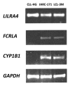
Figure 5. Gene expression difference observed by RT-PCR. Comparison between gene expression of LILRA4, FCRLA and CYP1B1 using RT-PCR in the CLL-HG cell line and 2 LCL lines IARC-171 and LCL-3M derived from normal B cells.
None
Figure 6. Multicolor immunofluorescence for EBNA-2 and LMP-1. From left to right: DAPI staining (Vectashield) for nuclear DNA; EBNA-2 staining with mouse anti-EBNA-2 mAb clone PE2, followed by goat anti-mouse IgG1-Alexa488; LMP-1 staining with anti-LMP-1 mAb clone S-12, followed by anti-mouse IgG-Alexa594; Merged image of EBNA-2 and LMP-1.

Similar articles

Cited by

References

    1. Klein U, Tu Y, Stolovitzky GA, Mattioli M, Cattoretti G, Husson H, et al. Gene expression profiling of B cell chronic lymphocytic leukemia reveals a homogeneous phenotype related to memory B cells. J Exp Med. 2001;194:1625–38. doi: 10.1084/jem.194.11.1625. - DOI - PMC - PubMed
    1. Rosenwald A, Alizadeh AA, Widhopf G, Simon R, Davis RE, Yu X, et al. Relation of gene expression phenotype to immunoglobulin mutation genotype in B cell chronic lymphocytic leukemia. J Exp Med. 2001;194:1639–47. doi: 10.1084/jem.194.11.1639. - DOI - PMC - PubMed
    1. Damle RN, Ghiotto F, Valetto A, Albesiano E, Fais F, Yan XJ, et al. B-cell chronic lymphocytic leukemia cells express a surface membrane phenotype of activated, antigen-experienced B lymphocytes. Blood. 2002;99:4087–93. doi: 10.1182/blood.V99.11.4087. - DOI - PubMed
    1. Griffin DO, Holodick NE, Rothstein TL. Human B1 cells in umbilical cord and adult peripheral blood express the novel phenotype CD20+ CD27+ CD43+ CD70. J Exp Med. 2011;208:67–80. doi: 10.1084/jem.20101499. - DOI - PMC - PubMed
    1. Kikushige Y, Ishikawa F, Miyamoto T, Shima T, Urata S, Yoshimoto G, et al. Self-renewing hematopoietic stem cell is the primary target in pathogenesis of human chronic lymphocytic leukemia. Cancer Cell. 2011;20:246–59. doi: 10.1016/j.ccr.2011.06.029. - DOI - PubMed

Publication types