. 2012 Mar 1;1(2):141-151.
doi: 10.4161/onci.1.2.18479.

T cells and T cell tumors efficiently generate antigen-specific cytotoxic T cell immunity when modified with an NKT ligand

Affiliations

T cells and T cell tumors efficiently generate antigen-specific cytotoxic T cell immunity when modified with an NKT ligand

Yeonseok Chung et al. Oncoimmunology. .

Abstract

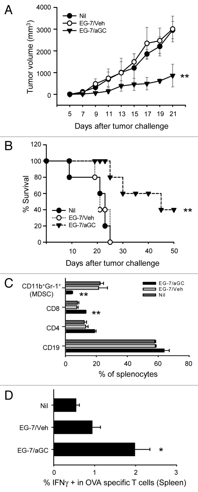
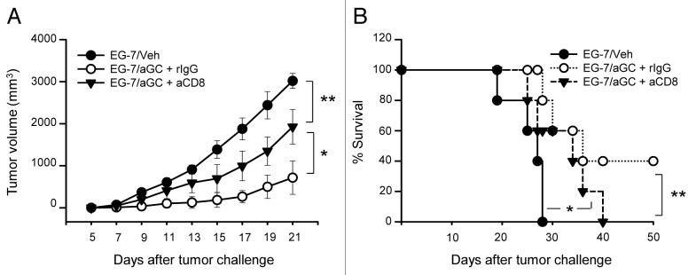
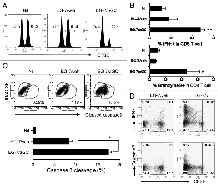
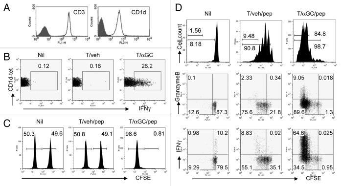
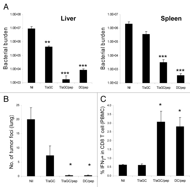
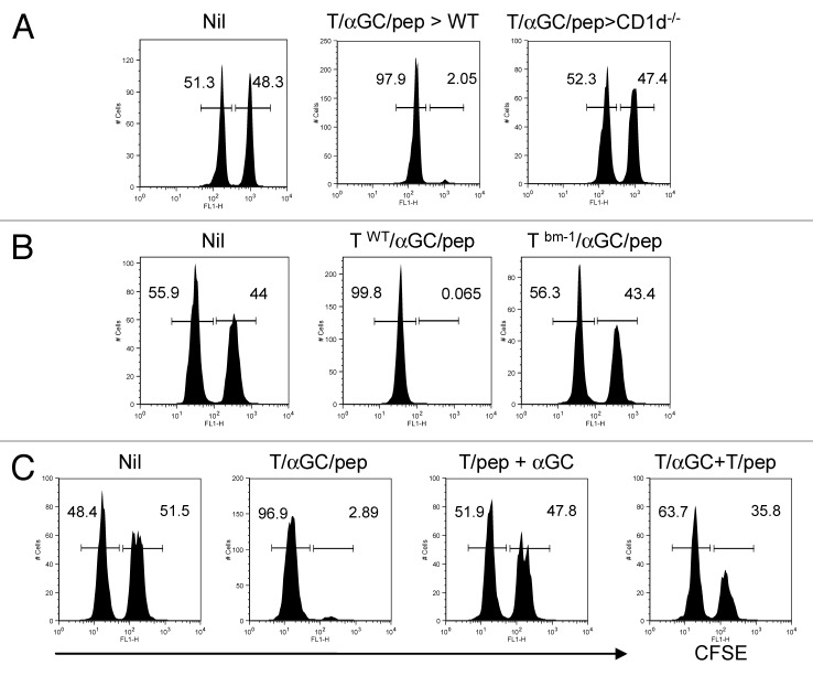
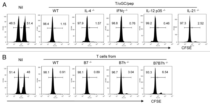
Various Invariant NKT (iNKT) cell ligands have been shown as potent adjuvants in boosting T cell reactivates to antigens on professional APC. Non-professional APC, such as T cells, also co-expressing MHC class I and CD1d, have been unattractive cell vaccine carriers due to their poor immunogenicity. Here, we report that T cells as well as T cell lymphoma can efficiently generate antigen-specific cytotoxic T lymphocytes (CTL) responses in mice in vivo, when formulated to present iNKT ligand α-galactosylceramide (αGC) on their surface CD1d. Vaccination with αGC-pulsed EG-7 T-cell lymphoma induced tumor-specific CTL response and suppressed the growth of EG-7 in a CD8 T cell-dependent manner. Injection of αGC-loaded CD4 T cells in mice efficiently activated iNKT cells in vivo. While T cells loaded with a class I-restricted peptide induced proliferation but not effector differentiation of antigen-specific CD8 T cells, injection of T cells co-pulsed with αGC strongly induced IFNγ and Granzyme B expression in T cells and complete lysis of target cells in vivo. Presentation of αGC and peptide on the same cells was required for optimal CTL response and vaccinating T cells appeared to directly stimulate both iNKT and cytotoxic CD8 T cells. Of note, the generation of this cytotoxic T cell response was independent of IL-4, IFNγ, IL-12, IL-21 and costimulation. Our data indicate that iNKT cell can license a non-professional APC to directly trigger antigen-specific cytotoxic T cell responses, which provides an alternative cellular vaccine strategy against tumors.


Figures

None
Figure 1. Vaccination with αGC-loaded EG-7 generated preventive antitumor activity. (A–D) EG-7 cells were co-cultured overnight with 1 μg/ml of αGC (EG-7/αGC) or vehicle followed by irradiation (50 Gy). C57BL/6 mice (n = 7 per group) were vaccinated with EG-7/veh, EG-7/αGC or untreated (Nil). One week later, all mice were subcutaneously injected with 1 × 10⁶ live EG-7 cells and the survival (A) and tumor size (B) were checked. (C) At the 3 weeks later, splenocytes were isolated from tumor bearing mice and analyzed by flow cytometer after staining with anti-CD11b, anti-Gr1, anti-CD19, anti-CD4 and anti-CD8 antibodies. (D) Splenocytes were restimulated with SIINFEKL in the presence of Golgi-Plug for 5 h before intracellular staining of IFNγ on OVA specific T cells. *p < 0.05, **p < 0.005, p values were calculated with 2-way ANOVA (A), Kaplan-Meier method (B) or Student's t-tests (C and D) in comparison with the EG-7/veh group.
None
Figure 2. CD8 T cells play a critical role in mediating antitumor activity. (A and B) C57BL/6 mice were vaccinated with EG-7/veh or EG-7/αGC on day -7 (n = 5 per group). EG-7/αGC vaccinated mice were intraperitoneally injected with CD8 depleting Ab (563.8) on day-3 and -1. On day 0, all mice were subcutaneously injected with 1 × 10⁶ live EG-7 cells and the tumor volume (A) and the survival (B) were checked. *p < 0.01, **p < 0.005, p values were calculated with 2-way ANOVA (A) or Kaplan-Meier method (B) in comparison with EG-7/veh and EG-7/αGC+rIgG or EG-7/αGC+rIgG and EG-7/αGC+anti-CD8 group.
None
Figure 3. Vaccination with αGC-loaded EG-7 generates OVA-specific cytotoxic T cell response in vivo. (A and B) EG-7 cells were cocultured overnight with 1 μg/ml of αGC (EG-7/αGC) or vehicle (0.5% polysorbate, EG-7/veh) followed by irradiation (50 Gy). C57BL/6 mice (n = 3 per group) were vaccinated with EG-7/veh, EG-7/αGC or left untreated (Nil). (A) One week later, an in vivo CTL assay for SIINFEKL was performed. CFSEhigh, peptide-pulsed target; CFSElow, peptide-unpulsed control. (B) One week after vaccination, CD8+ T cells were isolated and incubated with 1 µg/mL peptide-pulsed splenocytes that had been labeled with DDAO-SE for 2 h. The cells were fixed, permeabilized and stained with anti–cleaved caspase-3 mAb. The levels of cleaved caspase-3 in the DDAO-SE-positive cells were analyzed by flow cytometry. Data shown are representative FACS plots (upper panels) and mean ± SE (lower panel). *p < 0.05, in comparison with EG-7/veh group. (C) One week after the vaccination, splenocytes were isolated and restimulated with SIINFEKL for 5 h in the precence of Golgi-Plug before intracellular staining of granzyme B and IFNγ. (D) Ovalbumin-specific CD8 T cells were isolated, labeled with 10 µmol/L CFSE and i.v. transferred into their syngenic mice as described in Figure 1. On the following day, mice were i.v. injected with irradiated EG-7 cells manipulated in vitro with indicated conditions. Five days later, lymphoid cells from the spleen of the recipient mice were restimulated with SIINFEKL for 5 h before intracellular staining of granzyme B and IFNγ. Data are representative of at least two separate experiments. *p < 0.05, **p < 0.01, in comparison with non-treated (Nil) group.
None
Figure 4. Injection of conventional CD4 T cells coated with SIINFEKL and αGC induces a functional cytotoxic T cell response. (A) Lymphoid cells from spleen and lymph nodes of C57BL/6 mice were stained with FITC-conjugated anti-CD19, anti-NK1.1, anti-Gr-1, anti-CD11b, anti-CD11c, anti-CD8a, anti-I-Ab antibodies together with APC-conjugated CD4 Ab. Lineage negative and CD4+ cells were sorted by FACSAria. The sorted cells were stained with PE-conjugated anti-CD3 or anti-CD1d Ab. Filled histogram is isotype control. (B) Sorted CD4+ T cells were co-cultured with αGC (1 μg/ml). Cells were washed and intravenously injected into syngenic mice. Six hours later, lymphoid cells from spleen were stained with CD1d-tetramer and CD19 before intracellular IFNγ staining. CD1d-tetramer+CD19- cells were gated and analyzed. (C) The sorted CD4+ T cells were co-cultured with αGC (1 μg/ml) or vehicle (0.5% polysorbate) for 16 h including 1 h pulse with SIINFEKL. Cells were washed and intravenously injected into syngenic mice. One week later, syngenic lymphocytes were either loaded with 1 µmol/L peptides or left untouched before being labeled with CFSE at different concentrations (10 and 1 µmol/L, respectively). Equal numbers of the two populations were mixed and injected i.v. into mice. Eighteen to 24 h later, lymphoid cells from spleen and lymph nodes were analyzed to assess peptide-specific killing. (D) OT-I T cells were isolated using anti-CD8 microbeads and AutoMacs. These cells were labeled with 10 µmol/L CFSE and i.v. transferred into their syngenic mice. On the following day, mice were i.v. injected with T cells manipulated in vitro with indicated conditions. Forty-eight hours later, lymphoid cells from the spleen of the recipient mice were stained with phycoerythrin (PE)-conjugated anti-Vα2 antibody and then analyzed by flow cytometry. For intracellular Granzyme B and IFNγ staining, cells were restimulated with SIINFEKL for 5 h before intracellular staining of these molecules according to manufacturer’s instruction. Data are representative of at least two independent experiments.
None
Figure 5. Vaccination with T cell-based vaccine generates protective immunity against L. monocytogenes infection and tumor challenge. C57BL/6 mice (n = 3 mice per group) were vaccinated with the indicated cellular vaccine (day 0) before they were challenged with 5 × 10⁴ live L. monocytogenes expressing OVA (day 10). Three days after the bacterial challenge, bacterial burden in spleen and liver was measured (A). Data are a representative of three separate experiments. (B) C57BL/6 mice (n = 3 mice per group) were vaccinated with the indicated cellular vaccine (day 0). Ten days later, recipients were intravenously challenged with live 2 × 10⁵ B16-OVA. Two weeks after the tumor challenge, tumor foci in the lung were measured. (C) PBMC was isolated in mice vaccinated with the indicated cellular vaccine and restimulated with SIINFEKL in the presence of Golgi-Plug for 5 h before intracellular staining of IFNγ. Data are mean ± SE *p < 0.05, **p < 0.01, ***p < 0.001 in comparison with non-treated (Nil) group. Data are representative of two separate experiments.
None
Figure 6. Peptide and αGC on the same T cells are required for the optimal priming of CTL by iNKT-mediated T cell vaccine. (A) C57BL/6 (WT) or CD1d−/− mice were vaccinated with T cells co-pulsed with αGC and SIINFEKL (1 × 10⁶ per mouse). (B) T cells from WT or bm-1 mice were co-pulsed with αGC and SIINFEKL before being i.v. injected into WT mice. (C) C57BL/6 mice were vaccinated with T cells co-pulsed with αGC and SIINFEKL (1 × 10⁶ per mouse) or ‘a combination of T cells pulsed with SIINFEKL and of T cells pulsed with αGC’ (1 × 10⁶ each) or T cells pulsed with SIINFEKL plus free form of αGC (i.p.). CFSEhigh, peptide-pulsed target; CFSElow, peptide-unpulsed control. Data are a representative of at least two separate experiments.
None
Figure 7. Molecular requirements for efficient indication of CTL by iNKT-mediated T cell vaccine. (A) C57BL/6 mice (WT) or various cyokine-deficient mice with C57BL/6 background were vaccinated with T cells copulsed with αGC and SIINFEKL. (B) Pure CD4+ T cells were isolated from C57BL/6 (WT), B7−/−, B7h−/− or B7B7h−/− as described in Figure 1. Isolated T cells were co-pulsed with αGC and SIINFEKL ex vivo before injected into WT recipient. A week later, an in vivo CTL assay was performed. CFSEhigh, peptide-pulsed target; CFSElow, peptide-unpulsed control. Data are representative of three separate experiments.

Similar articles

Cited by

References

    1. Tupin E, Kinjo Y, Kronenberg M. The unique role of natural killer T cells in the response to microorganisms. Nat Rev Microbiol. 2007;5:405–17. doi: 10.1038/nrmicro1657. - DOI - PubMed
    1. Fujii S, Shimizu K, Smith C, Bonifaz L, Steinman RM. Activation of natural killer T cells by alpha-galactosylceramide rapidly induces the full maturation of dendritic cells in vivo and thereby acts as an adjuvant for combined CD4 and CD8 T cell immunity to a coadministered protein. J Exp Med. 2003;198:267–79. doi: 10.1084/jem.20030324. - DOI - PMC - PubMed
    1. Chung Y, Chang WS, Kim S, Kang CY. NKT cell ligand alpha-galactosylceramide blocks the induction of oral tolerance by triggering dendritic cell maturation. Eur J Immunol. 2004;34:2471–9. doi: 10.1002/eji.200425027. - DOI - PubMed
    1. Fujii S, Liu K, Smith C, Bonito AJ, Steinman RM. The linkage of innate to adaptive immunity via maturing dendritic cells in vivo requires CD40 ligation in addition to antigen presentation and CD80/86 costimulation. J Exp Med. 2004;199:1607–18. doi: 10.1084/jem.20040317. - DOI - PMC - PubMed
    1. Chang DH, Osman K, Connolly J, Kukreja A, Krasovsky J, Pack M, et al. Sustained expansion of NKT cells and antigen-specific T cells after injection of alpha-galactosyl-ceramide loaded mature dendritic cells in cancer patients. J Exp Med. 2005;201:1503–17. doi: 10.1084/jem.20042592. - DOI - PMC - PubMed

Publication types