. 2012 Mar 1;1(2):162-171.
doi: 10.4161/onci.1.2.18481.

Tgf-β1 produced by activated CD4(+) T Cells Antagonizes T Cell Surveillance of Tumor Development

Affiliations

Tgf-β1 produced by activated CD4(+) T Cells Antagonizes T Cell Surveillance of Tumor Development

Moses K Donkor et al. Oncoimmunology. .

Abstract

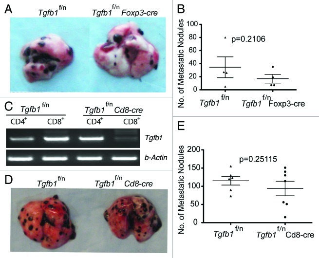
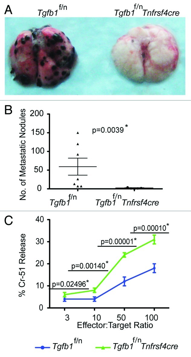
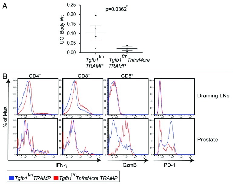
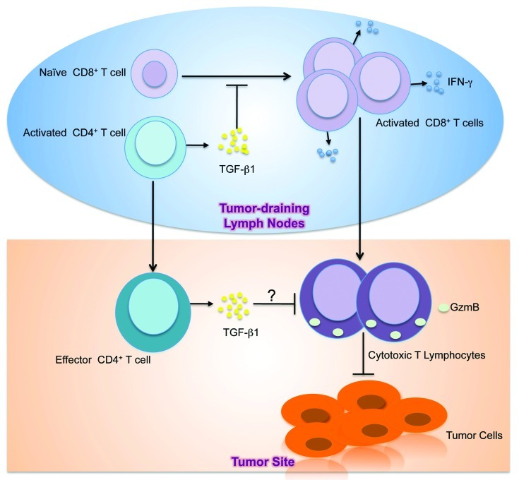
TGFβ1 is a regulatory cytokine with a crucial function in the control of T cell tolerance to tumors. Our recent study revealed that T cell-produced TGFβ1 is essential for inhibiting cytotoxic T cell responses to tumors. However, the exact TGFβ1-producing T cell subset required for tumor immune evasion remains unknown. Here we showed that deletion of TGFβ1 from CD8(+) T cells or Foxp3(+) regulatory T (Treg) cells did not protect mice against transplanted tumors. However, absence of TGFβ1 produced by activated CD4(+) T cells and Treg cells inhibited tumor growth, and protected mice from spontaneous prostate cancer. These findings suggest that TGFβ1 produced by activated CD4(+) T cells is a necessary requirement for tumor evasion from immunosurveillance.


Figures

None
Figure 1. Treg cell- or CD8+ T cell-derived TGFβ1 is dispensable for promoting tumor growth (A and B) B16-OVA melanoma cells were injected into age-matched Tgfb1f/n and Tgfb1f/nFoxp3-cre mice and pulmonary metastatic nodules assessed 15– 21 days later. (C) RT-PCR showing the deletion of Tgfb1 gene from FACS-sorted CD4+ and CD8+ T cells isolated from Tgfb1f/nCd8-cre mice and Tgfb1f/n control littermates (D and E) B16-OVA melanoma cells were injected into age-matched Tgfb1f/n and Tgfb1f/nCd8-cre mice and pulmonary metastatic nodules assessed 15–21 days later.
None
Figure 2. Deficiency of TGFβ1 in activated CD4+ T cells and Treg cells enhances tumor-specific CTL responses (A and B) B16-OVA melanoma cells were injected into age-matched Tgfb1f/n and Tgfb1f/nTnfrsf4-cre mice and pulmonary metastatic nodules assessed 15–21 days later. The p value between two groups of pulmonary metastatic nodules is shown (Students t-test) *Depicts statistically significant difference. (C) Tgfb1f/n and Tgfb1f/nTnfrsf4-cre mice were challenged intraperitoneally with 1 × 10⁶ El-4 tumor cells and the splenocytes were used as effectors against EL-4 targets at the indicated effector:target ratios. Chromium-51 (Cr-51) release assay 10 days after tumor challenge is shown. The p values between the two groups of number of metastatic nodules and percent Chromium-15 release are shown (Students t-test). *Depicts statistically significant difference.
None
Figure 3. Deficiency of TGF-β1 in activated CD4+ T cells and Treg cells inhibits tumor development (A) Tgfb1f/n-TRAMP and Tgfb1f/nTnfrsf4-cre-TRAMP mice were evaluated for tumor development at 8 months of age. The weights (Wt) of urogenital tracts (UG) normalized to body Wt ± s.e.m of Tgfb1f/n TRAMP (n = 4) and Tgfb1f/nTnfrsf4-cre TRAMP (n = 3) mice are shown. The p value between two groups of tumor burden is shown (Students t-test). *Depicts statistically significant difference. (B) Flow cytometric analysis examining the expression of interferon gamma (IFNγ), granzyme B (GzmB) and PD-1 proteins in CD4+ and CD8+ T cells in the draining lymph nodes and prostates of Tgfb1f/n TRAMP and Tgfb1f/nTnfrsf4cre-TRAMP mice. For IFNγ expression, T cells were re-stimulated in vitro for five hours with phorbol 12-myristate 13-acetate (PMA), ionomycin and GolgiStop. Representatives of two independent experiments are shown.
None
Figure 4. Model of TGFβ-mediated inhibition of antitumor T cell response. In the tumor-draining lymph nodes, CD4+ T cells become activated in response to tumor-associated antigens. Activated CD4+ T cells in the tumor draining lymph nodes secrete TGFβ1 that suppresses the activation, proliferation and differentiation of naïve tumor-specific CD8+ T cells into IFNγ-secreting T cells. Consequently, activated CD8+ T cells that migrate to the tumor site as effectors fail to elaborate CTL functions including GzmB expression. In addition, effector CD4+ T cells at the tumor site might also produce TGFβ1 to suppress the effector activities of CD8+ T cells. The net result is CTL dysfunction, which allows tumors to grow.

Similar articles

Cited by

References

    1. Burnet FM. The concept of immunological surveillance. Prog Exp Tumor Res. 1970;13:1–27. - PubMed
    1. Klein G, Klein E. Immune surveillance against virus-induced tumors and nonrejectability of spontaneous tumors: contrasting consequences of host versus tumor evolution. Proc Natl Acad Sci USA. 1977;74:2121–5. doi: 10.1073/pnas.74.5.2121. - DOI - PMC - PubMed
    1. Boon T, Coulie PG, Van den Eynde BJ, van der Bruggen P. Human T cell responses against melanoma. Annu Rev Immunol. 2006;24:175–208. doi: 10.1146/annurev.immunol.24.021605.090733. - DOI - PubMed
    1. Lee PP, Yee C, Savage PA, Fong L, Brockstedt D, Weber JS, et al. Characterization of circulating T cells specific for tumor-associated antigens in melanoma patients. Nat Med. 1999;5:677–85. doi: 10.1038/9525. - DOI - PubMed
    1. Savage PA, Vosseller K, Kang C, Larimore K, Riedel E, Wojnoonski K, et al. Recognition of a ubiquitous self antigen by prostate cancer-infiltrating CD8+ T lymphocytes. Science. 2008;319:215–20. doi: 10.1126/science.1148886. - DOI - PubMed

Publication types