. 2012 Jun 21:13:18.
doi: 10.1186/1471-2199-13-18.

PIM-1 kinase interacts with the DNA binding domain of the vitamin D receptor: a further kinase implicated in 1,25-(OH)2D3 signaling

Affiliations

PIM-1 kinase interacts with the DNA binding domain of the vitamin D receptor: a further kinase implicated in 1,25-(OH)2D3 signaling

Christina J Maier et al. BMC Mol Biol. .

Abstract

Background: The vitamin D3 receptor (VDR) is responsible for mediating the pleiotropic and, in part, cell-type-specific effects of 1,25-dihydroxyvitamin D3 (calcitriol) on the cardiovascular and the muscle system, on the bone development and maintenance, mineral homeostasis, cell proliferation, cell differentiation, vitamin D metabolism, and immune response modulation.

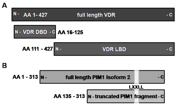
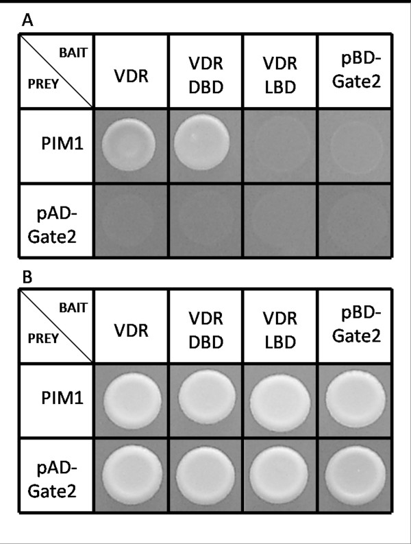
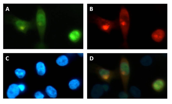
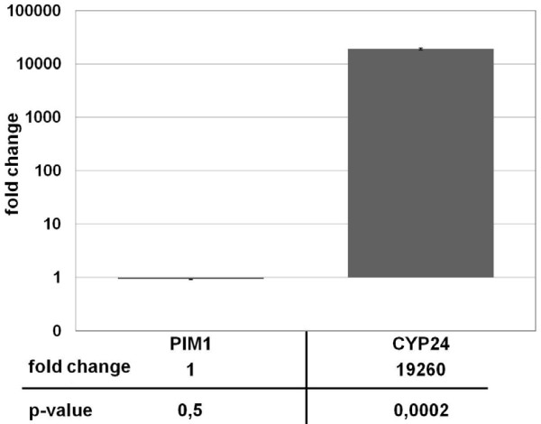
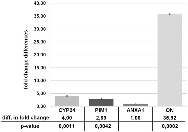
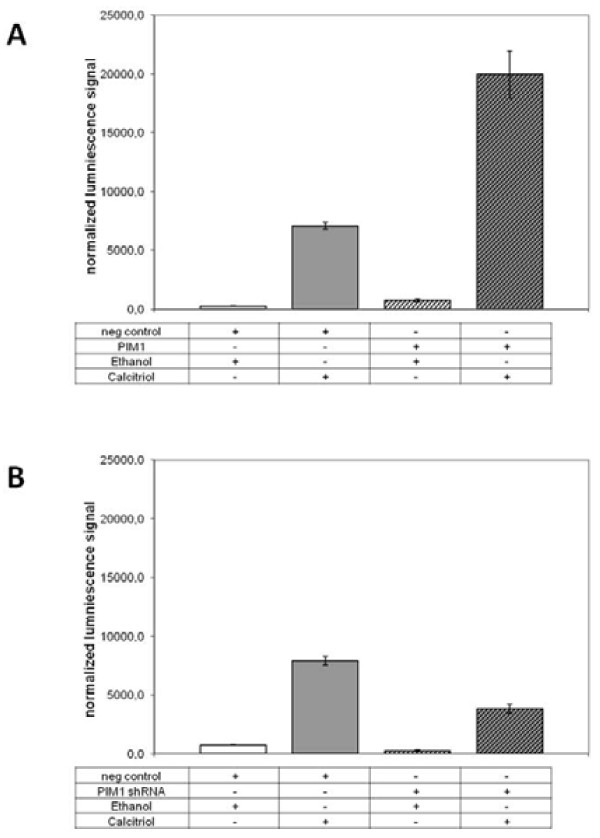
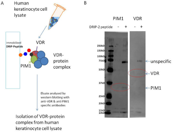
Results: Based on data obtained from genome-wide yeast two-hybrid screenings, domain mapping studies, intracellular co-localization approaches as well as reporter transcription assay measurements, we show here that the C-terminus of human PIM-1 kinase isoform2 (amino acid residues 135-313), a serine/threonine kinase of the calcium/calmodulin-regulated kinase family, directly interacts with VDR through the receptor's DNA-binding domain. We further demonstrate that PIM-1 modulates calcitriol signaling in HaCaT keratinocytes by enhancing both endogenous calcitriol response gene transcription (osteopontin) and an extrachromosomal DR3 reporter response.

Conclusion: These results, taken together with previous reports of involvement of kinase pathways in VDR transactivation, underscore the biological relevance of this novel protein-protein interaction.


Figures

Figure 1
Figure 1
Illustration of the used VDR/Pim-1 variants. (A) In addition to full-length VDR the DNA Binding Domain (DBD: AA 16–125) and the Ligand Binding Domain (LBD: AA 111–427) of the VDR were subjected for Y2H analysis as bait molecules. They were tested with (B) full-length PIM-1 isoform 2 as well as the truncated version of PIM-1 (AA 135–313).
Figure 2
Figure 2
Domain analysis of PIM-1 – VDR interaction. (A) Yeast cells co-transformed with PIM-1-pAD-Gate2 and VDR-Fullength-pBD-Gate2 grow directly on PPI selective medium as well as yeast cells co-transformed with PIM-1-pAD-Gate2 and DBD-pBD-Gate2. Cells co-transformed with PIM-1-pAD-Gate2 and LBD-pBD-Gate2, PIM-1-pAD-Gate2 and pBD-Gate2, pAD-Gate2 and VDR-Fullength-pBD-Gate2, pAD-Gate2 and DBD-pBD-Gate2, or pAD-Gate2 and LBD-pBD-Gate2 show no growth on PPI selective medium. (B) All combination show strong growth on medium selective for positive transformation.
Figure 3
Figure 3
PIM-1 and VDR co-localization in human keratinocytes. HaCaT cells were co-transfected with PIM-1-DsRed and VDR-YFP fusion constructs. (A) Image of the YFP-fluorescence of VDR. (B) Image of the red fluorescence of PIM-1. (C) Image of blue DAPI fluorescence. (D) Overlay of panels A, B and C showing colocalization of PIM-1 and VDR in the nucleus.
Figure 4
Figure 4
Real-Time analysis of PIM-1 trancription in human keratinocytes upon calcitriol stimulation. HaCAT cells were induced either with 0.2 μM calcitriol or with an equal amount of ethanol for 16 h. Levels of gene expression were analyzed by Real-Time PCR, and are shown as fold change differences between vehicle and calcitriol induction relative to the housekeeping gene ANXA1. The expression levels of PIM-1 show no significant differences between calcitriol and ethanol induction (p-value = 0,5), whereas CYP24A1 gene expression shows an enormous increase with calcitriol induction (p-value = 0.0002). Error bars represent the standard error of the mean (SEM) of n = 3 values.
Figure 5
Figure 5
Endogenous VDR target gene mRNA expression in PIM-1 overexpressing human keratinocytes. With PIM-1 overexpression the levels of CYP24A1, PIM-1, and OPN are increased after normalization with the housekeeping gene ANXA1. Results are shown as fold change of PIM-1 overexpression relative to cells transfected with the empty vector. The significant differences of PIM-1 overexpression compared to the empty control vector samples was considered as true by the low p-values. Error bars represent the SEM of n = 3 values.
Figure 6
Figure 6
PIM-1 dependent VDR-transactivation. (A) PIM-1 overexpression enhances calcitriol-induced extrachromosomal DR3 reporter response. The luminescence signal increased after induction of the transfected HaCaT cells with 0.2 μM calcitriol for 16 h but not following the addition of an equal amount of ethanol for the same time. The cells transfected with PIM-1 showed a 2.8-fold increase in response to calcitriol compared with the cells transfected with empty vector (pDEST26/neg control). Also, similar signal intensities were observed for vehicle-treated cells transfected with PIM-1 vs. vehicle-treated cells transfected with the empty vector. (B) PIM-1 knockdown through shRNA plasmid constructs reduces the calcitriol-induced extrachromosomal DR3 reporter response to the half compared with the cells transfected with the neg. control shRNA plasmid. Furthermore, about the similar decrease was observed by calcitriol-independent extrachromosomal DR3 reporter response. Vehicle-treated cells transfected with shRNA against PIM-1 vs. vehicle-treated cells transfected with the empty vector (pGFP-V-RS/neg. control) reduces the signal intensity. Error bars represent the SEM of n = 3 values.
Figure 7
Figure 7
Isolation of VDR-protein complex from human keratinocyte cell lysate. (A) Overview of the experimental procedure of the pull-down assay. (B) Western blot analysis with anti-PIM1 and anti-VDR antibody. PIM-1 and VDR were observed only in the lysate captured by the DRIP-2 peptide, while in the control lysate without peptide only unspecific lanes appeared. The expected sizes were for VDR about 48 kD, for PIM-1 about 34 kD and for the putative VDR/PIM-1 complex (unspecific band) respectively about 82 kD.

Similar articles

Cited by

References

    1. Carlberg C, Seuter S. The vitamin D receptor. Dermatol Clin. 2007;25:515–523. doi: 10.1016/j.det.2007.06.004. viii. - DOI - PubMed
    1. Carlberg C. Ligand-mediated conformational changes of the VDR are required for gene transactivation. J Steroid Biochem Mol Biol. 2004;89-90:227–232. - PubMed
    1. Privalsky ML. The role of corepressors in transcriptional regulation by nuclear hormone receptors. Annu Rev Physiol. 2004;66:315–360. doi: 10.1146/annurev.physiol.66.032802.155556. - DOI - PubMed
    1. Boyan BD, Wang L, Wong KL, Jo H, Schwartz Z. Plasma membrane requirements for 1alpha,25(OH)2D3 dependent PKC signaling in chondrocytes and osteoblasts. Steroids. 2006;71:286–290. doi: 10.1016/j.steroids.2005.09.018. - DOI - PubMed
    1. Wali RK, Baum CL, Sitrin MD, Brasitus TA. 1,25(OH)2 vitamin D3 stimulates membrane phosphoinositide turnover, activates protein kinase C, and increases cytosolic calcium in rat colonic epithelium. J Clin Invest. 1990;85:1296–1303. doi: 10.1172/JCI114567. - DOI - PMC - PubMed

MeSH terms

LinkOut - more resources