. 2012 Jun 21:12:14.
doi: 10.1186/1472-6807-12-14.

Structure of human aspartyl aminopeptidase complexed with substrate analogue: insight into catalytic mechanism, substrate specificity and M18 peptidase family

Affiliations

Structure of human aspartyl aminopeptidase complexed with substrate analogue: insight into catalytic mechanism, substrate specificity and M18 peptidase family

Apirat Chaikuad et al. BMC Struct Biol. .

Abstract

Background: Aspartyl aminopeptidase (DNPEP), with specificity towards an acidic amino acid at the N-terminus, is the only mammalian member among the poorly understood M18 peptidases. DNPEP has implicated roles in protein and peptide metabolism, as well as the renin-angiotensin system in blood pressure regulation. Despite previous enzyme and substrate characterization, structural details of DNPEP regarding ligand recognition and catalytic mechanism remain to be delineated.

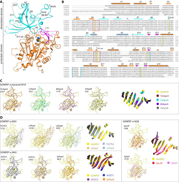
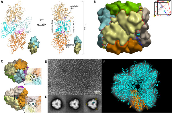
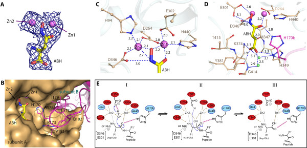
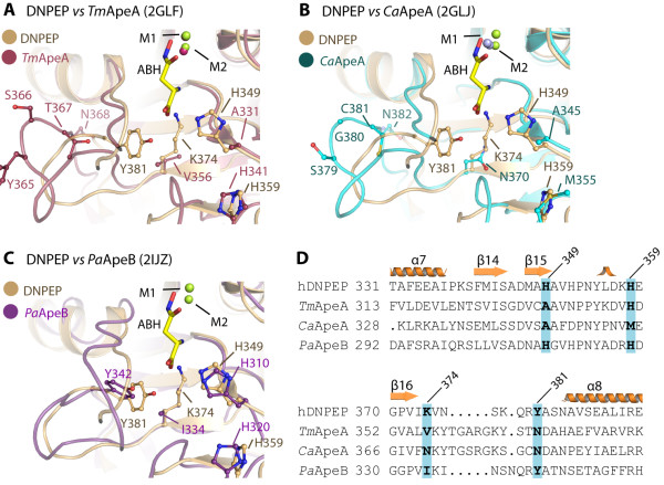
Results: The crystal structure of human DNPEP complexed with zinc and a substrate analogue aspartate-β-hydroxamate reveals a dodecameric machinery built by domain-swapped dimers, in agreement with electron microscopy data. A structural comparison with bacterial homologues identifies unifying catalytic features among the poorly understood M18 enzymes. The bound ligands in the active site also reveal the coordination mode of the binuclear zinc centre and a substrate specificity pocket for acidic amino acids.

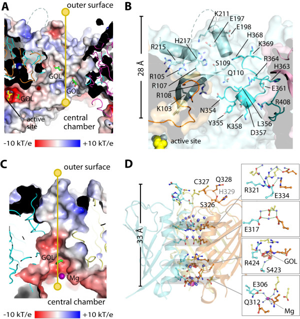
Conclusions: The DNPEP structure provides a molecular framework to understand its catalysis that is mediated by active site loop swapping, a mechanism likely adopted in other M18 and M42 metallopeptidases that form dodecameric complexes as a self-compartmentalization strategy. Small differences in the substrate binding pocket such as shape and positive charges, the latter conferred by a basic lysine residue, further provide the key to distinguishing substrate preference. Together, the structural knowledge will aid in the development of enzyme-/family-specific aminopeptidase inhibitors.


Figures

Figure 1
Figure 1
Overview of hDNPEP structure. (A) hDNPEP protomer organizes into the dimerization (blue) and proteolytic domains, the latter is further comprised of subdomain A (orange) and subdomain B (magenta). Zinc ions are shown as blue spheres and the ABH ligand as yellow sticks. (B) Sequence alignment of DNPEP (human, h; bovine, b) and M18 aminopeptidases from yeast Saccharomyces cerevisiae Lap4 (ScLAP4), yeast Ape4 (ScApe4) and two bacterial enzymes (Pseudomonas aeruginosa PaApeB and Thermotoga maritima TmApeA). Secondary structure elements, catalytic residues (yellow) and residues in the P1 substrate pocket (cyan) of hDNPEP are highlighted. Structural superimposition of hDNPEP with bacterial M18 APs (C) and with M20, M28 and M42 representatives from the MH clan (D) reveals highly conserved topology of the proteolytic domain. The superimposed structures include M18 APs: Thermotoga maritima ApeA (TmApeA), Clostridium acetobutylicum ApeA (CaApeA), Borrelia burgdorferi ApeA (BbApeA), Pseudomonas aeruginosa ApeB (PaApeB); M20 APs: Pseudomonas CPG2 (PsCPG2), Lactobacillus delbrueckii PepV (LdPepV), Legionella pneumophila DapE (LpDapE); M28; Aeromonas proteolytica LAP (ApLAP), Streptomyces griseus Ap (SgApS); and M42: Pyrococcus horikoshii TET1 and TET2 (PhTET1, PhTET2), Streptococcus pneumonia PepA (SpPepA). PDB IDs of all structures are given.
Figure 2
Figure 2
Dodecameric assembly. (A) A dimer building block of hDNPEP. (B) Surface representation of the arrangement of six dimers into a tetrahedron. The dimer building block, each coloured differently, is delineated by a red dotted line. The black-dotted line indicates the monomer-monomer interface within a dimer. Each dimer sits diagonally on six faces of a cubic box that encases the tetrahedron (red line, inset). Asterisks indicate positions of the narrow (yellow) and wide (blue) channels, which are located at 3-fold axes (arrow). (C) The openings of the narrow (top) and wide (bottom) channels. (D) Electron micrograph of negatively stained hDNPEP. (E) Examples of 2D classification images with a view down the wide channel, the 3-fold symmetry imposed in the right panel. (F) Fitting of hDNPEP crystal structure onto the 2D projection.
Figure 3
Figure 3
Architecture of wide (top) and narrow (bottom) channels. (A) Electrostatic surface of the wide channel with yellow line indicating the 28-Å route connecting the exterior and the central chamber. (B) Details of residues lining the wide channel, showing only one set of residues from one dimer. (C) Electrostatic surface along the 33-Å length of the narrow channel. (D) Details of residues lining the narrow channel. Bound glycerol (GOL) and magnesium (Mg) molecules are shown in stick and sphere, respectively.
Figure 4
Figure 4
Active site of hDNPEP. (A) |FO|- |Fc| omit map contoured at 3σ for zinc ions and ABH molecules. (B) Insertion of the β8-β9 loop from the neighboring subunit (magenta) completes the active site construction. Bonding interactions at (C) the binuclear metal catalytic centre and (D) the P1 substrate pocket in the hDNPEP structure. (E) Proposed catalytic mechanism for hDNPEP. The substrate peptide N-terminus is shown in both amine and its protonated form, which can engage in different interactions.
Figure 5
Figure 5
Structural comparison of the P1 pockets in hDNPEP and bacterial M18 members. (A-C) Three bacterial M18 AP structures (PDB ids in brackets) are superimposed onto hDNPEP, with particular focus on the P1 substrate pocket. This highlights variations not only in shape but also residue compositions for the P1 pocket, and may be correlated with different substrate specificities and enzyme activities among M18 enzymes. (D) A structure-based sequence alignment shows no conservation of four key residues of the hDNPEP P1 pocket among the bacterial M18 enzymes, in particular Lys374 likely to be a determinant for acidic amino acid preference.

Similar articles

Cited by

References

    1. Taylor A. Aminopeptidases: structure and function. FASEB J. 1993;7(2):290–298. - PubMed
    1. Mucha A, Drag M, Dalton JP, Kafarski P. Metallo-aminopeptidase inhibitors. Biochimie. 2010;92(11):1509–1529. doi: 10.1016/j.biochi.2010.04.026. - DOI - PMC - PubMed
    1. Li L, Wang J, Cooper MD. cDNA cloning and expression of human glutamyl aminopeptidase (aminopeptidase A) Genomics. 1993;17(3):657–664. doi: 10.1006/geno.1993.1386. - DOI - PubMed
    1. Banegas I, Prieto I, Vives F, Alba F, de Gasparo M, Segarra AB, Hermoso F, Duran R, Ramirez M. Brain aminopeptidases and hypertension. J Renin Angiotensin Aldosterone Syst. 2006;7(3):129–134. doi: 10.3317/jraas.2006.021. - DOI - PubMed
    1. Wright JW, Harding JW. Brain renin-angiotensin–a new look at an old system. Prog Neurobiol. 2011;95(1):49–67. doi: 10.1016/j.pneurobio.2011.07.001. - DOI - PubMed

Publication types

Associated data

LinkOut - more resources