. 2012 Aug;181(2):508-14.
doi: 10.1016/j.ajpath.2012.05.009. Epub 2012 Jun 19.

Activated B cells in the granulomas of nonhuman primates infected with Mycobacterium tuberculosis

Affiliations

Activated B cells in the granulomas of nonhuman primates infected with Mycobacterium tuberculosis

Jia Yao Phuah et al. Am J Pathol. 2012 Aug.

Erratum in

  • Am J Pathol. 2012 Nov;181(5):1889

Abstract

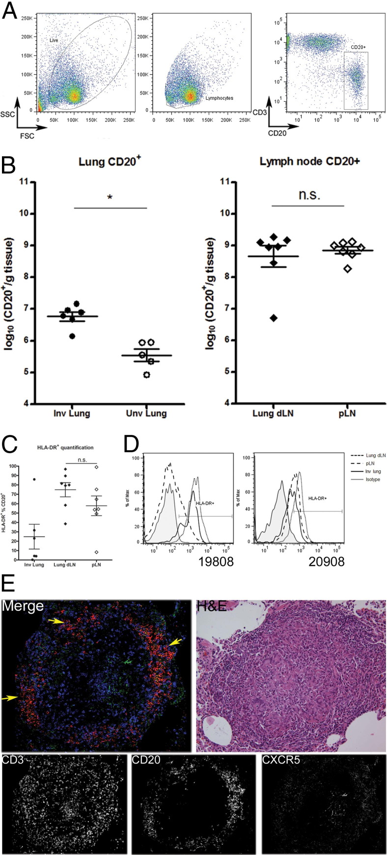
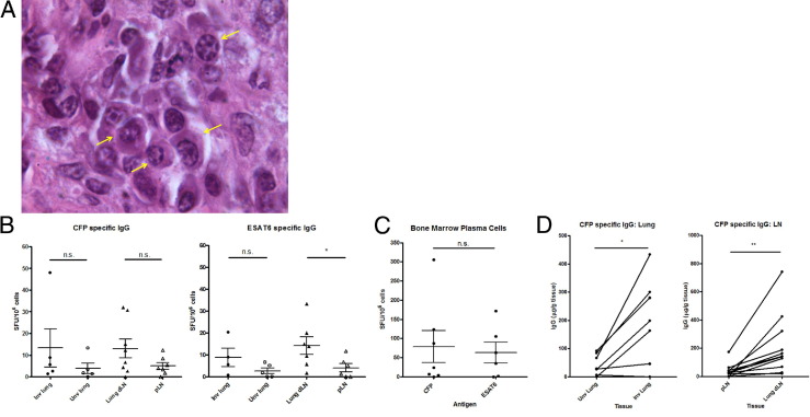
In an attempt to contain Mycobacterium tuberculosis, host immune cells form a granuloma as a physical and immunological barrier. To date, the contribution of humoral immunity, including antibodies and specific functions of B cells, to M. tuberculosis infection in humans remains largely unknown. Recent studies in mice show that humoral immunity can alter M. tuberculosis infection outcomes. M. tuberculosis infection in cynomolgus macaques recapitulates essentially all aspects of human tuberculosis. As a first step toward understanding the importance of humoral immunity to control of M. tuberculosis infection in primates, we characterized the B-cell and plasma-cell populations in infected animals and found that B cells are present primarily in clusters within the granuloma. The B-cell clusters are in close proximity to peripheral node addressin-positive cells and contain cells positive for Ki-67, a proliferation marker. Granuloma B cells also express CXCR5 and have elevated HLA-DR expression. Tissues containing M. tuberculosis bacilli had higher levels of M. tuberculosis-specific IgG, compared with uninvolved tissue from the same monkeys. Plasma cells detected within the granuloma produced mycobacteria-specific antibodies. Together, these data demonstrate that B cells are present and actively secreting antibodies specific for M. tuberculosis antigens at the site of infection, including lung granulomas and thoracic lymph nodes. These antibodies likely have the capacity to modulate local control of infection in tissues.


Figures

Figure 1
Figure 1
CD20+ B-cell clusters are present within macaque granulomas and have germinal center characteristics. A: A solid granuloma (merged image) with some caseum and a small mineralized focus was stained for CD20 (red), CD3 (green), and nuclei with DRAQ5 (blue) to show the distribution of immune cells with respect to granuloma morphology (H&E stain). CD20+ B cells and CD3+ T cells (depicted as CD20 and CD3 grayscale images, respectively) are found primarily within the lymphocytic cuff. B cells generally form clusters, but are also present as discrete cells, whereas T cells are usually dispersed. B: A caseous granuloma section with a mineralized core (H&E stain) (right panel) was stained for CD20 (red), PNAd (green), and Ki-67 (blue) to identify B cells, high endothelial venules, and proliferating cells, respectively. Two CD20+ clusters were imaged in detail for germinal center markers (boxed areas, left panel). C: A mature bronchus-associated lymphoid tissue cluster exhibits prominent Ki-67 staining within CD20+ cells in close association with PNAd+ high endothelial venules (arrow), reminiscent of B-cell follicles within lymph nodes. D: B-cell clusters within the lymphocytic cuff of the granuloma contain some Ki-67+ nuclei, with some sporadic PNAd staining. Merged images in C and D depict CD20 (red), PNAd (green), and Ki-67 (blue). Images shown are composites of 20 to 40 images. Original magnification: ×20 (B) or ×40 (C and D).
Figure 2
Figure 2
Activated B cells are present in mycobacteria-containing tissue. A: Lymphocytes were gated based on size [forward scatter (FSC)] and granularity [side scatter (SSC)]. B cells were further identified based on CD20+ expression. The fluorescence-activated cell sorting FACS plot depicts the cellular profile of a lymph node sample. B: Involved lung samples (Inv lung) have at least a 10-fold increase in CD20+ B cells, compared with uninvolved lung samples (Unv lung). B-cell numbers were similar within pLN and dLN. Each data point represents one tissue sample from one macaque. C: HLA-DR expression by CD20+ cells from dLN, although slightly elevated, was not significantly different, compared with cells from pLN. D: In contrast, analysis of tissues from two representative macaques (19808 and 20908) indicated elevated HLA-DR expression within CD20+ cells of dLN, compared with pLN of the same macaque. CD20+ from involved lung samples also stained positive for HLA-DR (uninvolved lung lacked sufficient B-cell numbers for analysis). E: Staining for CD3 (blue), CD20 (red), and CXCR5 (green) was done to determine whether B-cell clusters within the granuloma behave as ectopic germinal centers. CXCR5 was used to identify lymphocytes that are actively homing toward lymphoid tissue to identify germinal center reactions. Most of the B-cell clusters stained positive for CXCR5 (arrows), indicating activated CD20+ cells that are capable of homing toward lymphoid tissue. Data are expressed as means ± SEM (B and C). *P < 0.05. n = 7 (B and C). n.s., nonsignificant.
Figure 3
Figure 3
Granulomas contain plasma cells secreting mycobacteria-specific IgG. A: Plasma cells with classical histological features including clock-faced nuclei, perinuclear hof, and higher ratio of cytoplasm to nuclei were identified within macaque granulomas (arrows). B: Lung and lymph node samples were used in a plasma cell ELISPOT assay to quantitatively assess numbers of IgG-secreting plasma cells specific for CFP and ESAT6. Each data point represents a single sample from one macaque. Some groups have fewer than seven data points because not all samples yielded sufficient cells perform the ELISPOT assay. Significantly more ESAT6-specific plasma cells were detected within dLN, compared with pLN. ESAT6-specific IgG plasma-cell numbers within lung samples appeared to be greater within involved lung, but the difference was not statistically significant. C: Levels of both CFP and ESAT6 IgG-specific plasma cells within bone marrow from the sternum were similar. D: A pairwise comparison of CFP-specific IgG in tissue supernatants was done using ELISA. Each data point represents a single sample from one macaque. Each line represents a paired sample of uninvolved lung (Unv) and involved lung (Inv) or pLN and dLN. Involved lung or dLN contained higher levels of CFP-specific IgG, compared with uninvolved lung and pLN, respectively. Data are presented as means ± SEM. *P < 0.05; **P < 0.01. n = 7 (B and C); n = 14 (D).

References

    1. Wright A., Zignol M., WHO staff . WHO Press; Geneva: 2008. Anti-Tuberculosis Drug Resistance in the World. Fourth global report. The WHO/IUATLD Global Project on Anti-tuberculosis Drug Resistance Surveillance 2002–2007.
    1. Brewer T.F., Heymann S.J. To control and beyond: moving towards eliminating the global tuberculosis threat. J Epidemiol Community Health. 2004;58:822–825. - PMC - PubMed
    1. Maartens G., Wilkinson R.J. Tuberculosis. Lancet. 2007;370:2030–2043. - PubMed
    1. Saunders B.M., Britton W.J. Life and death in the granuloma: immunopathology of tuberculosis. Immunol Cell Biol. 2007;85:103–111. - PubMed
    1. Scanga C.A., Mohan V.P., Yu K., Joseph H., Tanaka K., Chan J., Flynn J.L. Depletion of CD4(+) T cells causes reactivation of murine persistent tuberculosis despite continued expression of interferon gamma and nitric oxide synthase 2. J Exp Med. 2000;192:347–358. - PMC - PubMed

Publication types

MeSH terms