. 2012 Sep 15;446(3):455-67.
doi: 10.1042/BJ20111961.

Novel atypical PKC inhibitors prevent vascular endothelial growth factor-induced blood-retinal barrier dysfunction

Affiliations

Novel atypical PKC inhibitors prevent vascular endothelial growth factor-induced blood-retinal barrier dysfunction

Paul M Titchenell et al. Biochem J. .

Abstract

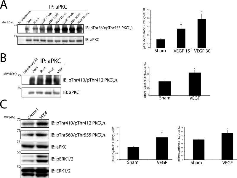
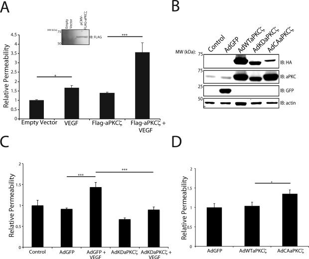
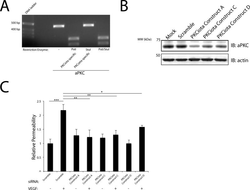
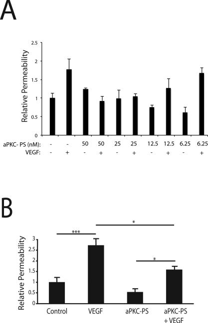
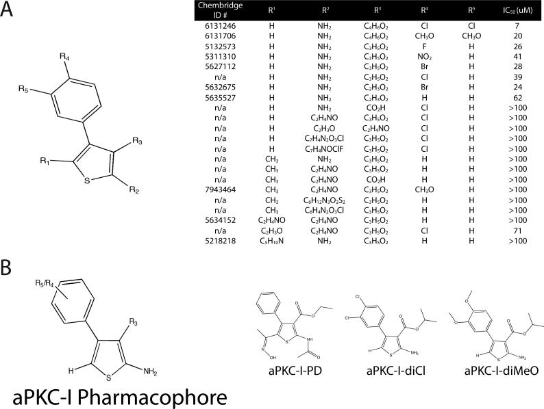
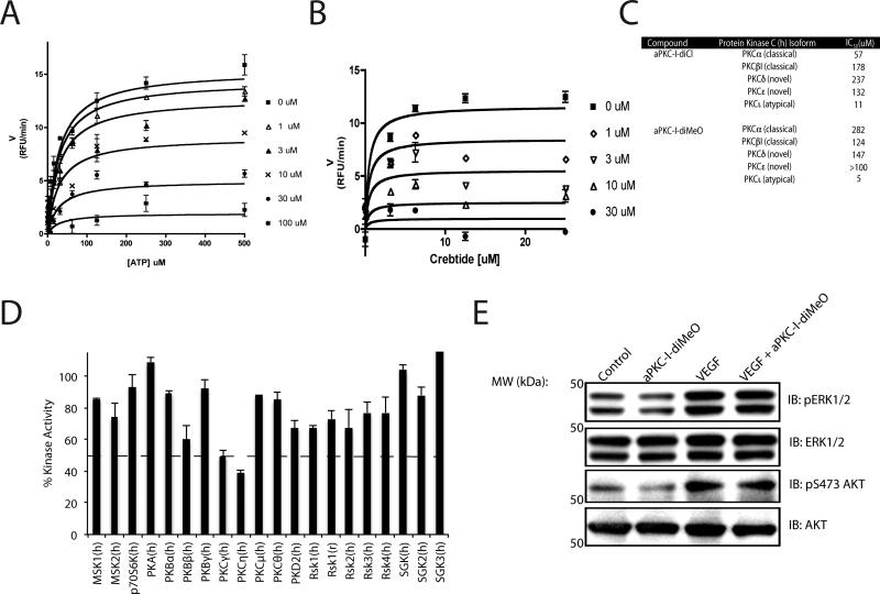
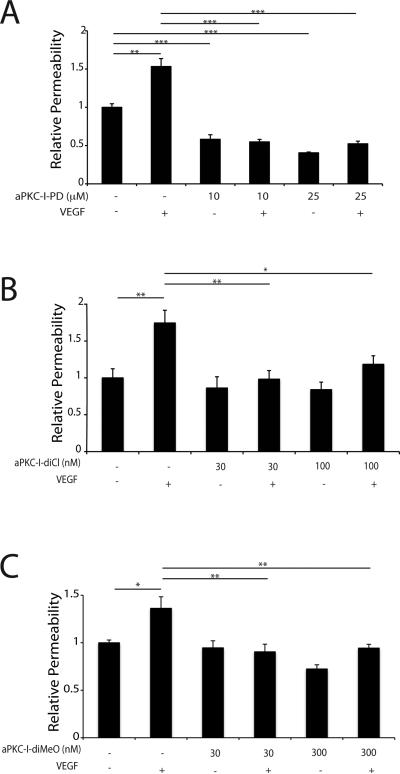
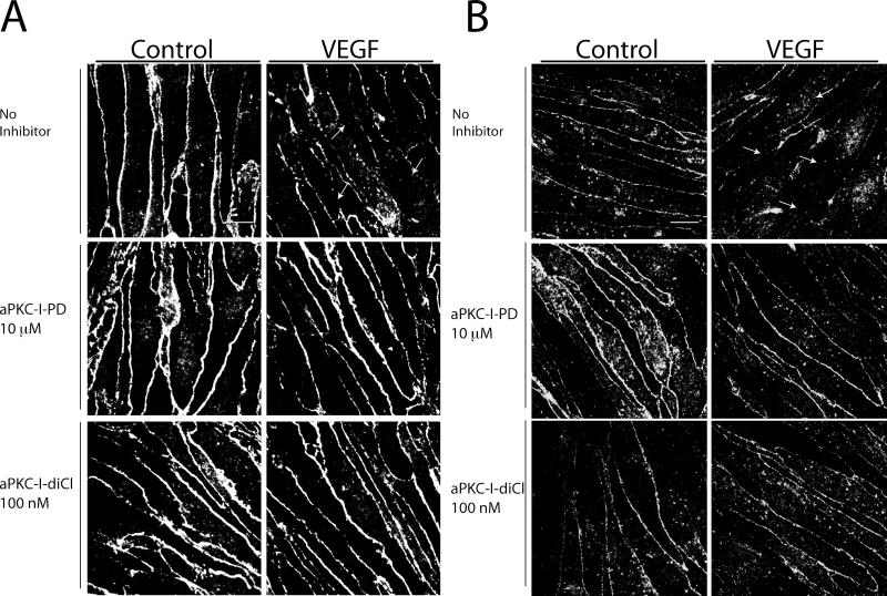
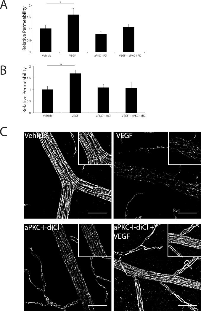
Pro-inflammatory cytokines and growth factors such as VEGF (vascular endothelial growth factor) contribute to the loss of the BRB (blood-retinal barrier) and subsequent macular oedema in various retinal pathologies. VEGF signalling requires PKCβ [conventional PKC (protein kinase C)] activity; however, PKCβ inhibition only partially prevents VEGF-induced endothelial permeability and does not affect pro-inflammatory cytokine-induced permeability, suggesting the involvement of alternative signalling pathways. In the present study, we provide evidence for the involvement of aPKC (atypical PKC) signalling in VEGF-induced endothelial permeability and identify a novel class of inhibitors of aPKC that prevent BRB breakdown in vivo. Genetic and pharmacological manipulations of aPKC isoforms were used to assess their contribution to endothelial permeability in culture. A chemical library was screened using an in vitro kinase assay to identify novel small-molecule inhibitors, and further medicinal chemistry was performed to delineate a novel pharmacophore. We demonstrate that aPKC isoforms are both sufficient and required for VEGF-induced endothelial permeability. Furthermore, these specific, potent, non-competitive, small-molecule inhibitors prevented VEGF-induced tight junction internalization and retinal endothelial permeability in response to VEGF in both primary culture and in rodent retina. The results of the present study suggest that aPKC inhibition with 2-amino-4-phenyl-thiophene derivatives may be developed to preserve the BRB in retinal diseases such as diabetic retinopathy or uveitis, and the BBB (blood-brain barrier) in the presence of brain tumours.


Figures

Fig. 1
Fig. 1. VEGF activates aPKC isoforms in both the rodent retina and in primary retinal endothelial cells
(A) VEGF was intra-vitreally injected for the indicated time and retinas were excised from Sprague-Dawley rats. aPKC was immunoprecipitated where indicated and immunoblotted for the autophosphorylation residue, pThr560/Thr555. (B) VEGF was intra-vitreally injected for 15 min and retinas were excised from Sprague-Dawley rats. aPKC was immunoprecipitated where indicated and immunoblotted for the pThr410/Thr412. Successful immunoprecipiation was verified with immunoblotting for aPKC. Quantitation of the results (mean ± SEM) from three independent experiments and are expressed relative to the control with a total of n≥8. *, p<0.05, **p<0.01. (C) BREC were treated with VEGF (50 ng/ml) and lysates were subjected to immunoblotting for pThr410/Thr412 and pThr560/Thr555 PKCζ/ι, aPKC, pERK1/2, and ERK1/2 as described (methods). Quantitation of the results are expressed as the mean relative to the control, error bars represent ± SEM. n≥8. *, p<0.05, ** p<0.01.
Fig. 2
Fig. 2. aPKC kinase activity is both sufficient and required for VEGF-induced retinal endothelial permeability in primary culture
(A) Primary BREC were transfected with the plasmid pCMV-FLAG-aPKCζ or empty vector where indicated and grown to confluence on 0.4 μm Transwell filters.Immunoblot analysis utilizing Flag antibody showed successful expression of transgene. After 24 h serum deprivation, cells on filters were treated with 50 ng/ml VEGF as indicated. Permeability to 70 kDa RITC-Dextran tracer was measured over 4 hour time course. (B) BREC were transduced with recombinant adenoviruses and an immunoblot utilizing HA tag was performed to demonstrate successful transduction. Total aPKC content was monitored to demonstrate extent of transgene overexpression compared to control. (C) BREC were grown on transwells as above and infected with AdKDaPKCζ 6 h prior to a 24 h serum deprivation. Permeability to 70 kDa RITC-Dextran tracer was measured over 4 hour time course following VEGF treatment. (D) BREC were grown on transwells as above and infected with AdWTaPKCζ and AdCAaPKCζ. Permeability to 70 kDa RITC-Dextran tracer was measured over 4-hour time course following VEGF treatment. All results are expressed as the mean relative to the control with a total of n≥8, error bars represent ± SEM. Average Po values for Control and AdGFP were 2.0×10⁻⁶ (cm/s) and 1.47×10⁻⁶ (cm/s). ***, p<0.001 *, p<0.05.
Fig. 3
Fig. 3. PKCι mediates VEGF-induced retinal endothelial permeability in BREC
(A) PCR was performed using homologous primers for aPKCζ/ι that contained a unique restriction enzyme site to differentiate between the isoforms (Pst1 PKCι) and (Stu1 PKCζ). The amplicon generated was then subjected to restriction enzyme digestion. (B) BREC were transfected with either 100 nM Scramble or PKCι Constructs A/C/D and subjected to immunoblot analysis for aPKC. Actin served as a loading control. (C) BREC were transfected with either Scramble or with PKCι Construct A/C/D for 72 h. Permeability to 70 kDa RITC-Dextran tracer was measured over 4-hour time course following VEGF treatment. The results are expressed as the mean, relative to the control, error bars represent ± SEM. n≥3. Average Po values for Scr were 6×10⁻⁷ (cm/s) *, p<0.05, **,p<0.01, ***,p<0.001.
Fig. 4
Fig. 4. aPKC peptide inhibitor blocks the VEGF-induced increase in endothelial permeability
(A) BREC were grown to confluence on 0.4 μm Transwell filters then stepped-down for 24 h. BREC were treated for 30 min with the indicated concentration of aPKC peptide inhibitor (aPKC-PS) prior to 30 min treatment with 50 ng/ml VEGF where indicated. Permeability of the monolayer to 70kDa RITC-Dextran was measured. n≥4. (B) BREC were treated with 50 nM aPKC peptide inhibitor for 30 min prior to 30 min treatment with 50 ng/ml VEGF. n≥4. Permeability was measured as in (A). The results are expressed as the mean relative to the control, error bars represent ± SEM. ***, p<0.001; *, p<0.05.
Fig. 5
Fig. 5. Identification of novel, small molecule phenyl-thiophene inhibitors of aPKC isoforms
(A) Structure-activity relationships (SAR) were performed on phenyl-thiophene derivatives and IC50 determined using an in vitro luminescence based kinase assay against PKCζ using 200 ng/ml PKCζ, 25 uM CREBtide substrate, and 0.1 uM ATP. IC50 values were calculated using Prism Software with values fitted to a sigmoidal dose-response using variable slope. (n=6). (B) Pharmacophore of aPKC inhibitor noting essential substitutions and the structure of aPKC-I-PD, aPKC-I-diCl, and aPKC-I-diCl compounds are displayed where indicated. Compounds with Chembridge ID numbers (#) are provided and n/a (not applicable) applies to compounds synthesized in collaboration with Apogee, Inc. (methods).
Fig. 6
Fig. 6. Mechanism of action and specificity characterization of aPKC-Is
(A) ATP competition assay using ADP quest to measure initial velocities with 500 ng/ml PKCζ and excess CREBtide substrate or (B) with excess ATP. Ki was determined as described in methods. (C) IC50 profiling was performed with aPKC-I-diCl and aPKC-I-diMeO utilizing a radiolabeled kinase assay at Km app for ATP. (D) aPKC-I-diMeO at 100 uM (10-fold Ki) was screened against 20 kinases of the AGC super-familiy at Km app for ATP using a radiolabeled kinase assay. (E) BREC were pretreated for 30 min with 0.3 uM aPKC-I-diMeO and then stimulated with VEGF (50ng/ml) for 15 min. Lysates were subjected to immunoblotting for pERK1/2, ERK1/2, pS473-AKT, and AKT as described (methods).
Fig. 7
Fig. 7. Phenyl-thiophene inhibitors of aPKC isoforms prevent VEGF-induced retinal endothelial permeability in primary culture
BREC were grown to confluence on 0.4 μm Transwell filters then serum deprived for 24 h. (A) BREC were treated for 30 min with aPKC-I-PD (B) or aPKC-I-diCl (C) or aPKC-I-diMeO prior to 30 min treatment with 50 ng/ml VEGF where indicated. Permeability of the monolayer to 70kDa RITC-Dextran was measured. The results are expressed as the mean relative to the control with a total of n≥8, error bars represent ± SEM. Average Po value in (A) was 2.43×10⁻⁶ (cm/s), (B) 2.49×10⁻⁷ (cm/s) and (C) 1.46×10⁻⁶ (cm/s). ***, p<0.001; **, p<0.01; *, p<0.05.
Fig. 8
Fig. 8. Novel small molecule inhibitors of aPKC isoforms prevent the effect of VEGF on breakdown of the retinal endothelial tight junction complex
BREC were grown to confluence on coverslips and treated with 10 μM aPKC-I-PD, or 100 nM aPKC-I-diCl 30 min prior to 50 ng/ml VEGF. Cells were fixed 60 min after VEGF treatment and stained with primary antibodies against (A) ZO-1 or (B) occludin. Cells were imaged with confocal microscopy and are shown as the max projection of serial stacks. Images are representative of several similar fields. Scale bar = 10 μm.
Fig. 9
Fig. 9. aPKC-Is block the VEGF induction of retinal vascular permeability in vivo
Sprague-Dawley rats were injected intra-vitreally with (A) aPKC-I-PD at 25 μM or (B) aPKC-I-diCl at 25 μM with or without 50 ng VEGF per eye and compared to vehicle injection. After 3 h, rats received a femoral vein injection of 45 mg/kg Evans blue. After 2 h, animals were perfused with citrate/paraformaldehyde buffer for 2 min, retinas removed, dried and Evans blue extracted with formamide. Evans blue was quantified on a spectrophotometer and normalized to plasma levels measured pre-perfusion. Permeability was calculated and expressed as μl plasma/g dry weight/h circulation. The results are expressed as the mean relative to the control, error bars represent ± SEM. n≥8 per group, * p<0.05. (C) Retina flat mounts were prepared as described in Materials and Methods and immuno-stained for occludin. Images are representative of several fields and are shown as the max projection of serial stacks obtained from confocal imaging. Scale bar = 50 μm.

References

    1. Antonetti DA, Barber AJ, Bronson SK, Freeman WM, Gardner TW, Jefferson LS, Kester M, Kimball SR, Krady JK, LaNoue KF, Norbury CC, Quinn PG, Sandirasegarane L, Simpson IA. Diabetic retinopathy: seeing beyond glucose-induced microvascular disease. Diabetes. 2006;55:2401–2411. - PubMed
    1. Gardner TW, Larsen M, Girach A, Zhi X. Diabetic macular oedema and visual loss: relationship to location, severity and duration. Acta Ophthalmologica. 2009;87:709–713. - PubMed
    1. Sander B, Thornit DN, Colmorn L, Strom C, Girach A, Hubbard LD, Lund-Andersen H, Larsen M. Progression of diabetic macular edema: correlation with blood retinal barrier permeability, retinal thickness, and retinal vessel diameter. Investigative ophthalmology & visual science. 2007;48:3983–3987. - PubMed
    1. Antonetti DA, Klein R, Gardner TW. Diabetic retinopathy. N. Eng. J. Med. 2012;366:1227–1239. - PubMed
    1. Penn JS, Madan A, Caldwell RB, Bartoli M, Caldwell RW, Hartnett ME. Vascular endothelial growth factor in eye disease. Prog. Retin. Eye. Res. 2008;27:331–371. - PMC - PubMed

Publication types

MeSH terms