

Diabetes-impaired wound healing is improved by matrix therapy with heparan sulfate glycosaminoglycan mimetic OTR4120 in rats
- PMID: 22721969
- PMCID: PMC3447910
- DOI: 10.2337/db11-1329
Diabetes-impaired wound healing is improved by matrix therapy with heparan sulfate glycosaminoglycan mimetic OTR4120 in rats
Abstract
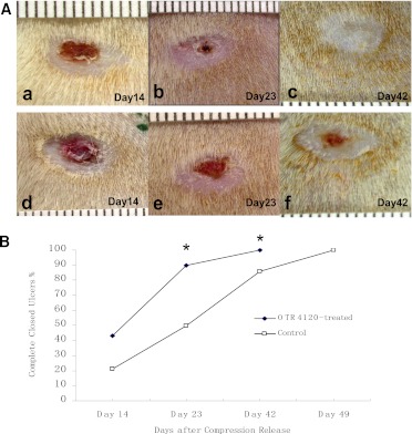
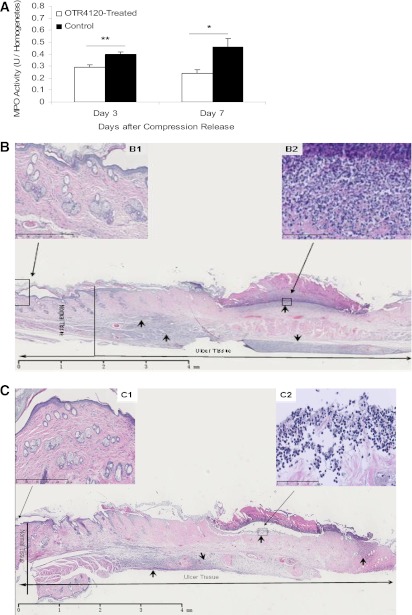
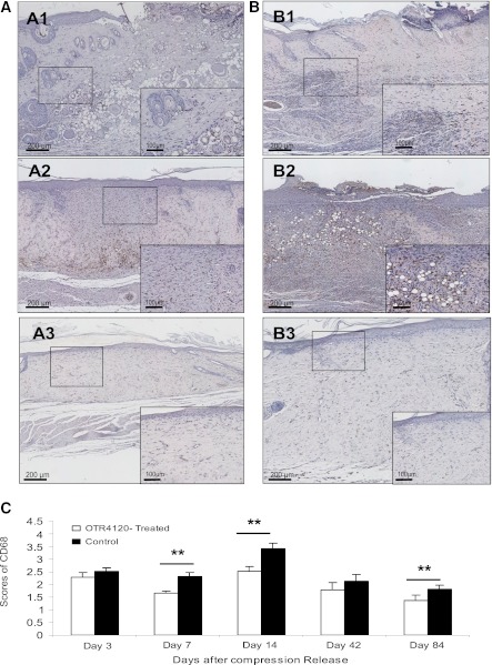
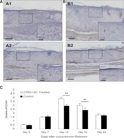
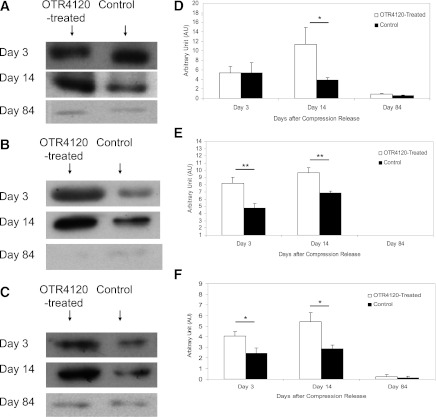
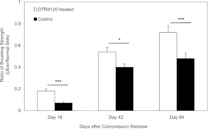
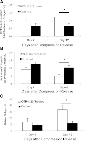
Wound healing in diabetes is frequently impaired, and its treatment remains a challenge. We tested a therapeutic strategy of potentiating intrinsic tissue regeneration by restoring the wound cellular environment using a heparan sulfate glycosaminoglycan mimetic, OTR4120. The effect of OTR4120 on healing of diabetic ulcers was investigated. Experimental diabetes was induced by intraperitoneal injection of streptozotocin. Seven weeks after induction of diabetes, rats were ulcerated by clamping a pair of magnet disks on the dorsal skin for 16 h. After magnet removal, OTR4120 was administered via an intramuscular injection weekly for up to 4 weeks. To examine the effect of OTR4120 treatment on wound heal-ing, the degree of ulceration, inflammation, angiogenesis, and collagen synthesis were evaluated. We found that OTR4120 treatment significantly reduced the degree of ulceration and the time of healing. These effects were associated with reduced neutrophil infiltration and macrophage accumulation and enhanced angiogenesis. OTR4120 treatment also increased the collagen content with an increase of collagen type I biosynthesis and reduction of collagen type III biosynthesis. Moreover, restoration of the ulcer biomechanical strength was significantly enhanced after OTR4120 treatment. This study shows that matrix therapy with OTR4120 improves diabetes-impaired wound healing.
Figures

Similar articles
-
Heparan sulfate glycosaminoglycan mimetic improves pressure ulcer healing in a rat model of cutaneous ischemia-reperfusion injury.Wound Repair Regen. 2011 Jul-Aug;19(4):505-14. doi: 10.1111/j.1524-475X.2011.00704.x. Epub 2011 Jun 7. Wound Repair Regen. 2011. PMID: 21649786
-
Stimulated neovascularization, inflammation resolution and collagen maturation in healing rat cutaneous wounds by a heparan sulfate glycosaminoglycan mimetic, OTR4120.Wound Repair Regen. 2009 Nov-Dec;17(6):840-52. doi: 10.1111/j.1524-475X.2009.00548.x. Wound Repair Regen. 2009. PMID: 19903305
-
The Four-Herb Chinese Medicine Formula Tuo-Li-Xiao-Du-San Accelerates Cutaneous Wound Healing in Streptozotocin-Induced Diabetic Rats through Reducing Inflammation and Increasing Angiogenesis.J Diabetes Res. 2016;2016:5639129. doi: 10.1155/2016/5639129. Epub 2015 Dec 29. J Diabetes Res. 2016. PMID: 27057551 Free PMC article.
-
RGTA OTR4120, a heparan sulfate mimetic, is a possible long-term active agent to heal burned skin.J Biomed Mater Res A. 2007 Jan;80(1):75-84. doi: 10.1002/jbm.a.30874. J Biomed Mater Res A. 2007. PMID: 16958049
-
Systematic review and meta-analysis of mouse models of diabetes-associated ulcers.BMJ Open Diabetes Res Care. 2020 May;8(1):e000982. doi: 10.1136/bmjdrc-2019-000982. BMJ Open Diabetes Res Care. 2020. PMID: 32467222 Free PMC article.
Cited by
-
Heparan Sulfate Mimetics: A New Way to Optimize Therapeutic Effects of Hydrogel-Embedded Mesenchymal Stromal Cells in Colonic Radiation-Induced Damage.Sci Rep. 2019 Jan 17;9(1):164. doi: 10.1038/s41598-018-36631-6. Sci Rep. 2019. PMID: 30655576 Free PMC article.
-
ReGeneraTing Agents (rgta®) technology combined with antibiotics improves outcomes for infections in the upper limb.Clin Case Rep. 2021 Jan 27;9(3):1083-1091. doi: 10.1002/ccr3.3645. eCollection 2021 Mar. Clin Case Rep. 2021. PMID: 33768787 Free PMC article.
-
RNA-Binding Proteins HuB, HuC, and HuD are Distinctly Regulated in Dorsal Root Ganglia Neurons from STZ-Sensitive Compared to STZ-Resistant Diabetic Mice.Int J Mol Sci. 2019 Apr 22;20(8):1965. doi: 10.3390/ijms20081965. Int J Mol Sci. 2019. PMID: 31013625 Free PMC article.
-
Engineering microparticles based on solidified stem cell secretome with an augmented pro-angiogenic factor portfolio for therapeutic angiogenesis.Bioact Mater. 2022 Apr 2;17:526-541. doi: 10.1016/j.bioactmat.2022.03.015. eCollection 2022 Nov. Bioact Mater. 2022. PMID: 35846945 Free PMC article.
-
Pyroptosis and inflammasomes in diabetic wound healing.Front Endocrinol (Lausanne). 2022 Aug 5;13:950798. doi: 10.3389/fendo.2022.950798. eCollection 2022. Front Endocrinol (Lausanne). 2022. PMID: 35992142 Free PMC article. Review.
References
-
- Lioupis C. Effects of diabetes mellitus on wound healing: an update. J Wound Care 2005;14:84–86 - PubMed
-
- Lobmann R, Ambrosch A, Schultz G, Waldmann K, Schiweck S, Lehnert H. Expression of matrix-metalloproteinases and their inhibitors in the wounds of diabetic and non-diabetic patients. Diabetologia 2002;45:1011–1016 - PubMed
-
- Schultz GS, Wysocki A. Interactions between extracellular matrix and growth factors in wound healing. Wound Repair Regen 2009;17:153–162 - PubMed
Publication types
MeSH terms
Substances
LinkOut - more resources
Full Text Sources
Other Literature Sources
