

Functional connectivity networks in nonbothersome tinnitus
- PMID: 22722065
- PMCID: PMC4049138
- DOI: 10.1177/0194599812451414
Functional connectivity networks in nonbothersome tinnitus
Abstract
Objective: To assess functional connectivity in cortical networks in patients with nonbothersome tinnitus compared with a normal healthy nontinnitus control group by measuring low-frequency (<0.1 Hz) spontaneous blood oxygenation level-dependent (BOLD) signals at rest.
Design: Case-control.
Setting: Academic medical center.
Participants: Nonbothersome, idiopathic subjective tinnitus for at least 6 months (n = 18) and a normal healthy nontinnitus control group (n = 23).
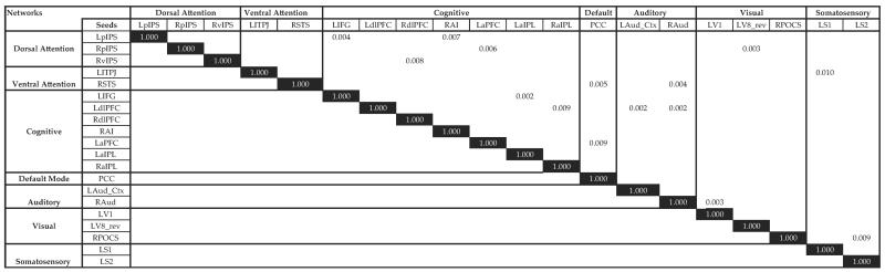
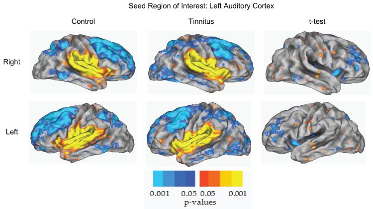
Main outcome measure: Functional connectivity differences in 58 a priori selected seed regions of interest encompassing cortical loci in the default mode, attention, auditory, visual, somatosensory, and cognitive networks.
Results: The median age of the 18 subjects was 54 years (interquartile range [IQR], 52-57), 66% were male, 90% were white, median Tinnitus Handicap Inventory (THI) score was 8 (IQR, 4-14), and a median Beck Depression Index score was 1 (IQR, 0-5). The median age for the control group was 46 years (IQR, 39-54), and 52% were male. Of the 58 seeds analyzed, no regions had significantly different functional connectivity among the nonbothersome tinnitus group when compared with the control group.
Conclusion: Among nonbothersome tinnitus patients, the tinnitus percept does not appear to alter the functional connectivity of the auditory cortex or other key cortical regions. Trial Registration ClinicalTrials.gov Identifier: NCT01049828.
Figures
References
-
- Gatehouse S. The role of non-auditory factors in measured and self-reported disability. Acta Otolaryngol Suppl. 1990;476:249–256. - PubMed
-
- Jacobson GP, Calder JA, Newman CW, Peterson EL, Wharton JA, Ahmad BK. Electrophysiological indices of selective auditory attention in subjects with and without tinnitus. Hear Res. 1996;97:66–74. - PubMed
-
- Wilson PH, Henry J, Bowen M, Haralambous G. Tinnitus reaction questionnaire: psychometric properties of a measure of distress associated with tinnitus. J Speech Hear Res. 1991;34:197–201. - PubMed
-
- Tyler RS, Baker LJ. Difficulties experienced by tinnitus sufferers. J Speech Hear Disord. 1983;48:150–154. - PubMed
-
- Jastreboff PJ, Jastreboff MM. Tinnitus retraining therapy: a different view on tinnitus [Review] ORL J Otorhinolaryngol Relat Spec. 2006;68:23–29. - PubMed
Publication types
MeSH terms
Associated data
Grants and funding
LinkOut - more resources
Full Text Sources
Other Literature Sources
Medical
