Case Reports
. 2012 Dec;41(12):1631-6.
doi: 10.1007/s00256-012-1460-z. Epub 2012 Jun 22.

The double suprascapular foramen: unique anatomical variation and the new hypothesis of its formation

Affiliations
Case Reports

The double suprascapular foramen: unique anatomical variation and the new hypothesis of its formation

Michał Polguj et al. Skeletal Radiol. 2012 Dec.

Abstract

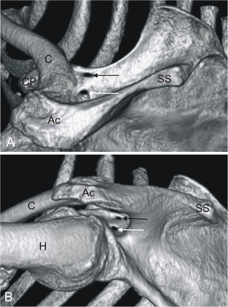
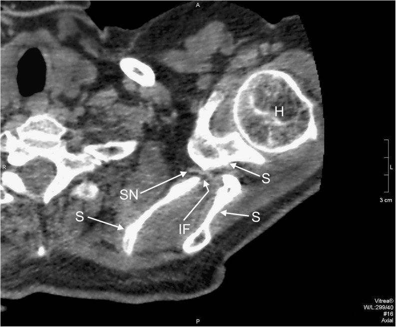
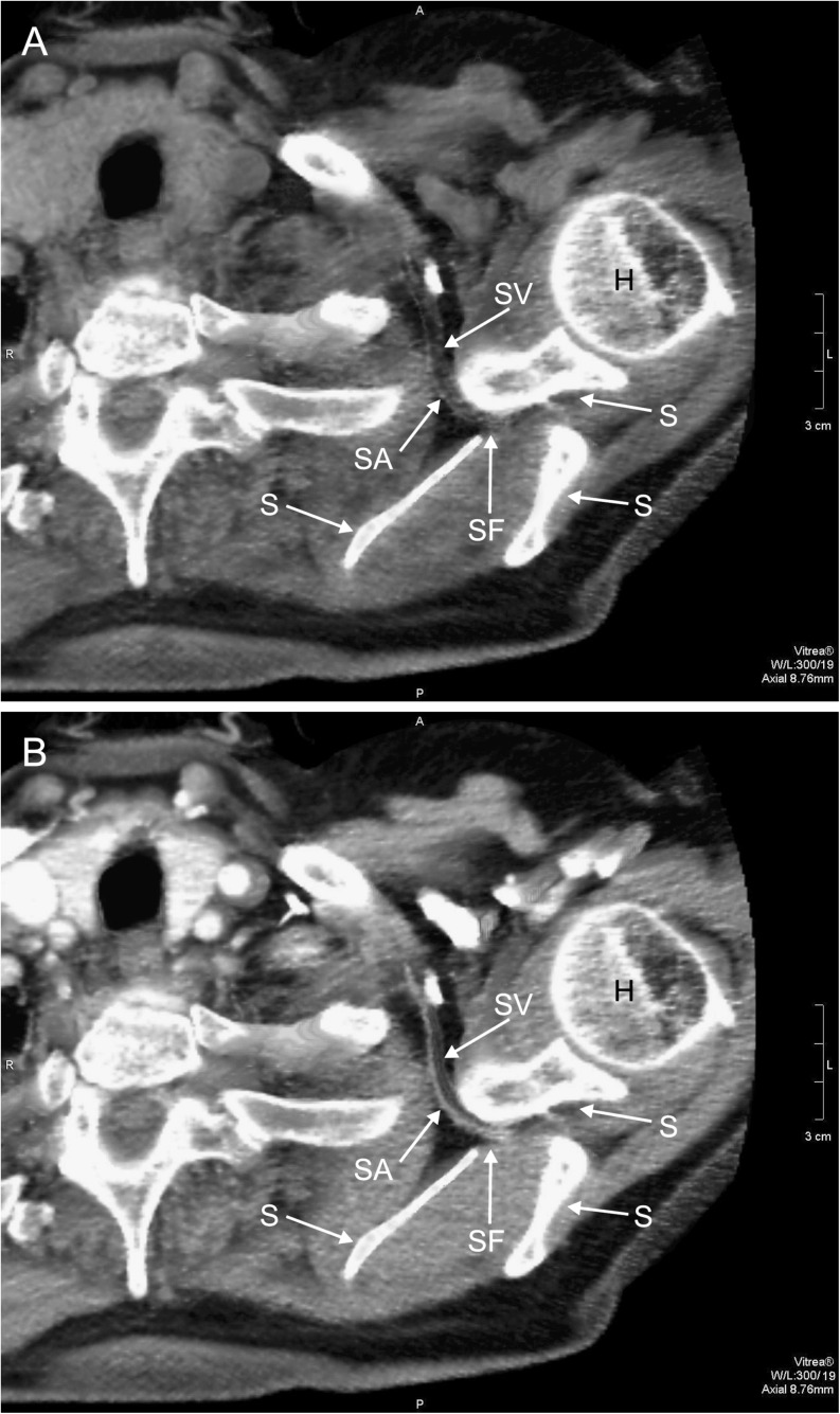
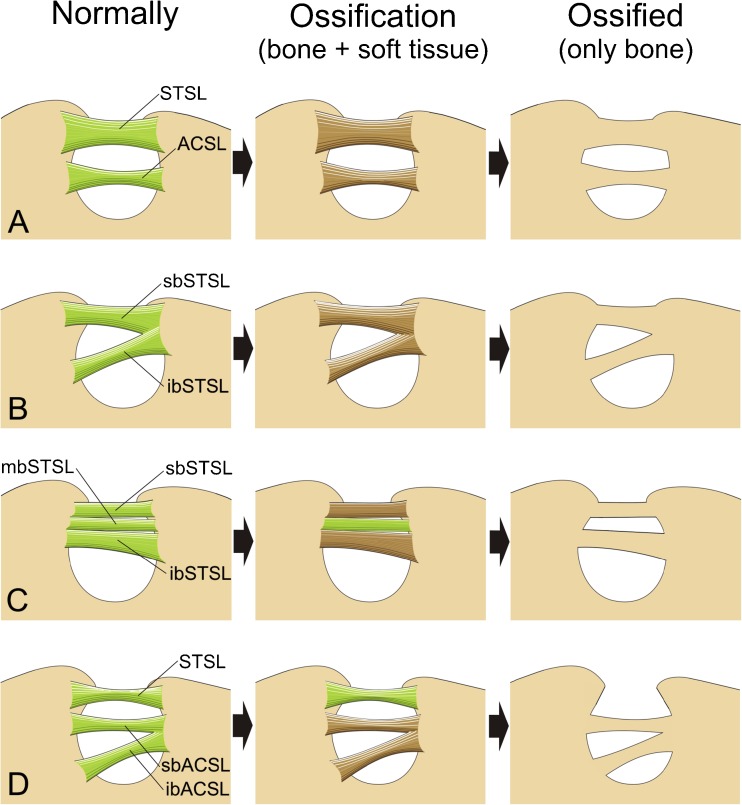
A unique anatomical variation of the suprascapular notch was discovered in one scapula from 610 analyzed by three-dimensional CT reconstruction. Two bony bridges were found, converting it into a double suprascapular foramen, in the left upper extremity of an 56-year-old Caucasian female. This variation might be a risk factor for suprascapular nerve entrapment. Suprascapular nerve running through inferior suprascapular foramen was discovered. Suprascapular vessels passed through superior suprascapular foramen (artery lay medially and vein laterally). A new hypothesis of double suprascapular foramen formation (mechanism of creation) is presented based on recent anatomical findings (e.g., the discovery in 2002 of the anterior coracoscapular ligament). Knowledge of the anatomical variations described in this study should be helpful in arthroscopic and open procedures at the suprascapular region and also confirms the safety of operative decompression for the suprascapular nerve.


Figures

Fig. 1
Fig. 1
Three-dimensional volume rendering (VR) MDCT demonstrating double suprascapular foramen: a supero-posterior view; b infero-posterior view. → black arrow superior suprascapular foramen, → white arrow inferior suprascapular foramen, Ac acromion, C clavicle, CP coracoid process, H humerus, SS scapular spine
Fig. 2
Fig. 2
Dual-phase helical CT, transverse scan on the level of the inferior suprascapular foramen: H humerus, IF inferior suprascapular foramen, SN suprascapular nerve, S scapula
Fig. 3
Fig. 3
Dual-phase helical CT, transverse scan on the level of the superior suprascapular foramen: A without contrast material, B after injection of contrast medium. H humerus, SF superior suprascapular foramen, SA suprascapular artery, SV suprascapular vein, S scapula
Fig. 4
Fig. 4
Schematic arrangements demonstrating hypotheses of double suprascapular notch formation: a first hypothesis, b second hypothesis, c third hypothesis, d fourth hypothesis. STSL superior transverse scapular ligament, sbSTSL superior band of superior transverse scapular ligament, mbSTSL middle band of superior transverse scapular ligament, ibSTSL inferior band of superior transverse scapular ligament, ACSL anterior coracoscapular ligament, sbACSL superior band of anterior coracoscapular ligament, ibACSL inferior band of anterior coracoscapular ligament

References

    1. Barwood SA, Burkhart SS, Lo IK. Arthroscopic suprascapular nerve release at the suprascapular notch in a cadaveric model: an anatomic approach. Arthroscopy. 2007;23(2):221–225. doi: 10.1016/j.arthro.2006.11.011. - DOI - PubMed
    1. Rengachary SS, Burr D, Lucas S, Hassanein KM, Mohn MP, Matzke H. Suprascapular entrapment neuropathy: a clinical, anatomical, and comparative study. Part 1: clinical study. Neurosurgery. 1979;5:441–446. doi: 10.1227/00006123-197910000-00006. - DOI - PubMed
    1. Romeo AA, Rotenberg DD, Bach BR., Jr Suprascapular neuropathy. J Am Acad Orthop Surg. 1999;7(6):358–367. - PubMed
    1. Zehetgruber H, Noske H, Lang T, Wurnig C. Suprascapular nerve entrapment. A meta-analysis. Int Orthop. 2002;26(6):339–343. doi: 10.1007/s00264-002-0392-y. - DOI - PMC - PubMed
    1. Bayramoğlu A, Demiryürek D, Tüccar E, Erbil M, Aldur MM, Tetik O, Doral MN. Variations in anatomy at the suprascapular notch possibly causing suprascapular nerve entrapment: an anatomical study. Knee Surg Sports Traumatol Arthrosc. 2003;11(6):393–398. doi: 10.1007/s00167-003-0378-3. - DOI - PubMed

Publication types