. 2012;36(1):50-7.
doi: 10.1159/000339285. Epub 2012 Jun 19.

Iron sucrose impairs phagocytic function and promotes apoptosis in polymorphonuclear leukocytes

Affiliations

Iron sucrose impairs phagocytic function and promotes apoptosis in polymorphonuclear leukocytes

Hirohito Ichii et al. Am J Nephrol. 2012.

Abstract

Background: With the recent implementation of bundling reimbursement policy, the use of intravenous (IV) iron preparations for the management of anemia in the end-stage renal disease (ESRD) population has dramatically increased. Iron overload increases the risk of infections in individuals with or without kidney disease. IV iron administration in ESRD patients impairs bacteriocidal capacity of polymorphonuclear leukocytes (PMNs) against Escherichia coli. These preparations consist of an elemental iron core and a carbohydrate shell. In addition to the iron core, the carbohydrate shell may affect PMNs. We therefore examined the effect of iron sucrose, a commonly used preparation, on phagocytic capacity of PMNs from a group of normal individuals against Gram-positive (Staphylococcus aureus) and Gram-negative (E. coli) bacteria.

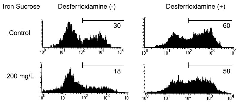
Methods: Iron sucrose was added to heparinized blood samples at pharmacologically-relevant concentrations and incubated for 4 and 24 h at 37°C to simulate in vivo condition. Blood samples mixed with equal volume of saline solution served as controls. To isolate the effects of the carbohydrate shell, blood samples were co-treated with the iron chelator, desferrioxamine.

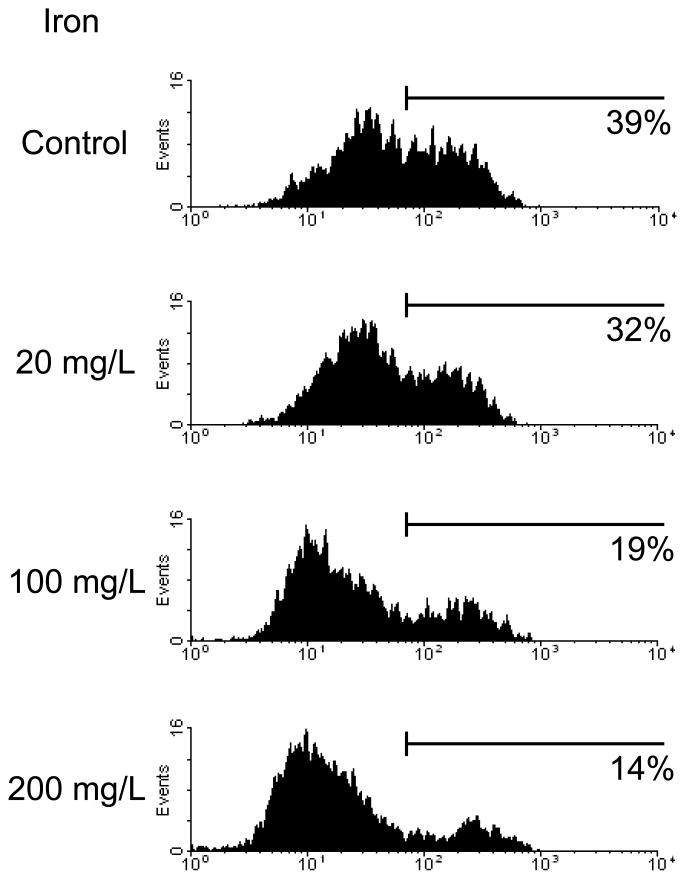
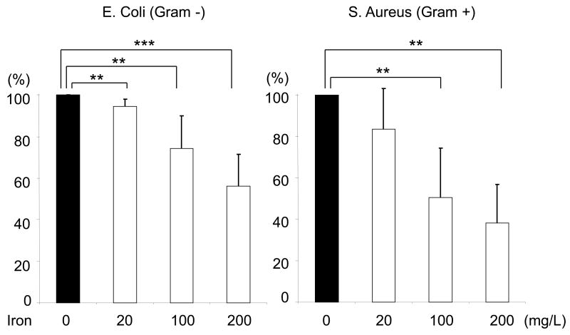
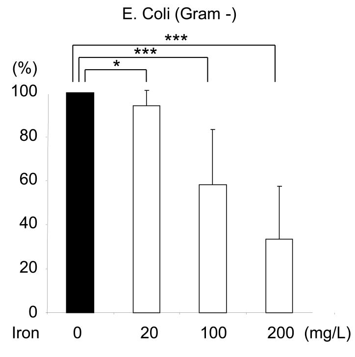
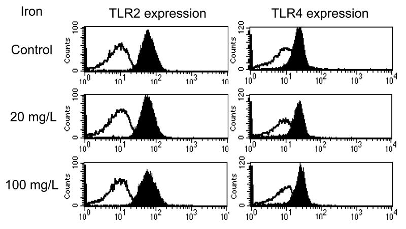
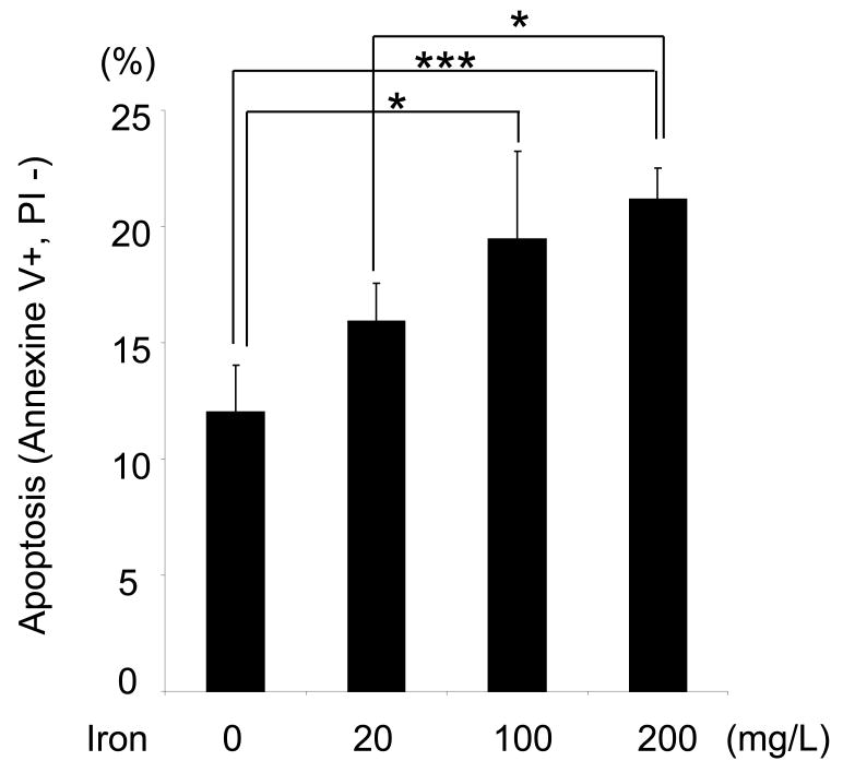
Results: Iron sucrose caused significant PMN apoptosis and dose-dependent suppression of phagocytic function against both Gram-positive and Gram-negative bacteria. These abnormalities were prevented by desferrioxamine which precluded contribution of the carbohydrate shell to the PMN dysfunction.

Conclusions: At pharmacologically-relevant concentrations, iron sucrose promotes apoptosis and inhibits phagocytic activities of PMNs. The deleterious effect of iron sucrose is mediated by its elemental iron core, not its carbohydrate shell, and as such may be shared by other IV iron preparations.


Conflict of interest statement

Conflict of Interest: None of the authors have any conflicts of interest associated with the work presented in this manuscript.

Figures

Figure 1
Figure 1. Effect of Iron Sucrose on phagocytic function in PMNs
The iron sucrose was added to heparinized blood samples from each healthy subject at various concentrations, 0, 20, 100, 200 mg/L and incubated for 4 hr and 24 hr at 37° C. Gram positive (Staphylococcus Aureus) and Gram negative (Escherichia Coli) bacteria were used for the evaluation of phagocytic function in PMNs. The phagocytic function against Gram positive and negative bacteria was evaluated by flow cytometry. A: Representive FACS data for phagocytic function against gram positive bacteria. B: Phagocytic function against Gram negative and positive bacteria after 4 hours incubation with iron sucrose. C: Phagocytic function after 24 hours incubation with iron sucrose. * P<0.05, ** P< 0.01, *** P<0.005.
Figure 2
Figure 2. Prevention of Iron Sucrose induced phagocytic dysfunction with desferrioxiamine in PMNs
To exclude the possible effect of the complex carbohydrate shell from that of the elemental iron core of the drug, blood samples were simultaneously treated with iron sucrose and the iron chelator, desferrioxamine.
Figure 3
Figure 3. Effect of Iron Sucrose on Toll-like eceptor expression in PMNs
The iron sucrose was added to heparinized blood samples from each healthy subject at various concentrations. The toll-like receptor 2 and 4 expressions in PMNs were examined using flow cytometry after 4 or 24 hours incubation with Iron Sucrose. Black histogram: anti- human TLR2 or 4 (+). White histogram: negative control.
Figure 4
Figure 4. Effect of Iron Sucrose on PMNs' viability
The iron sucrose was added to heparinized blood samples from each healthy subject at various concentrations. Annexine-V (+) PI (-) population was evaluated as apoptotic PMNs using flow cytometry after 24 hours incubation with iron sucrose. * P<0.05, *** P<0.005.

References

    1. Girndt M, Sester U, Sester M, et al. Impaired cellular immune function in patients with end-stage renal failure. Nephrol Dial Transplant. 1999;14:2807–2810. - PubMed
    1. United States Renal Data System: USRDS. Annual Data Report National Institutes of Health Diabetes and Digestive and Kidney Diseases. Bethesda. MD: 1998.
    1. Sarnak MJ, Jaber BL. Mortality caused by sepsis in patients with end-stage renal disease compared with the general population. Kidney Int. 2000;58:1758–1764. - PubMed
    1. Girndt M, Sester M, Sester U, et al. Molecular aspects of T- and B-cell function in uremia. Kidney Int Suppl. 2001;78:S206–211. - PubMed
    1. Sester U, Sester M, Hauk M, et al. T-cell activation follows Th1 rather than Th2 pattern in haemodialysis patients. Nephrol Dial Transplant. 2000;15:1217–1223. - PubMed

Publication types

MeSH terms