
Novel mutations target distinct subgroups of medulloblastoma

Giles Robinson et al. Nature. .

Abstract

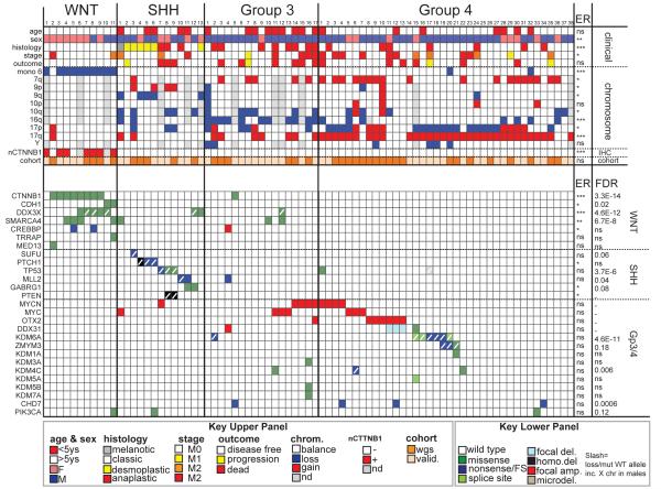
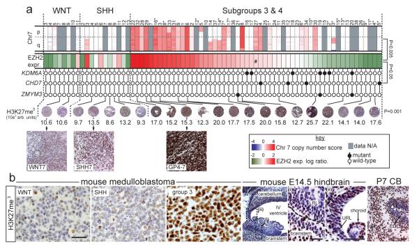
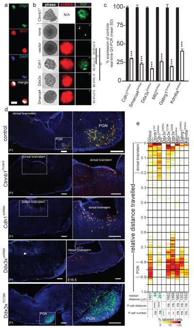
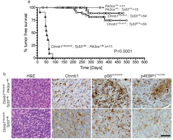
Medulloblastoma is a malignant childhood brain tumour comprising four discrete subgroups. Here, to identify mutations that drive medulloblastoma, we sequenced the entire genomes of 37 tumours and matched normal blood. One-hundred and thirty-six genes harbouring somatic mutations in this discovery set were sequenced in an additional 56 medulloblastomas. Recurrent mutations were detected in 41 genes not yet implicated in medulloblastoma; several target distinct components of the epigenetic machinery in different disease subgroups, such as regulators of H3K27 and H3K4 trimethylation in subgroups 3 and 4 (for example, KDM6A and ZMYM3), and CTNNB1-associated chromatin re-modellers in WNT-subgroup tumours (for example, SMARCA4 and CREBBP). Modelling of mutations in mouse lower rhombic lip progenitors that generate WNT-subgroup tumours identified genes that maintain this cell lineage (DDX3X), as well as mutated genes that initiate (CDH1) or cooperate (PIK3CA) in tumorigenesis. These data provide important new insights into the pathogenesis of medulloblastoma subgroups and highlight targets for therapeutic development.


Figures

Figure 1
Figure 1. The genomic landscape of medulloblastoma
Top: clinical, histologic, gross chromosomal, nuclear CTNNB1 (nCTNNB1), and cohort (discovery or validation) details of 79 medulloblastomas by subgroup. Below: genetic alterations detected in 27 genes of particular interest. Color key at bottom. ANOVA (continuous) or Fisher’s exact (categorical) p-value is shown right. False discovery estimates (FDR) of each mutation are shown right. ***=P<0.0005; **=P<0.005; *=P<0.05; ns=not significant.
Figure 2
Figure 2. Deregulation of H3K27me3 in subgroup-3 and 4 human and mouse medulloblastoma
(a) Top row, SNP profiles of chromosome 7 copy number in medulloblastomas (samples as Figure 1; *=subgroup-3 cases). Second row, expression of EZH2. Subgroup-3 and 4 tumours are ordered left to right by expression level, #=median expression point (Bonferroni corrected p-value of EZH2 expression vs. chromosome 7 gain). Third row, mutation status of KDM6A, CHD7 and ZMYM3 (p-value, Fisher’s exact mutations vs. EZH2 expression). Fourth row, H3K27me3 immunohistochemistry (numbers=colorimetry, p-value ANOVA). (b) H3K27me3 expression (right) in mouse Blbp-Cre ; Ctnnb1+/lox(Ex3) ; Tp53flx/flx (WNT), Ptch1+/−; Tp53−/− (SHH) and Myc ; Ink4c−/− (group 3) medulloblastomas and (left) developing hindbrain. high power views of E14.5 (i) LRL and (ii) upper rhombic lip (URL). IGL=internal granule layer, EGL=external germinal layer. Scale bar=50μm. White arrows in P7 cerebellum pinpoint H3K27me3 cells in the EGL.
Figure 3
Figure 3. Genes mutated in WNT-subgroup medulloblastomas regulate LRLPs
(a) Isolated Olig3+/Wnt1+ LRLPs were transduced in (b) with mutant Ctnnb1 (above hashed line) or the indicated shRNA-Red Fluorescence Protein construct (below hashed line). LRLPs were also transduced (+) or not (−) with a Tcf-Lef-enhanced green fluorescence (Tcf) reporter. Numbers right report clonal % 2′ to 3′ passage neurosphere formation (+SD). (c) Knock-down of genes targeted by shRNA relative to control transduced cells. (d) Immunofluorescence of P1 mouse hindbrains electroporated in utero at E14.5 with GFP (to control for equivalence of electroporation between embryos control) and the indicated construct. High-power views of indicated areas are shown right. Cells targeted by Ddx3x-shRNA are present 48 hours post electroporation but ablated by P1. Scale=200μm. (e) Heatmap reporting the distribution of GFP+/RFP+ cells in eletroporated mice at P1. Median distance migrated by cells, and p-values of migration distance and cell number relative to controls is shown (****, p<0.00005; ***, p<0.0005; **, p<0.005; *, p<0.05. Red and green text reports significant increase or decrease, respectively relative to control).
Figure 4
Figure 4. Pik3caE545K accelerates but does not initiate WNT-subgroup medulloblastoma
Tumour free survival of mice of the indicated genotype. All mice carry the Blbp-cre allele. Log Rank P<0.0001. (b) Hematoxylin and eosin and immunohistochemical stains of indicated tumors. Scale=50 μm.

References

    1. CBTRUS . Statistical Report: Primary Brain Tumors in the United States, 1995-1999. Central Brain Tumor Registry of the United States; Hinsdale, IL: 2006. 2002.
    1. Taylor MD, et al. Molecular subgroups of medulloblastoma: the current consensus. Acta Neuropathol. 2012;123:465–472. Epub 2011 Dec 2012. - PMC - PubMed
    1. Schuller U, et al. Acquisition of granule neuron precursor identity is a critical determinant of progenitor cell competence to form Shh-induced medulloblastoma. Cancer Cell. 2008;14:123–134. - PMC - PubMed
    1. Yang ZJ, et al. Medulloblastoma can be initiated by deletion of Patched in lineage-restricted progenitors or stem cells. Cancer Cell. 2008;14:135–145. - PMC - PubMed
    1. Gibson P, et al. Subtypes of medulloblastoma have distinct developmental origins. Nature. 2010;468:1095–1099. - PMC - PubMed

Publication types

MeSH terms

Substances