. 2013 Feb;48(2):222-37.
doi: 10.1007/s00535-012-0619-7. Epub 2012 Jun 22.

Butein inhibits ethanol-induced activation of liver stellate cells through TGF-β, NFκB, p38, and JNK signaling pathways and inhibition of oxidative stress

Affiliations

Butein inhibits ethanol-induced activation of liver stellate cells through TGF-β, NFκB, p38, and JNK signaling pathways and inhibition of oxidative stress

Agnieszka Szuster-Ciesielska et al. J Gastroenterol. 2013 Feb.

Abstract

Background: Butein has been reported to prevent and partly reverse liver fibrosis in vivo; however, the mechanisms of its action are poorly understood. We, therefore, aimed to determine the antifibrotic potential of butein.

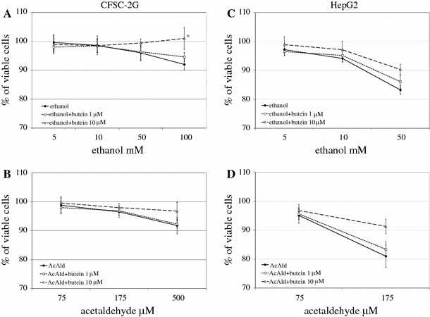
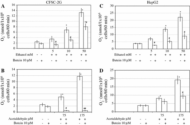
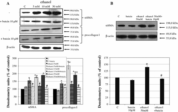
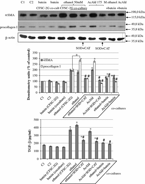
Methods: We assessed the influence of the incubation of hepatic stellate cells (HSCs) and hepatoma cells (HepG2) with butein on sensitivity to ethanol- or acetaldehyde-induced toxicity; the production of reactive oxygen species (ROS); the expression of markers of HSC activation, including smooth muscle α-actin (α-SMA) and procollagen I; and the production of transforming growth factor-β1 (TGF-β1), metalloproteinases-2 and -13 (MMP-2and MMP-13), and tissue inhibitors of metalloproteinases (TIMPs). The influence of butein on intracellular signals in HSCs; i.e., nuclear factor-κB (NFκB), c-Jun N-terminal kinase (JNK), and p38 mitogen-activated protein kinase (p38 MAPK) induced by ethanol was estimated.

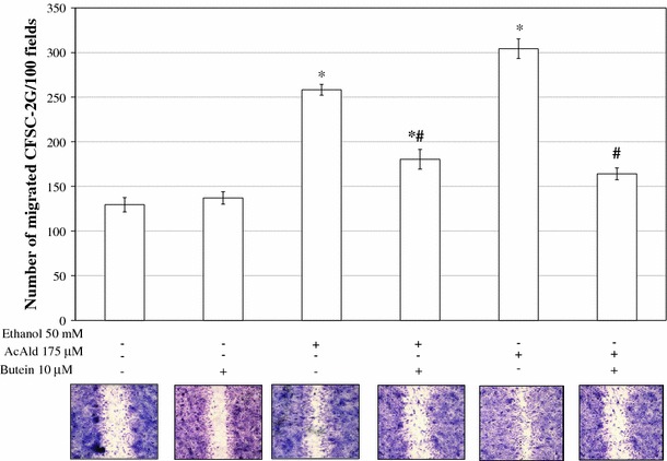
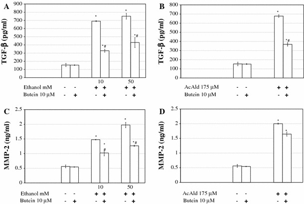
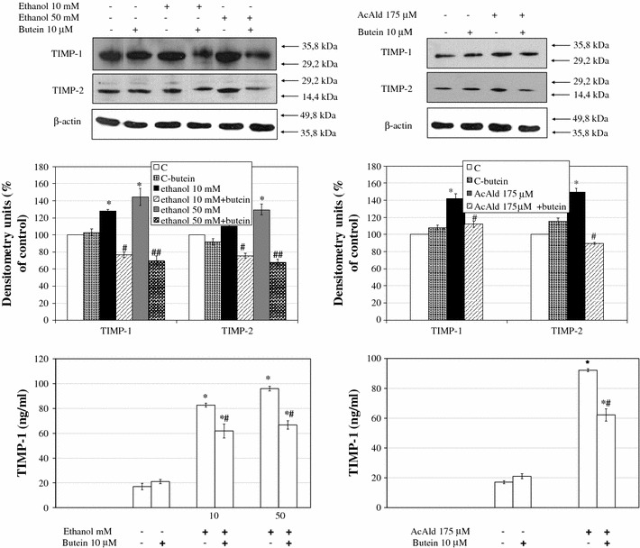
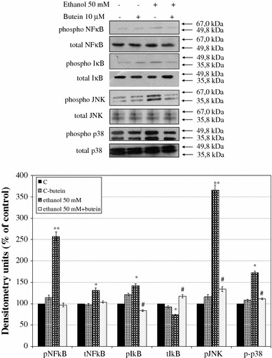
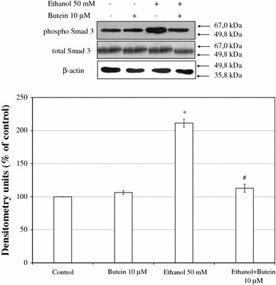
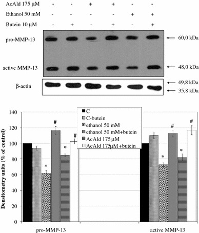
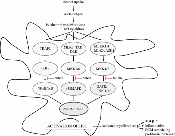
Results: Butein protected HSCs and HepG2 cells against ethanol toxicity by the inhibition of ethanol- or acetaldehyde-induced production of ROS when cells were incubated separately or in co-cultures; butein also inhibited HSC activation measured as the production of α-SMA and procollagen I. As well, butein downregulated ethanol- or acetaldehyde-induced HSC migration and the production of TGF-β, TIMP-1, and TIMP-2; decreased the activity of MMP-2; and increased the activity of MMP-13. In ethanol-induced HSCs, butein inhibited the activation of the p38 MAPK and JNK transduction pathways as well as significantly inhibiting the phosphorylation of NF κB inhibitor (IκB) and Smad3.

Conclusions: The results indicated that butein inhibited ethanol- and acetaldehyde-induced activation of HSCs at different levels, acting as an antioxidant and inhibitor of ethanol-induced MAPK, TGF-β, and NFκB/IκB transduction signaling; this result makes butein a promising agent for antifibrotic therapies.


Figures

Fig. 1a-d
Fig. 1a-d
The influence of butein on ethanol- and acetaldehyde-induced toxicity in hepatic stellate cells (HSCs; CFSC-2G) and HepG2 cells. Cells were preincubated in medium with 1 or 10 μM butein for 24 h. Subsequently, ethanol or acetaldehyde at the indicated concentrations was added. After 24 h of incubation, the toxicity was determined by the MTT method. Values are means ± SD of results from four independent experiments each with eight separate cell cultures. *Statistically significant at p ≤ 0.05 in comparison to cells incubated with ethanol alone (Wilcoxon test)
Fig. 2
Fig. 2
Preincubation of HSCs and HepG2 cells for 24 h with 10 μM butein inhibits ethanol-induced (a, c) and acetaldehyde-induced (b, d) superoxide anion production. Cells were preincubated with butein for 24 h, after which an assay for superoxide anion was performed in which nanomoles of O2 per 1 × 106 cells per 60 min were calculated. Results are expressed as means ± SD of four independent experiments each with eight separate cell cultures. *Significantly different from respective controls (cells incubated without ethanol, acetaldehyde, and butein or treated only with butein), p ≤ 0.05. #Statistically significant at p ≤ 0.05 in comparison to cells treated with ethanol or acetaldehyde alone. Butein significantly changed both the ethanol and acetaldehyde effects, p ≤ 0.01 (two-way analysis of variance [ANOVA])
Fig. 3
Fig. 3
Incubation of HSCs with 10 μM butein induces quiescence of cells activated by ethanol. HSCs were preincubated with butein for 24 h before treatment with ethanol (a) or first activated by ethanol for 24 h and incubated with butein for the next 24 h (b; lane C control). Markers of HSC activation such as α-smooth muscle actin (α-SMA) and procollagen I were measured by western blot. β-Actin expression served as the loading control. On the right, the arrows indicate the position of the molecular weight markers used in the experiments. Representative blots are shown. Experiments were done in triplicate (each with four separate cell cultures), and the bars represent means ± SD. *Significantly different from respective control (cells incubated without ethanol); p ≤ 0.05. #Statistically significant difference, at p ≤ 0.05, in comparison to cells treated with ethanol alone. +Significantly different from the lower ethanol concentration (5 mM), ++(10 mM); p ≤ 0.01. Butein significantly changed the ethanol effect, p ≤ 0.01 (two-way ANOVA)
Fig. 4
Fig. 4
Butein inhibits HSC activation stimulated by co-cultures of HSCs with HepG2 cells treated with ethanol or acetaldehyde. HSCs were seeded in 6-well plastic plates. At the same time HepG2 cells were seeded into tissue culture inserts with a membrane (pore diameter 0.4 μm) and incubated for 24 h at 37 °C. After that, HepG2 cells in the inserts were treated with ethanol or acetaldehyde for 3 h at 37 °C, washed, and moved into cultures of stellate cells in the wells of plastic plates in which the medium was supplemented with antioxidant enzymes such as superoxide dismutase (SOD) (240 U/ml) and catalase (CAT) (40 U/ml). Appropriate controls were also prepared (C1 HSCs only, C2 HSCs with HepG2 cells without any chemicals). Cells were co-cultured for 24 h at 37 °C and HSCs were collected for the measurement of α-SMA and procollagen I expression. In co-culture supernatants transforming growth factor-β (TGF-β) was also determined (enzyme-linked immunosorbent assay [ELISA] method). Experiments were done in triplicate (each with three separate cell cultures), and the bars represent means ± SD. *Significantly different from respective controls (cells incubated without ethanol or acetaldehyde); p ≤ 0.05. #Statistically significant at p ≤ 0.05 in comparison to co-culture where HepG2 cells had been treated with ethanol or acetaldehyde alone. aSignificantly different in comparison to ethanol treatment in CFSC-2G only, p ≤ 0.05. Butein significantly changed the ethanol or acetaldehyde effect, p ≤ 0.01 (two-way ANOVA)
Fig. 5
Fig. 5
Butein inhibits motility of HSCs. A wound healing assay was performed on HSCs grown to a confluent cell layer in which a wound was scraped to remove a linear area of cells. The cultures were treated with 10 μM butein for 24 h and then 50 mM ethanol or 175 μM acetaldehyde was added, and the cells were allowed 24 h to migrate. Representative images of different conditions are shown. The experiment was repeated five times. *Statistically significant at p ≤ 0.05 in comparison to respective controls (cells not treated or treated only with butein). #Statistically significant at p ≤ 0.05 in comparison to cells treated with ethanol or acetaldehyde alone (Wilcoxon test)
Fig. 6a, b
Fig. 6a, b
Preincubation of HSCs with 10 μM butein inhibits ethanol- and acetaldehyde-induced production of TGF-β and matrix metalloproteinase-2 (MMP-2). The cells were preincubated with 10 μM butein for 24 h and subsequently induced to produce TGF-β and MMP-2 by the addition of ethanol at the indicated concentrations or by the addition of 175 μM acetaldehyde. The levels of TGF-β and MMP-2 were measured by ELISA and are shown as the means ± SD of three independent experiments each with four separate cell cultures. *Significantly different from respective controls (cells not treated or treated only with butein), p ≤ 0.01. #Statistically significant at p ≤ 0.05 in comparison to cells treated with ethanol or acetaldehyde alone. Butein significantly changed the ethanol or acetaldehyde effect, p ≤ 0.01 (two-way ANOVA)
Fig. 7
Fig. 7
The effect of preincubation of HSCs with 10 μM butein on parameters related to extracellular matrix remodeling induced by ethanol. Western blot analyses for tissue inhibitor of metalloproteinase-1 (TIMP-1) and TIMP-2 were performed on cell lysates derived from cells preincubated for 24 h with 10 μM butein and subsequently incubated for 24 h with the indicated ethanol and acetaldehyde concentrations. The upper panels show representative blots from three independent experiments each with four separate cell cultures, the middle panels show densitometry analysis of bands, and the lower panels show the TIMP-1 ELISA assay. *Significantly different from respective controls (C cells not treated, C-butein treated only with butein), p ≤ 0.01. #Statistically significant in comparison to cells treated with ethanol or acetaldehyde alone, # p ≤ 0.05, ## p ≤ 0.001. Butein significantly changed the ethanol (p ≤ 0.01) and acetaldehyde (p ≤ 0.1) effect (two-way ANOVA)
Fig. 8
Fig. 8
The effect of preincubation of HSCs with 10 μM butein on the phosphorylation of nuclear factor κB (NFκB), nuclear factor κB inhibitor (ΙκΒ), c-Jun N-terminal kinase (JNK), and p38 mitogen-activated protein kinase (MAPK; p38). The cells were preincubated with 10 μM butein for 24 h and subsequently exposed or not to 50 mM ethanol for 20 min. The relative densitometry readings (mean ± SD) from three independent experiments each with four separate cell cultures are shown in the lower panel. The upper panel shows representative blots of total (t) and phosphorylated (p) forms of NFκB, IκB, JNK, and p38 MAPK. *Significantly different from respective controls (cells not treated or treated only with butein), *p ≤ 0.05, **p ≤ 0.001. #Statistically significant at p ≤ 0.05 in comparison to cells treated with ethanol alone (Wilcoxon test)
Fig. 9
Fig. 9
The effect of preincubation of HSCs with 10 μM butein on the phosphorylation of Smad3. The cells were preincubated with 10 μM butein for 24 h and then exposed to 50 mM ethanol for 24 h. The amounts of phosphorylated and total Smad3 (used as loading control) were measured by western blotting (lower panel). The upper panel shows representative blots. Each figure is representative of three independent experiments each with four separate cell cultures. Band intensities were measured, and the ratio of phosphorylated Smad3 in the absence of butein and ethanol was used as a control (100 %). The values shown are means ± SD. *Significantly different from respective controls (cells not treated or treated only with butein), p ≤ 0.05. #Statistically significant at p ≤ 0.05 in comparison to cells treated with ethanol (Wilcoxon test)
Fig. 10
Fig. 10
Preincubation of HSCs with 10 μM butein restores the production of MMP-13 decreased by ethanol or acetaldehyde. Western blot analysis for MMP-13 was performed on cell lysates derived from cells preincubated for 24 h with 10 μM butein and subsequently incubated for 24 h with the indicated ethanol or acetaldehyde concentrations. The representative western blots are shown in the upper panel. Each bar in the lower panel represents the mean ± SD from four independent experiments. *Significantly different from respective controls (cells not treated or treated only with butein), p ≤ 0.01. #Statistically significant at p ≤ 0.05 in comparison to cells treated with ethanol or acetaldehyde alone. Butein significantly changed the ethanol and acetaldehyde effect, p ≤ 0.01 (two-way ANOVA)
Fig. 11
Fig. 11
Possible mechanisms of the antifibrotic effects of butein on hepatic stellate cells treated with ethanol. Butein attenuates oxidative stress and ? the phosphorylation of kinases p38 and JNK as well as Smad3. In effect, it downregulates ECM remodeling and the production of profibrotic proteins. ASK1, apoptosis signal-regulating kinase 1; DLK, dual leucine zipper-bearing kinase; ECM, extracellular matrix; IκB, nuclear factor kappaB inhibitor; IKKs, IkappaB kinases; JNK, c-Jun N-terminal kinase; MAPK, mitogen-activated protein kinase; MEKK, mitogen-activated protein kinase/extracellular signal-regulated kinase kinase; MKK, mitogen-activated protein kinase kinase; MLK3, mixed-lineage kinase 3; NFκB, nuclear factor kappaB; SAPK, stress-activated protein kinase; TAK, Triticum aestivum kinase; TRAF2, tumor necrosis factor (TNF)-receptor-associated factor 2

Similar articles

Cited by

References

    1. Bataller R, Brenner DA. Liver fibrosis. J Clin Invest. 2005;115:209–218. - PMC - PubMed
    1. Senoo H. Structure and function of hepatic stellate cells. Med Electron Microsc. 2004;37:3–15. doi: 10.1007/s00795-003-0230-3. - DOI - PubMed
    1. Gao B, Bataller R. Alcoholic liver disease: pathogenesis and new therapeutic targets. Gastroenterology. 2011;141:1572–1585. doi: 10.1053/j.gastro.2011.09.002. - DOI - PMC - PubMed
    1. Siegmund SV, Dooley S, Brenner DA. Molecular mechanisms of alcohol-induced hepatic fibrosis. Dig Dis. 2005;23:264–274. doi: 10.1159/000090174. - DOI - PubMed
    1. Friedman SL, Yamasaki G, Wong L. Modulation of transforming growth factor beta receptors of rat lipocytes during the hepatic wound healing response. Enhanced binding and reduced gene expression accompany cellular activation in culture and in vivo. J Biol Chem. 1994;269:10551–10558. - PubMed

Publication types

MeSH terms