. 2012 Dec;33(11):2062-7.
doi: 10.3174/ajnr.A3151. Epub 2012 Jun 21.

The shrunken, bright cerebellum: a characteristic MRI finding in congenital disorders of glycosylation type 1a

Affiliations

The shrunken, bright cerebellum: a characteristic MRI finding in congenital disorders of glycosylation type 1a

P Feraco et al. AJNR Am J Neuroradiol. 2012 Dec.

Abstract

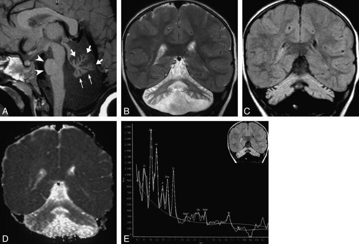
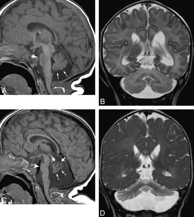
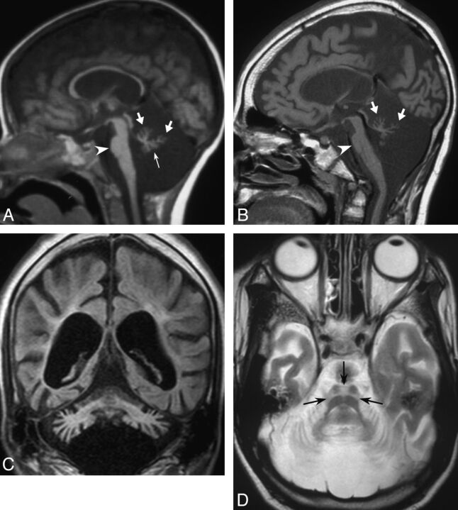
CDG-1a is an early-onset neurodegenerative disease with selective hindbrain involvement and highly variable clinical presentation. We retrospectively reviewed the clinical records and MR imaging studies of 5 children (3 boys and 2 girls aged 12 days to 2 years at presentation) with molecularly confirmed CDG-1a. The cerebellum was hypoplastic at presentation in 4 cases, progressive bulk loss involved the cerebellum and the pons in all cases, and the cerebellar cortex and subcortical white matter were hyperintense on T2-weighted and FLAIR images in all. We conclude that CDG-1a likely results from a combination of cerebellar hypoplasia and atrophy. Cerebellar volume loss with diffuse T2/FLAIR hyperintensity seems to be a peculiar association in the field of cerebellar atrophies, and may be useful to address the differential diagnosis.


Figures

Fig 1.
Fig 1.
Typical MR imaging findings in CDG-1a; case 4 at age 2 years. A, Sagittal T1-weighted image shows shrunken appearance of the vermis, with commensurate folial volume loss and fissural enlargement involving the anterior lobe and declive (thick arrows) and flattened inferior vermis (thin arrows). The pontine protuberance is small (arrowheads). B, Coronal T2-weighted and (C) FLAIR images show shrunken, hyperintense hemispheric cerebellar cortex bilaterally; the cerebellar white matter is also mildly hyperintense compared with supratentorial white matter, resulting in a “bright cerebellum” appearance. D, Coronal ADC map shows the cerebellum is hyperintense compared with the supratentorial brain, consistent with facilitated diffusion. E, 1H-MRS findings; single-voxel PRESS technique at TE 23 ms. Upper right coronal FLAIR image shows placement of a 18 × 18 × 18 mm voxel on right cerebellar hemisphere. Corresponding spectrum shows markedly reduced NAA peak, mildly reduced Cho, and elevated inositol (mI and sI) resonances. There is a high alpha-Glx peak at 3.8 ppm, perhaps in relation to hepatic dysfunction in this patient. Glx indicates glutamine/glutamate complex.
Fig 2.
Fig 2.
Evolution of findings in CDG-1a; case 1 at age 12 days (A and B) and 11 months (C and D). At presentation, sagittal T1-weighted image (A) shows mild hypoplasia of the inferior vermis (thin arrows); the pons and superior vermis appear normal. Coronal T2-weighted image (B) shows normal cerebellar hemispheres. At 11-month follow-up, sagittal T1-weighted image (C) shows considerable volume loss of the vermis. Notice, in particular, atrophic involution of the anterior lobe and declive (thick arrows), with corresponding fissural enlargement, whereas the inferior vermis retains a flattened appearance (thin arrows), with not so large fissures. The pontine protuberance is also diminished in size (arrowhead). Coronal T2-weighted image (D) also shows reduced size of both cerebellar hemispheres. The cerebellar cortex is hyperintense.
Fig 3.
Fig 3.
Long term follow-up in CDG-1a; case 3 at age 2 years (A) and 16 years (B–D). At presentation, sagittal T1-weighted image (A), performed 20 years ago on a 0.5T MR unit, shows markedly small cerebellar vermis, with larger fissures superiorly (thick arrows) compared with a flattened appearance inferiorly (thin arrow). The pons is small (arrowhead). After 14 years, sagittal T1-weighted image (B) shows further volume loss involving both the superior (thick arrows) and inferior vermis. The pontine protuberance is further reduced (arrowhead), and the corpus callosum is markedly thinned. Coronal FLAIR image (C) shows hyperintense, atrophic cerebellum, supratentorial atrophy with white matter gliosis, and ventricular enlargement. Axial T2-weighted image (D) shows the pontine “hot cross bun” sign (arrows) and cerebellar cortical atrophy.

References

    1. Jaeken J, Artigas J, Barone R, et al. . Phosphomannomutase deficiency is the main cause of carbohydrate deficient glycoprotein syndrome with type I isoelectrofocusing pattern of serum sialotransferrins. J Inherit Metab Dis 1997; 20: 447– 49 - PubMed
    1. Sparks SE, Krasnewich DM. PMM2-CDG (CDG-Ia). In: Pagon RA, Bird TD, Dolan CR, et al., eds. GeneReviews [Internet]. Seattle: University of Washington; 1993- 2005 Aug 15 [updated 2011 Apr 21].
    1. Pearl PL, Krasnewich D. Neurologic course of congenital disorders of glycosylation. J Child Neurol 2001; 16: 409– 13 - PubMed
    1. Antoun H, Villeneuve N, Gelot A, et al. . Cerebellar atrophy: an important feature of carbohydrate deficient glycoprotein syndrome type 1. Pediatr Radiol 1999; 29: 194– 98 - PubMed
    1. Jensen PR, Hansen FJ, Skovby F. Cerebellar hypoplasia in children with the carbohydrate-deficient glycoprotein syndrome. Neuroradiology 1995; 37: 328– 30 - PubMed