. 2012 Oct;11(10):863-85.
doi: 10.1074/mcp.M111.014845. Epub 2012 Jun 21.

Exosomes as biomarker enriched microvesicles: characterization of exosomal proteins derived from a panel of prostate cell lines with distinct AR phenotypes

Affiliations

Exosomes as biomarker enriched microvesicles: characterization of exosomal proteins derived from a panel of prostate cell lines with distinct AR phenotypes

Elham Hosseini-Beheshti et al. Mol Cell Proteomics. 2012 Oct.

Abstract

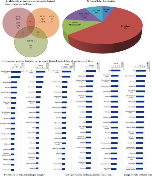
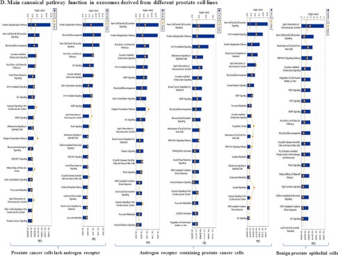
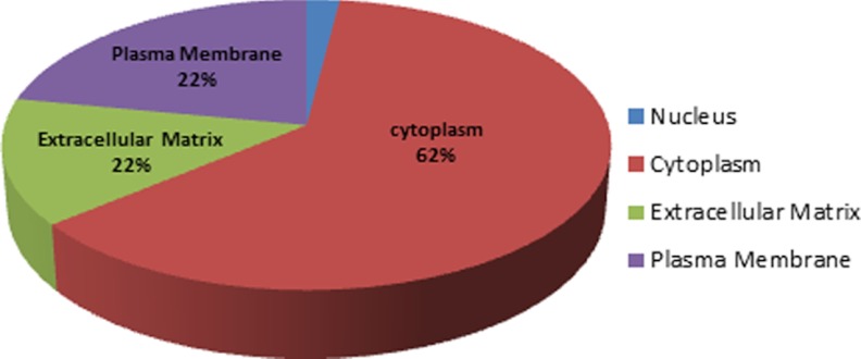
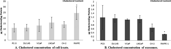
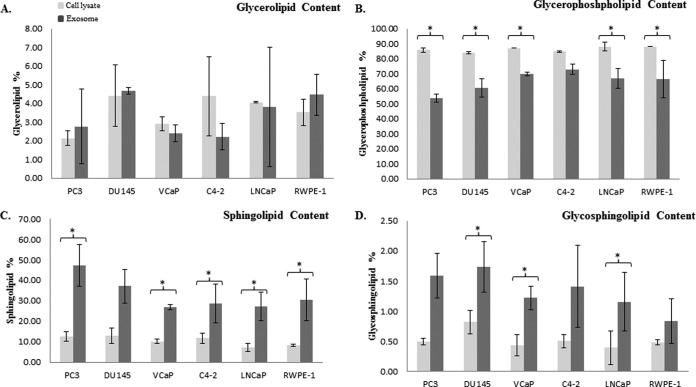
Prostate cancer is the leading type of cancer diagnosed in men. In 2010, ~217,730 new cases of prostate cancer were reported in the United States. Prompt diagnosis of the disease can substantially improve its clinical outcome. Improving capability for early detection, as well as developing new therapeutic targets in advanced disease are research priorities that will ultimately lead to better patient survival. Eukaryotic cells secrete proteins via distinct regulated mechanisms which are either ER/Golgi dependent or microvesicle mediated. The release of microvesicles has been shown to provide a novel mechanism for intercellular communication. Exosomes are nanometer sized cup-shaped membrane vesicles which are secreted from normal and cancerous cells. They are present in various biological fluids and are rich in characteristic proteins. Exosomes may thus have potential both in facilitating early diagnosis via less invasive procedures or be candidates for novel therapeutic approaches for castration resistance prostate cancer. Because exosomes have been shown previously to have a role in cell-cell communication in the local tumor microenvironment, conferring activation of numerous survival mechanisms, we characterized constitutive lipids, cholesterol and proteins from exosomes derived from six prostate cell lines and tracked their uptake in both cancerous and benign prostate cell lines respectively. Our comprehensive proteomic and lipidomic analysis of prostate derived exosomes could provide insight for future work on both biomarker and therapeutic targets for the treatment of prostate cancer.


Figures

Fig. 1.
Fig. 1.
Mechanism involved in exosome formation and trafficking in the microenvironment.
Fig. 2.
Fig. 2.
Transmission Electron Microscopy (TEM). TEM images of exosomes derived from different androgen independent and androgen sensitive prostate cancer cell lines including PC3, DU145, VCaP, LNCaP and C4–2 versus benign epithelial prostate cell line RWPE-1. Exosomes were negatively stained with 2% uracyl acetate after removing the extra moisture. Cup-shaped structures, with 30–100 nm size were identified as being exosomes.
Fig. 3.
Fig. 3.
Western blot analysis for exosome markers in exosomes and corresponding cell lysate samples. A, Exosomes have been purified based on their unique size and density by ultracentrifugation with 30% sucrose-deuterium. Twenty-5 μg of total protein associated with purified exosomes derived from six different prostate cell lines were analyzed by Western blotting using different exosome markers. B, Twenty-five μg of total protein associated with cell lysates of six different prostate cell lines were analyzed by Western blotting using different exosome markers.
Fig. 4.
Fig. 4.
Confocal microscopy. A, B, Confocal microscopy was used to visualize purified DU145 derived exosomes which were stained with Cell TrackerTM Orange CMTMR teramethylrhodamine. PC3, VCaP, LNCaP, C4–2 and RWPE-1 cells (104) were cultured on each chamber slide and incubated for 12 h with purified-stained exosomes. Confocal micrograph clearly demonstrates that transferred DU145 derived exosomes are not only attached to the cell membrane of host cells but have actually been taken up by these cells and are present in their cytoplasm. C, Confocal microscopy was also used to visualize freshly isolated exosomes derived from a CLUGFP stably over-expressing LNCaP cell line, which contains CLUGFP, being taken up by PC3 (AR-ve) and LNCaP (AR+ve) PCa cell lines after overnight incubation. Both cell lines were further fixed and stained with DAPI and E-Cadherin prior to imaging of the cells by confocal microscopy.
Fig. 5.
Fig. 5.
Proteomic analysis of different prostate cancer cell lines. A, Venn diagram describing the mutuality of proteins in exosomes derived from the benign epithelial prostate cell line (RWPE-1) versus five different prostate cancer cell lines categorized based on androgen sensitivity (PC3, DU145 and VCaP, LNCaP, C4–2). Numbers in ( ) are representative of the total number of proteins present in each cell lines, Numbers in () are representative of the total number of proteins present in each cell lines, Numbers in [ ] are representative of proteins present in either designated category and not present in any other undesignated category. Numbers denoted with * are the mutual proteins present in all cell lines in each category. B, Pie chart showing the subcellular localization of proteins found in exosomes derived from six different prostate cell lines. C, Bar chart indicating the cellular function of proteins found within exosomes determined using Ingenuity software. D, Predicted top canonical pathways are represented by the identified exosomal proteins.
Fig. 5.
Fig. 5.
Proteomic analysis of different prostate cancer cell lines. A, Venn diagram describing the mutuality of proteins in exosomes derived from the benign epithelial prostate cell line (RWPE-1) versus five different prostate cancer cell lines categorized based on androgen sensitivity (PC3, DU145 and VCaP, LNCaP, C4–2). Numbers in ( ) are representative of the total number of proteins present in each cell lines, Numbers in () are representative of the total number of proteins present in each cell lines, Numbers in [ ] are representative of proteins present in either designated category and not present in any other undesignated category. Numbers denoted with * are the mutual proteins present in all cell lines in each category. B, Pie chart showing the subcellular localization of proteins found in exosomes derived from six different prostate cell lines. C, Bar chart indicating the cellular function of proteins found within exosomes determined using Ingenuity software. D, Predicted top canonical pathways are represented by the identified exosomal proteins.
Fig. 6.
Fig. 6.
Biomarker proteins: Subcellular localization. Pie chart showing the subcellular localization of biomarkers found in exosomes derived from prostate cancer cell lines; PC3, DU145, VCaP, LNCaP, and C4–2.
Fig. 7.
Fig. 7.
Cholesterol concentration. The bar diagrams show the cholesterol concentration of A, lysates of PC3, DU145 and VCaP, LNCaP, C4–2, and RWPE-1 cells B, exosomes derived from the six different prostate cell lines. Cholesterol results were normalized to protein concentration of each sample and expressed as μg Cholesterol/μg Protein. * indicate significantly difference (p < 0.05).
Fig. 8.
Fig. 8.
Exosome lipidomic data. The lipid content of four major lipid classes was measured in PC3, DU145 and VCaP, LNCaP, C4–2 and RWPE-1 cells and compared with their derived exosomes, using LC-MS. The bar diagrams are representative of A, glycerolipid, B, glycerophospholipid, C, sphingolipid, D, glycosphingolipid in cell lysates and exosomes. Relative amounts of each lipid group were calculated as (sum of all AUC's for a particular lipid group) ÷ (sum of AUC's for all lipid groups) ×100%. * denotes a significant difference (p < 0.05) between exosomes and their corresponding cells. No significant differences were seen between the cell lysates and exosomes of respective cell lines in any of lipid class.

Similar articles

Cited by

References

    1. Jemal A., Siegel R., Ward E., Murray T., Xu J., Smigal C., Thun M. J. (2006) Cancer statistics, 2006. CA Cancer J. Clin. 56, 106–130 - PubMed
    1. Chan J. M., Holick C. N., Leitzmann M. F., Rimm E. B., Willett W. C., Stampfer M. J., Giovannucci E. L. (2006) Diet after diagnosis and the risk of prostate cancer progression, recurrence, and death (United States). Cancer Causes Control 17, 199–208 - PubMed
    1. Bruchovsky N., Klotz L. H., Sadar M., Crook J. M., Hoffart D., Godwin L., Warkentin M., Gleave M. E., Goldenberg S. L. (2000) Intermittent androgen suppression for prostate cancer: Canadian Prospective Trial and related observations. Mol Urol 4, 191–199; discussion 201 - PubMed
    1. Gleave M. E., Goldenberg S. L., Chin J. L., Warner J., Saad F., Klotz L. H., Jewett M., Kassabian V., Chetner M., Dupont C., Van Rensselaer S. (2001) Randomized comparative study of 3 versus 8-month neoadjuvant hormonal therapy before radical prostatectomy: biochemical and pathological effects. J Urol. 166, 500–506; discussion 506–507 - PubMed
    1. Lan C. Y., Huang H., Liu J. H. (2008) [Prognostic value of serum CA(125) level change during chemotherapy post-surgery in patients with advanced epithelial ovarian carcinoma]. Zhonghua Fu Chan Ke Za Zhi 43, 732–736 - PubMed

MeSH terms