. 2012 Aug 15;303(4):G490-7.
doi: 10.1152/ajpgi.00120.2012. Epub 2012 Jun 21.

Elevated lipopolysaccharide in the colon evokes intestinal inflammation, aggravated in immune modulator-impaired mice

Affiliations

Elevated lipopolysaccharide in the colon evokes intestinal inflammation, aggravated in immune modulator-impaired mice

Eunok Im et al. Am J Physiol Gastrointest Liver Physiol. .

Abstract

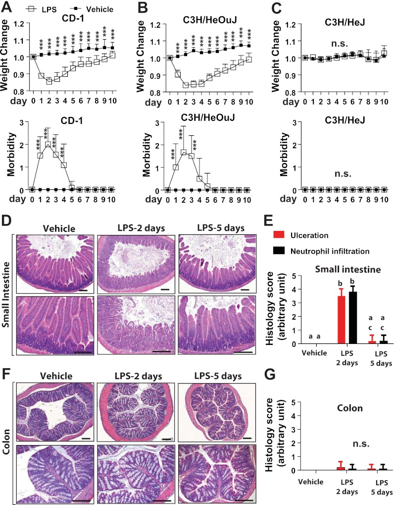
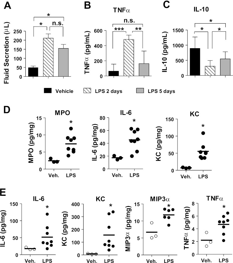
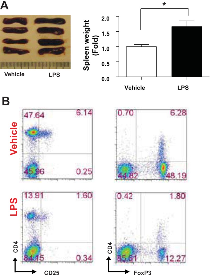
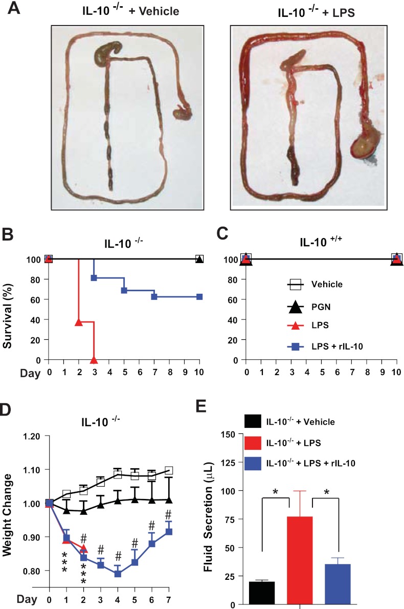
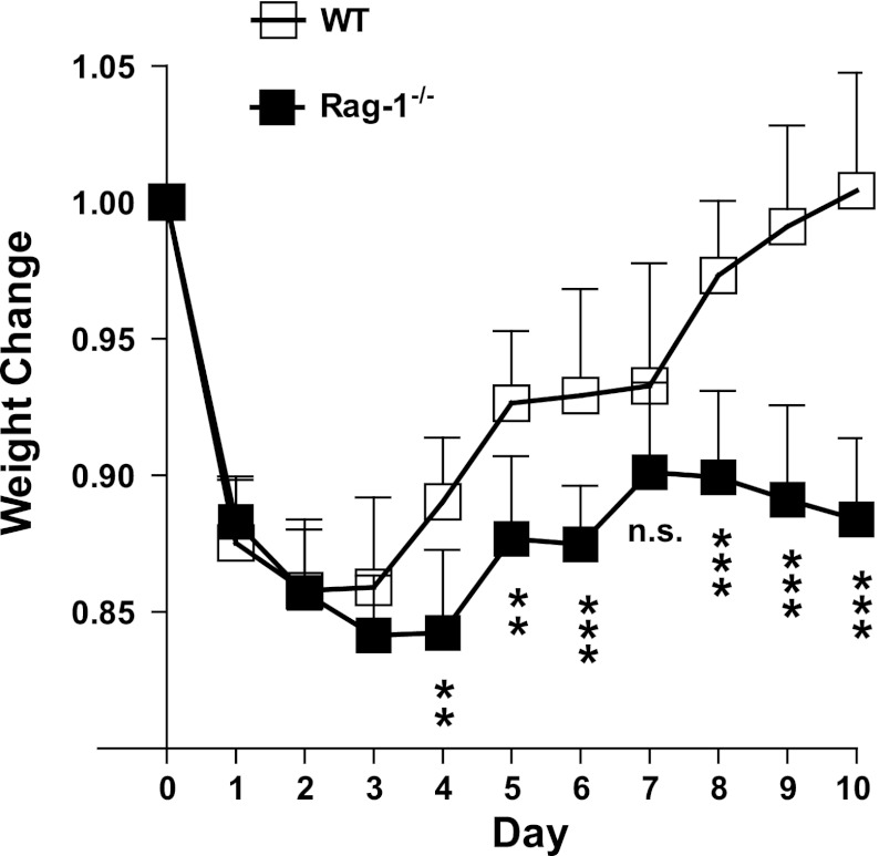
Frequency of gram-negative bacteria is markedly enhanced in inflamed gut, leading to augmented LPS in the intestine. Although LPS in the intestine is considered harmless and, rather, provides protective effects against epithelial injury, it has been suggested that LPS causes intestinal inflammation, such as necrotizing enterocolitis. Therefore, direct effects of LPS in the intestine remain to be studied. In this study, we examine the effect of LPS in the colon of mice instilled with LPS by rectal enema. We found that augmented LPS on the luminal side of the colon elicited inflammation in the small intestine remotely, not in the colon; this inflammation was characterized by body weight loss, increased fluid secretion, enhanced inflammatory cytokine production, and epithelial damage. In contrast to the inflamed small intestine induced by colonic LPS, the colonic epithelium did not exhibit histological tissue damage or inflammatory lesions, although intracolonic LPS treatment elicited inflammatory cytokine gene expression in the colon tissues. Moreover, we found that intracolonic LPS treatment substantially decreased the frequency of immune-suppressive regulatory T cells (CD4(+)/CD25(+) and CD4(+)/Foxp3(+)). We were intrigued to find that LPS-promoted intestinal inflammation is exacerbated in immune modulator-impaired IL-10(-/-) and Rag-1(-/-) mice. In conclusion, our results provide evidence that elevated LPS in the colon is able to cause intestinal inflammation and, therefore, suggest a physiological explanation for the importance of maintaining the balance between gram-negative and gram-positive bacteria in the intestine to maintain homeostasis in the gut.


Figures

Fig. 1.
Fig. 1.
Intracolonic LPS treatment induces transient inflammation in the small intestine, and not in the colon, in a Toll-like receptor 4 (TLR4)-dependent manner. A–C: body weight change (×100%) and morbidity (scored as described in materials and methods) in CD-1, C3H/HeOuJ, and C3H/HeJ mice intracolonically treated with LPS (50 μg/mouse, 1 enema each day during the experimental period) or vehicle (LPS-free saline solution), determined as previously described (17, 30). Values are means ± SD; n = 28 LPS-treated and 33 vehicle-treated CD-1 mice, 12 LPS-treated and 12 vehicle-treated C3H/HeOuJ mice, and 13 LPS-treated and 12 vehicle-treated C3H/HeJ mice. Data were compared by 2-way ANOVA (with treatment and time as factors) followed by Bonferroni's multiple-comparison t-test to assess differences between groups: ***P < 0.001 vs. corresponding LPS-treated mice at the same time point. ns, Not significant. D and F: low-magnification (top) and high-magnification (bottom) images of hematoxylin-eosin-stained paraffin-embedded sections of small intestine (jejunum) and colon from CD-1 mice treated with vehicle or LPS for 2 and 5 days. Scale bars, 200 μm. Images are representative of all animals from each group. E and G: histology scores for sections shown in D and F. Values are means ± SD, n = 10/group. Data were compared by ANOVA (treatment and histological index as factors) followed by Bonferroni's multiple-comparison t-test to assess differences between groups: means without a common letter (a, b, c) are significantly different (P < 0.001).
Fig. 2.
Fig. 2.
Intracolonic LPS treatment increases intestinal fluid secretion and induces differential expression of inflammatory mediators. A: volume of fluid collected from small intestine of C57BL/6 mice intracolonically treated with vehicle or LPS for 2 and 5 days (n = 4/group). B and C: ELISA of TNFα and IL-10 in secreted intestinal fluid (n = 8/group). Values are means ± SD. *P < 0.05; **P < 0.01; ***P < 0.0001 (Mann-Whitney U-test). D and E: ELISA of myeloperoxidase (MPO) and inflammatory cytokine [IL-6, KC, macrophage inflammatory protein 3α (MIP3α), and TNFα] expression in total tissue extracts from small intestine (D) and colon (E) of C57BL/6 mice intracolonically treated with vehicle (Veh, n = 3) or LPS (n = 8) for 2 days. Values were normalized by total protein concentration. *P < 0.05 (Mann-Whitney U-test).
Fig. 3.
Fig. 3.
Regulatory T (Treg) cell population was reduced by intracolonic LPS treatment. A: photograph of enlarged spleen from C57BL/6 mice treated with vehicle or LPS for 2 days (left) and spleen weight (right). Values are means ± SD, n = 4/group. *P < 0.05 (Mann-Whitney U-test). B: flow cytometry of CD4+/CD25+ or CD4+/Foxp3+ Treg cells isolated from spleens shown in A and double-stained with fluorescence-conjugated antibodies (CD25-phycoerythrin and CD4-allophycocyanin) and then with Alexa Fluor 488-conjugated FoxP3 antibody. Numbers in quadrants indicate percentage of each cell subset. Data are representative of 3 independent experiments.
Fig. 4.
Fig. 4.
LPS-induced intestinal inflammation is exaggerated in IL-10−/− mice. A: photographs of the entire gastrointestinal tract of IL-10−/− mice on a C57BL/6 background intracolonically treated with vehicle or LPS (25 μg/mouse) for 2 days. B and C: Kaplan-Meier survival plots. B: significant difference in survival rate of IL-10−/− mice treated with vehicle (n = 15), LPS (n = 16), LPS + recombinant mouse IL-10 (rIL-10, 0.1 μg/mouse, n = 16), or peptidoglycan (PGN, 25 μg/mouse, n = 12). Significant survival difference was determined by log-rank test followed by Bonferroni's multiple-comparison method: P < 0.0001 (vehicle vs. LPS); P < 0.0001 (LPS vs. LPS + rIL-10). C: no change in survival of IL-10+/+ mice treated with vehicle, LPS, LPS + rIL-10, or PGN (n = 12/group). D: body weight change (×100%) in IL-10−/− mice intracolonically treated with vehicle (n = 4), PGN (n = 16), LPS (n = 20), or LPS + rIL-10 (n = 10). No LPS-treated IL-10−/− mice survived more than 2 days. Data were compared by 2-way ANOVA followed by Bonferroni's multiple-comparison t-test: #P < 0.001 vs. vehicle; ***P < 0.001 vs. vehicle. E: volume of intestinal fluid obtained from entire length of the small intestine of IL-10−/− mice treated with vehicle, LPS, or LPS + rIL-10 for 2 days. Values are means ± SD (n = 4/group). *P < 0.05 (Mann-Whitney U-test).
Fig. 5.
Fig. 5.
Rag-1−/− mice do not recover from LPS-induced weight loss. Rag-1−/− (n = 11) and wild-type [WT (C57BL/6)] mice (n = 11) were intracolonically treated with LPS(n = 4/group), and body weight changes (×100%) were monitored. Data were compared by 2-way ANOVA (with treatment and time as factors) followed by Bonferroni's multiple-comparison t-test: **P < 0.01; ***P < 0.001 vs. WT.

References

    1. Abreu MT, Arnold ET, Thomas LS, Gonsky R, Zhou Y, Hu B, Arditi M. TLR4 and MD-2 expression is regulated by immune-mediated signals in human intestinal epithelial cells. J Biol Chem 277: 20431–20437, 2002 - PubMed
    1. Cario E, Podolsky DK. Differential alteration in intestinal epithelial cell expression of Toll-like receptor 3 (TLR3) and TLR4 in inflammatory bowel disease. Infect Immun 68: 7010–7017, 2000 - PMC - PubMed
    1. Chan KL, Wong KF, Luk JM. Role of LPS/CD14/TLR4-mediated inflammation in necrotizing enterocolitis: pathogenesis and therapeutic implications. World J Gastroenterol 15: 4745–4752, 2009 - PMC - PubMed
    1. Choi YJ, Im E, Chung HK, Pothoulakis C, Rhee SH. TRIF mediates Toll-like receptor 5-induced signaling in intestinal epithelial cells. J Biol Chem 285: 37570–37578, 2010 - PMC - PubMed
    1. Choi YJ, Im E, Pothoulakis C, Rhee SH. TRIF modulates TLR5-dependent responses by inducing proteolytic degradation of TLR5. J Biol Chem 285: 21382–21390, 2010 - PMC - PubMed

Publication types

MeSH terms