. 2012 Sep 1;303(5):G546-60.
doi: 10.1152/ajpgi.00431.2011. Epub 2012 Jun 21.

Hepatocyte-specific ablation of spermine/spermidine-N1-acetyltransferase gene reduces the severity of CCl4-induced acute liver injury

Affiliations

Hepatocyte-specific ablation of spermine/spermidine-N1-acetyltransferase gene reduces the severity of CCl4-induced acute liver injury

Kamyar Zahedi et al. Am J Physiol Gastrointest Liver Physiol. .

Abstract

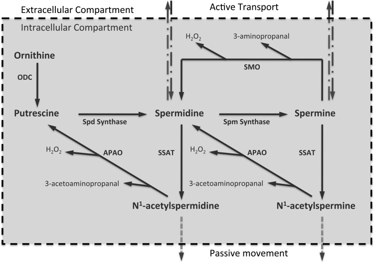
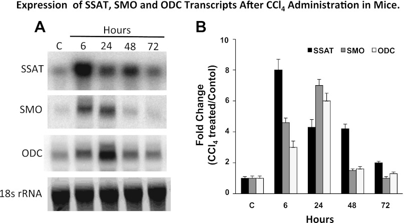
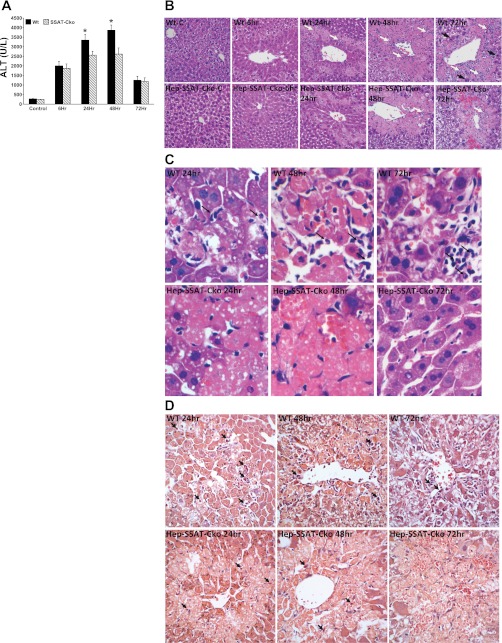
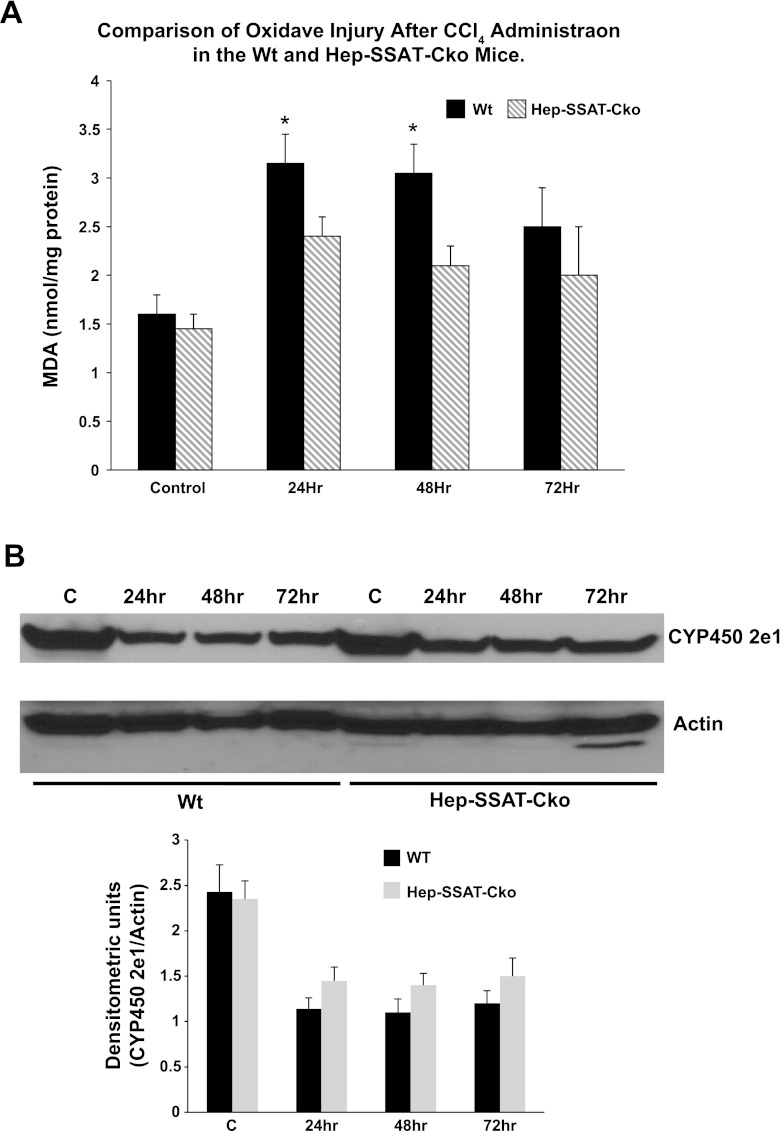
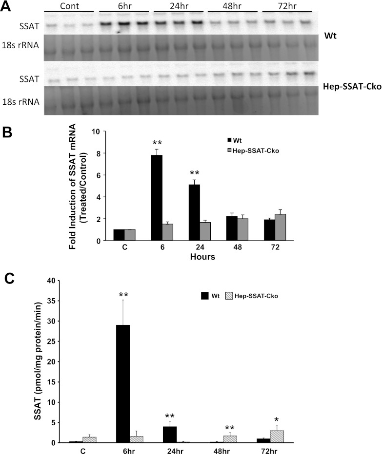
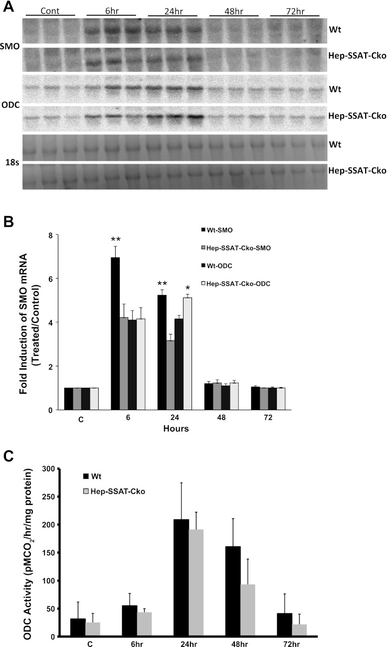
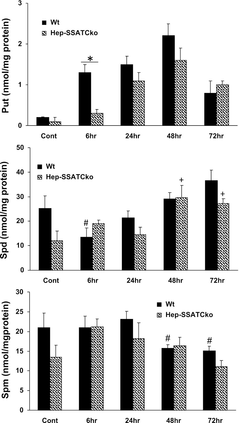
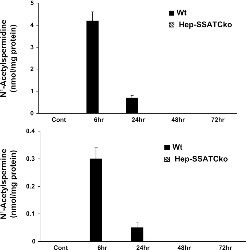
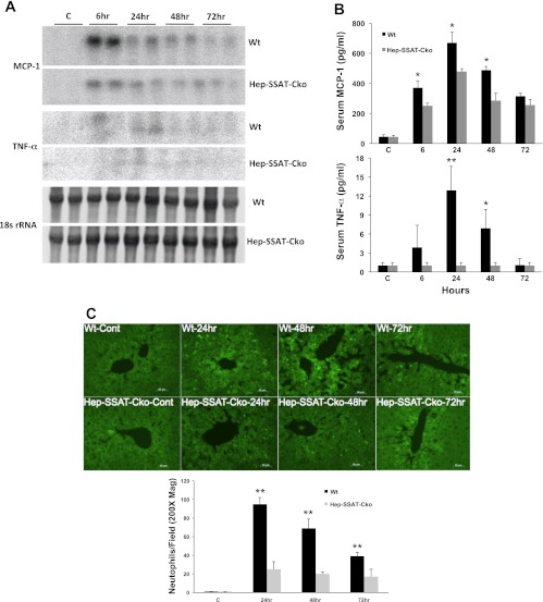
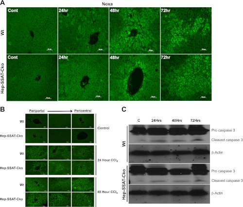
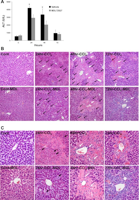
Activation of spermine/spermidine-N(1)-acetyltransferase (SSAT) leads to DNA damage and growth arrest in mammalian cells, and its ablation reduces the severity of ischemic and endotoxic injuries. Here we have examined the role of SSAT in the pathogenesis of toxic liver injury caused by carbon tetrachloride (CCl(4)). The expression and activity of SSAT increase in the liver subsequent to CCl(4) administration. Furthermore, the early liver injury after CCl(4) treatment was significantly attenuated in hepatocyte-specific SSAT knockout mice (Hep-SSAT-Cko) compared with wild-type (WT) mice as determined by the reduced serum alanine aminotransferase levels, decreased hepatic lipid peroxidation, and less severe liver damage. Cytochrome P450 2e1 levels remained comparable in both genotypes, suggesting that SSAT deficiency does not affect the metabolism of CCl(4). Hepatocyte-specific deficiency of SSAT also modulated the induction of cytokines involved in inflammation and repair as well as leukocyte infiltration. In addition, Noxa and activated caspase 3 levels were elevated in the livers of WT compared with Hep-SSAT-Cko mice. Interestingly, the onset of cell proliferation was significantly more robust in the WT compared with Hep-SSAT Cko mice. The inhibition of polyamine oxidases protected the animals against CCl(4)-induced liver injury. Our studies suggest that while the abrogation of polyamine back conversion or inhibition of polyamine oxidation attenuate the early injury, they may delay the onset of hepatic regeneration.


Figures

Fig. 1.
Fig. 1.
Regulation of cellular polyamine homeostasis. The cellular levels of polyamines are tightly regulated through import, export, synthesis, and catabolism. The initial step in polyamine synthesis is the decarboxylation of ornithine by ODC to form Put. Sequential addition of aminopropyl residues to Put and Spd leads to the formation of Spd and Spm. Polyamines are degraded through back conversion (SSAT and APAO cascade) and oxidation of Spm by SMO. Oxidation of acetylated polyamines by APAO and Spm by SMO generate cytotoxic molecules such as 3-aminopropanal, 3-acetoaminopropanal, and H2O2. SSAT, spermine/spermidine-N1-acetyltransferase; ODC, ornithine decarboxylase; Put, putrescine; Spd, spermidine; Spm, spermine; APAO, N1-acetylpolyamine oxidase; SMO, Spm oxidase.
Fig. 2.
Fig. 2.
Targeting strategy and characterization of hepatocyte-specific SSAT knockout mice. A: structure of LoxP-SSAT construct and the gene ablation strategy are depicted (top and bottom). B: disruption of SSAT gene was confirmed by examining the genomic DNA from the tail (top) and liver (bottom) of Alb-SSAT-Cko (Hep-SSAT-Cko) and their Cre-deficient (SSAT-CkoNeo−/Flp−) or wild-type (Wt, WT) littermates. C: examination of SSAT mRNA expression in the livers of hepatocyte-specific SSAT knockout mice (Hep-SSAT-Cko), WT, and SSAT-Cko Neo−/Flp− mice.
Fig. 3.
Fig. 3.
Effect of carbon tetrachloride (CCl4) treatment on the hepatic expression of SSAT, SMO, and ODC mRNAs. Liver RNA samples (15 μg/well of total RNA) from control and CCl4-treated animals were size fractionated and subjected to Northern blot analysis to assess the expression of SSAT, SMO, and ODC transcripts. A: Northern blot analysis results indicate that all three transcripts are upregulated in the livers of CCl4-treated animals. B: densitometric analysis of the Northern blot analysis results were normalized against the intensity of their corresponding 18s rRNA band. These results were used to compare the induction of each transcript in the livers of CCl4- to that of vehicle-treated animals. The induction values are presented as fold increase over control. The blots are representative of 3 independent studies.
Fig. 4.
Fig. 4.
Assessing the effect of hepatocyte-specific SSAT deficiency on the severity of CCl4-induced liver injury. WT and Hep-SSAT-Cko were given intraperitoneal injections of CCl4 or vehicle. Animals (n = 6/treatment group/genotype) were killed at timed intervals after treatment. A: serum alanine aminotransferase (ALT) levels of control and CCl4-treated WT and Hep-SSAT-Cko mice (n = 6/group) were compared following the protocol outlined in materials and methods. *P < 0.05 WT compared with SSAT-Cko. B: liver histology (magnification, ×400) of control and injured animals from both genotypes were compared. Infiltrating neutrophils (small white arrows), chronic inflammatory cells (large white arrows) and mitotic bodies (large black arrows) are marked. C: liver histology of CCl4-treated WT and Hep-SSAT-Cko mice was examined at ×600 magnification (black arrows mark the infiltrating neutrophils). D: livers of CCl4-treated WT and Hep-SSAT-Cko mice were subjected to chloroacetate estrase staining to identify the infiltrating neutrophils (black arrows).
Fig. 5.
Fig. 5.
Comparison of the number of proliferating cells in the livers of WT and Hep-SSAT-Cko mice after CCl4-induced liver injury. Expression of proliferating cell nuclear antigen (PCNA), a marker of proliferating cells, was compared in the livers of WT and Hep-SSAT-Cko animals after CCl4-induced liver injury.
Fig. 6.
Fig. 6.
Comparison of the severity of oxidative injury in WT and Hep-SSAT-Cko mice. A: onset of oxidative damage in the liver of WT and Hep-SSAT-Cko animals (n = 5/group/genotype) after CCl4-induced hepatotoxic injury was compared by determining the extent of lipid peroxidation as measured by the malondialdehyde (MDA) levels in the liver extracts. *Levels were significantly higher in WT vs. Hep-SSAT-Cko at 24 h and 48 h. B: adaptive downregulation of liver CYP2e1 was compared in WT and Hep-SSAT-Cko mice. CYP2e1 and actin signals from Western blot analyses of liver extracts from control and CCl4-treated WT and Hep-SSAT-Cko mice were subjected to densitometric analysis to compare the effect of CCl4-induced liver injury on the expression of CYP2e1. The blots are representative and the densitometry studies are the average ± SD of 3 independent samples.
Fig. 7.
Fig. 7.
Comparison of the time course of expression and activity of SSAT in the livers of control and CCl4-treated WT and Hep-SSAT-Cko mice. A: liver RNA from control and CCl4-treated WT and Hep-SSAT-Cko mice (n = 3/group) was size fractionated and subjected to Northern blot analysis to compare the expression levels of SSAT transcript. B: intensity of the SSAT mRNA bands were normalized against their corresponding 18s rRNA bands. These results were used to compare the induction of SSAT transcript in the livers of CCl4- or vehicle-treated WT and Hep-SSAT-Cko animals. The induction values are presented as fold increase over control. C: enzymatic activity of SSAT was measured and compared in the livers of time-matched control and CCl4-treated WT and Hep-SSAT-Cko mice (n = 3/group). *P < 0.05 is considered significant; **P < 0.01.
Fig. 8.
Fig. 8.
Comparison of the time course of expression of SMO and ODC in the livers of vehicle and CCl4-treated WT and Hep-SSAT-Cko mice. A: liver RNA from control and CCl4-treated WT and Hep-SSAT-Cko mice (n = 3/group) were subjected to Northern blot analysis to compare the expression levels of SMO and ODC transcripts. B: intensity of SMO and ODC mRNA bands for each sample was determined by densitometry and normalized against the corresponding 18s rRNA band. The induction values are presented as fold increase over control. C: ODC activity was measured in the livers of vehicle and CCl4-treated WT and Hep-SSAT-Cko animals. *P < 0.05 is considered significant; **P < 0.01.
Fig. 9.
Fig. 9.
Comparison of the polyamine content in the livers of control and CCl4-treated WT and Hep-SSAT-Cko mice. The content of Put, Spd, and Spm (top, middle, and bottom, respectively) in the livers of control and CCl4-treated WT and Hep-SSAT-Cko mice were measured by HPLC (results are means ± SE of n = 3/treatment group/genotype). Hepatic content of each polyamine was compared in CCl4-treated and control samples as well as time-matched samples from both genotypes. Polyamine levels are expressed as pmol/mg of protein. P > 0.05 is considered significant. *A significant difference between WT and Hep-SSAT-Cko. #Significant differences between vehicle-treated controls and CCl4-treated WT animals. +Significant differences between vehicle-treated controls and CCl4-treated Hep-SSAT-Cko mice.
Fig. 10.
Fig. 10.
Comparison of the acetylated polyamine content in the livers of vehicle and CCl4-treated WT and Hep-SSAT-Cko mice. The content of N1-acetylspermidine and N1-acetylspermine (top and bottom, respectively) in the livers of control (Cont) and CCl4-treated WT and Hep-SSAT-Cko mice were measured by HPLC (results are means ± SE of n = 3/treatment group/genotype). Hepatic levels of acetylated polyamines were compared in CCl4-treated and control samples as well as time-matched samples from both genotypes. Polyamine levels are expressed as pmol/mg of protein.
Fig. 11.
Fig. 11.
Comparison of the inflammatory response of WT and Hep-SSAT-Cko animals subjected to CCl4-induced liver injury. A: liver RNA form control and CCl4-treated WT and Hep-SSAT-Cko mice (n = 2/group) was subjected to Northern blot analysis to compare the monocyte chemotactic protein-1 (MCP-1) and TNF-α levels. Data is representative of 2 independent studies. B: serum levels of MCP-1 and TNF-α were compared in control and CCl4-treated WT and Hep-SSAT-Cko mice (n = 3/group). *P < 0.05 is considered significant; **P < 0.01. C: infiltration of neutrophils in livers of vehicle and CCl4-treated mice was assessed by immunofluorescent microscopy (magnification, ×200). The number of neutrophils was determined in 10 independent fields (magnification, ×200), and the results were expressed as means ± SD. **P < 0.01.
Fig. 12.
Fig. 12.
Comparison of Noxa and activated caspase 3 levels in the livers of vehicle and CCl4-treated WT and Hep-SSAT-Cko animals. A: expression of Noxa was assessed by immunofluorescent staining of liver sections from control and CCl4-treated WT and Hep-SSAT-Cko mice (magnification, ×200). B: activated/cleaved caspase 3 levels in the livers of control and CCl4-treated WT and Hep-SSAT-Cko mice were examined by immunofluorescent microscopy (magnification, ×200). C: levels of pro and cleaved caspase 3 were examined in the liver extracts of vehicle and CCl4-treated WT and Hep-SSAT-Cko mice. Equal loading was confirmed by determining the actin levels in the size-fractionated extracts. The blots are representative of 3 independent samples.
Fig. 13.
Fig. 13.
Examining the role of polyamine oxidases downstream of SSAT in CCl4-induced liver injury. A: serum ALT levels of control and CCl4-treated mice injected with MDL72527 or vehicle (n = 6/group) were compared following the protocol outlined in the materials and methods. *P < 0.05 is considered significant. Results are means ± SE of 3 independent samples. B: liver histology (magnification, ×200) of control and injured animals from vehicle and MDL72527-treated groups were compared. White arrows, infiltrating cells; black arrows, areas of necrosis. C: liver histology (magnification, ×400) of control and injured animals from vehicle and MDL72527-treated animals were compared. White arrows, infiltrating cells; black arrows, steatotic vesicles.

References

    1. Alhonen L, Parkkinen JJ, Keinanen T, Sinervirta R, Herzig KH, Janne J. Activation of polyamine catabolism in transgenic rats induces acute pancreatitis. Proc Natl Acad Sci USA 97: 8290–8295, 2000 - PMC - PubMed
    1. Alhonen L, Rasanen TL, Sinervirta R, Parkkinen JJ, Korhonen VP, Pietila M, Janne J. Polyamines are required for the initiation of rat liver regeneration. Biochem J 362: 149–153, 2002 - PMC - PubMed
    1. Bansal MB, Kovalovich K, Gupta R, Li W, Agarwal A, Radbill B, Alvarez CE, Safadi R, Fiel MI, Friedman SL, Taub RA. Interleukin-6 protects hepatocytes from CCl4-mediated necrosis and apoptosis in mice by reducing MMP-2 expression. J Hepatol 42: 548–556, 2005 - PMC - PubMed
    1. Barone S, Okaya T, Rudich S, Petrovic S, Tenrani K, Wang Z, Zahedi K, Casero RA, Lentsch AB, Soleimani M. Distinct and sequential upregulation of genes regulating cell growth and cell cycle progression during hepatic ischemia-reperfusion injury. Am J Physiol Cell Physiol 289: C826–C835, 2005 - PubMed
    1. Baskaya MK, Rao AM, Dogan A, Donaldson D, Gellin G, Dempsey RJ. Regional brain polyamine levels in permanent focal cerebral ischemia. Brain Res 744: 302–308, 1997 - PubMed

Publication types