Comparative Study
. 2012 Jul 15;189(2):988-1001.
doi: 10.4049/jimmunol.1103031. Epub 2012 Jun 20.

Cross-species transcriptional network analysis defines shared inflammatory responses in murine and human lupus nephritis

Affiliations
Comparative Study

Cross-species transcriptional network analysis defines shared inflammatory responses in murine and human lupus nephritis

Celine C Berthier et al. J Immunol. .

Abstract

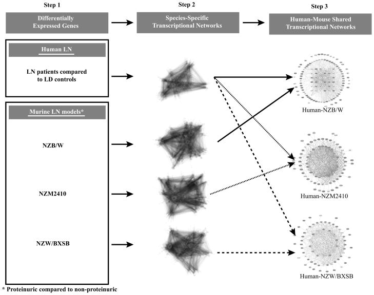
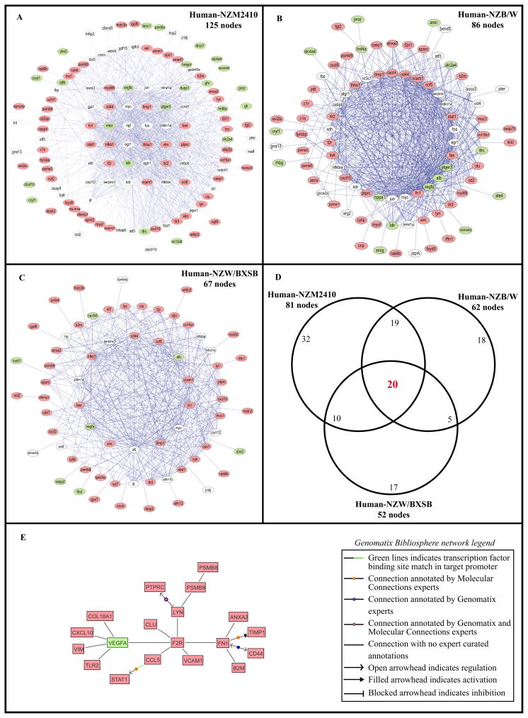
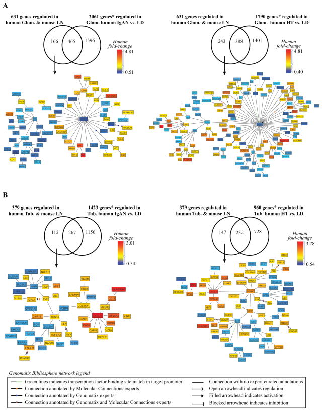
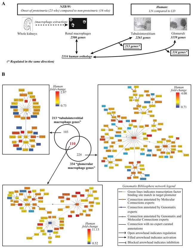
Lupus nephritis (LN) is a serious manifestation of systemic lupus erythematosus. Therapeutic studies in mouse LN models do not always predict outcomes of human therapeutic trials, raising concerns about the human relevance of these preclinical models. In this study, we used an unbiased transcriptional network approach to define, in molecular terms, similarities and differences among three lupus models and human LN. Genome-wide gene-expression networks were generated using natural language processing and automated promoter analysis and compared across species via suboptimal graph matching. The three murine models and human LN share both common and unique features. The 20 commonly shared network nodes reflect the key pathologic processes of immune cell infiltration/activation, endothelial cell activation/injury, and tissue remodeling/fibrosis, with macrophage/dendritic cell activation as a dominant cross-species shared transcriptional pathway. The unique nodes reflect differences in numbers and types of infiltrating cells and degree of remodeling among the three mouse strains. To define mononuclear phagocyte-derived pathways in human LN, gene sets activated in isolated NZB/W renal mononuclear cells were compared with human LN kidney profiles. A tissue compartment-specific macrophage-activation pattern was seen, with NF-κB1 and PPARγ as major regulatory nodes in the tubulointerstitial and glomerular networks, respectively. Our study defines which pathologic processes in murine models of LN recapitulate the key transcriptional processes active in human LN and suggests that there are functional differences between mononuclear phagocytes infiltrating different renal microenvironments.


Figures

Figure 1
Figure 1. Analytical strategy of cross-species shared tubulointerstitial transcriptional networks using the Tool for Approximate LargE graph matching (TALE)
Individual transcriptional networks were generated using the literature-based Genomatix Bibliosphere software and were overlapped using TALE to define cross-species shared transcriptional networks.
Figure 2
Figure 2. Human-mouse shared tubulointerstitial transcriptional networks
Networks sharing the most connections between human LN and (A) NZM2410 (nephritic vs. young control) (125 nodes), (B) NZB/W (36 wks with established nephritis vs. 23 wks pre-nephritic) (86 nodes), and (C) NZW/BXSB (nephritic vs. prenephritic) (67 nodes). (D) represents the overlap of the nodes between the three comparisons, using only the nodes regulated in the same direction in both species in each network (81/125 nodes in the NZM2410, 62/86 nodes in the NZB/W and 52/67 nodes in the NZW/BXSB). Legend: Each node represents a gene; each edge (blue line) represents a connection between two nodes. The nodes having more than 20 connections, between 2 and 19 connections and only one connection are displayed in the inner layer, the middle layer and the outside layer, respectively. In red, green and white are respectively the nodes up-regulated in both species, down-regulated in both species and discordantly regulated among species. (E) represents the transcriptional Genomatix Bibliosphere network from the 20 overlapping nodes of the TALE results.
Figure 3
Figure 3. Human-NZB/W mouse LN comparison with IGAN and HT
A. Glomerular compartment. B. Tubulointerstitial compartment. The figures display the transcriptional networks (Genomatix Bibliosphere) obtained from the genes that were co-cited in PubMed abstracts in the same sentence linked to a function word (B2 filter) (145 of166 genes (A-left panel), 220 of 243 genes (A-right panel), 104 of 112 genes (B-left panel), 138 of 147 genes (Bright panel)). LN regulated transcripts were mapped into the transcriptional networks and included in the comparison * q-value<0.05 and fold-change ≥ 1.2 for the up-regulated genes and ≤ 0.8 for the down-regulated genes.
Figure 4
Figure 4. Renal LN F4/80hi intrinsic functional analysis
A. Analytical strategy of renal LN “macrophage” functional analysis (q-value <0.05, fold-change ≥1.2 for the up-regulated genes and ≤0.8 for the down-regulated genes). B. Overlap of the defined tubulointerstitial and glomerular “macrophage genes”. Transcriptional networks generated using the literature-based Genomatix Bibliosphere software from the 103 tubulointerstitial-restricted genes, the 224 glomerular-restricted genes and the 110 genes shared by both compartments and mouse F4/80hi intrinsic macrophages (q-value<0.05, fold-change ≥1.2 for the up-regulated genes and ≤0.8 for the down-regulated genes). Respectively, the pictures display the 42/103, 78/110 and 120/224 genes that were co-cited in PubMed abstracts in the same sentence linked to a function word (B2 filter).
Figure 5
Figure 5. Localization of macrophage/DC markers in human lupus nephritis
Immunohistochemistry for CD68 (A, D, G), S100 (B, E) and DC-SIGN (C, F, H) was performed on consecutive sections from a pre-transplant biopsy (A–C), and biopsies with lupus nephritis class IV (ISN/RPS 2003 classification, original magnifications X200 in A–F, X400 in G and H). Scattered CD68 and lower numbers of S100 positive cells were found in the tubulointerstitium (arrow) and occasionally in glomerular capillaries (arrowhead) in pretransplant biopsies (A, B). A low number of DC-SIGN positive cells were present in the tubulointerstitium (C, arrow). In contrast prominent numbers of CD68 and DC-SIGN positive cells where present in biopsies from patients with lupus nephritis (D–G). Consecutive sections demonstrate a prominent number of CD68 positive cells in glomerular capillaries (arrowheads in D and G), but also a prominent accumulation of CD68 positive cells in the tubulointerstitium (arrows). DC-SIGN positive cells were restricted to the tubulointerstitium (arrow in H), but no DC-SIGN was expressed on glomerular CD68 positive cells.

References

    1. Doria A, Iaccarino L, Ghirardello A, Zampieri S, Arienti S, Sarzi-Puttini P, Atzeni F, Piccoli A, Todesco S. Long-term prognosis and causes of death in systemic lupus erythematosus. Am J Med. 2006;119:700–706. - PubMed
    1. Cook RJ, Gladman DD, Pericak D, Urowitz MB. Prediction of short term mortality in systemic lupus erythematosus with time dependent measures of disease activity. J Rheumatol. 2000;27:1892–1895. - PubMed
    1. Ward MM. Changes in the incidence of end-stage renal disease due to lupus nephritis, 1982–1995. Arch Intern Med. 2000;160:3136–3140. - PubMed
    1. Ward MM. Changes in the incidence of endstage renal disease due to lupus nephritis in the United States, 1996–2004. J Rheumatol. 2009;36:63–67. - PMC - PubMed
    1. Grande JP. Experimental models of lupus nephritis. Contrib Nephrol. 2011;169:183–197. - PubMed

Publication types

MeSH terms

Associated data