Review
. 2011 Dec;84 Spec No 3(Spec Iss 3):S258-68.
doi: 10.1259/bjr/74240815.

Imaging congenital heart disease in adults

Affiliations
Review

Imaging congenital heart disease in adults

P J Kilner. Br J Radiol. 2011 Dec.

Abstract

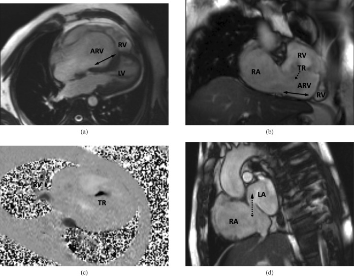
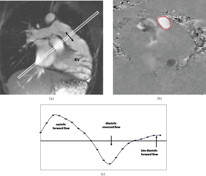
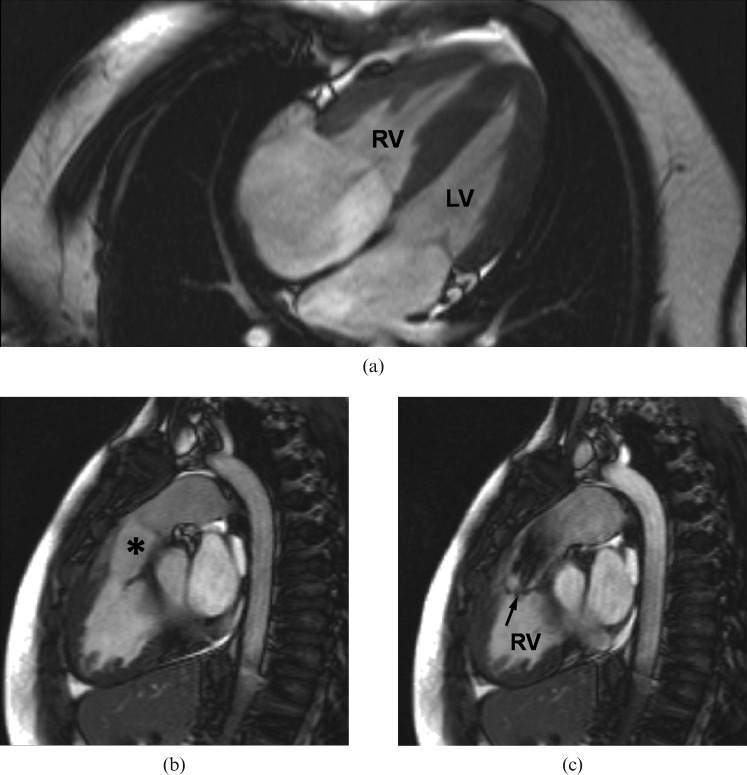
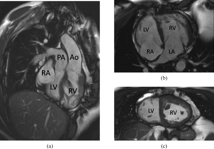
Transthoracic echocardiography is the first-line modality for cardiovascular imaging in adults with congenital heart disease (ACHD). The windows of access that are possible with transthoracic echocardiography are, however, rarely adequate for all regions of interest. The choice of further imaging depends on the clinical questions that remain to be addressed. The strengths of MRI include comprehensive access and coverage, providing imaging of all parts of the right ventricle, the pulmonary arteries, pulmonary veins and aorta. Cine images and velocity maps are acquired in specifically aligned planes, with stacks of cines or dynamic contrast angiography providing more comprehensive coverage. Tissues can be characterised if necessary, and MRI provides relatively accurate measurements of biventricular function and volume flow. These parameters are important in the assessment and follow-up of adults after repairs for tetralogy of Fallot or transposition of the great arteries and after Fontan operations. The superior spatial resolution and rapid acquisition of CT are invaluable in selected situations, including the visualisation of anomalous coronary or aortopulmonary collateral arteries, the assessment of luminal patency after stenting and imaging in patients with pacemakers. Ionising radiation is, however, a concern in younger patients who may need repeated investigation. Adults with relatively complex conditions should ideally be imaged in a specialist ACHD centre, where dedicated echocardiographic and cardiovascular MRI services are a necessary facility. General radiologists should be aware of the nature and pathophysiology of congenital heart disease, and should be alert for previously undiagnosed cases presenting in adulthood, including cases of atrial septal defect, aortic coarctation, patent ductus arteriosus, double-chambered right ventricle and congenitally corrected transposition.


Figures

Figure 1
Figure 1
Features of Ebstein anomaly in a 43-year-old who has not had surgery shown by MRI. (a) Four chamber and (b) oblique coronal cines show the atrialised part of the right ventricle (ARV) relative its functional part (RV, right ventricle). The double-headed arrows indicate the extent of apical displacement of the septal and inferior insertions of the tricuspid valve. The dotted arrow indicates the direction of tricuspid regurgitation (TR). (c) Flow (dark) through a velocity mapping plane located to transect the jet (dimensions about 5×14 mm) of severe TR. (d) An atrial short axis cine shows a widely patent foramen ovale with bidirectional flow, mainly from the right atrium (RA) to the left atrium (LA).
Figure 2
Figure 2
Pulmonary regurgitation measured by MRI through-plane velocity mapping in repaired tetralogy of Fallot. (a) Cine imaging aligned with the right ventricular outflow tract showed no effective pulmonary valve. Mapping of velocities through (b) a plane transecting the main pulmonary artery showed (c) systolic forward flow, diastolic reversed flow and late diastolic forward flow at the time of atrial systole. Absence of effective valve action typically results in a regurgitant fraction of ∼35–45%, only occasionally exceeding 50%, for example if exacerbated by branch pulmonary artory stenosis or otherwise increased resistance distal to compliant arterial segments. RV, right ventricle.
Figure 3.MRI
Figure 3.MRI
MRI features of a double-chambered right ventricle (RV), or subinfundibular stenosis, in a 16-year-old who has not had surgery. (a) The RV appears hypertrophied in a four-chamber view, whereas (b) the infundibular region (*) and the pulmonary valve above it are unobstructed. (c) The level of obstruction is seen in a systolic short axis image. The jet from the hypertrophied part of the RV to the infundibular cavity is arrowed. LV, Left ventricle.
Figure 4
Figure 4
Unoperated “congenitally corrected” transposition of the great arteries shown by MRI. (a,b) Both the atrio-ventricular and the ventriculo-arterial connections are discordant. Note the expected apical displacement of the septal insertion of the tricuspid valve of the right ventricle (RV) relative to that of the mitral valve of the left ventricle (LV), and the hypertrophied muscle of the systemic RV. (c) In the mid short axis image, the left ventricular cavity can be identified as the one on the smoother, less trabeculated, side of the ventricular septum. PA, pulmonary artery; Ao, aorta.

References

    1. Hoffman JI, Kaplan S. The incidence of congenital heart disease. J Am Coll Cardiol 2002;39:1890–900 - PubMed
    1. Perloff JK, Warnes CA. Challenges posed by adults with repaired congenital heart disease. Circulation 2001;103:2637–43 - PubMed
    1. Silversides CK, Marelli A, Beauchesne L, Dore A, Kiess M, Salehian O, et al. Canadian Cardiovascular Society 2009 consensus conference on the management of adults with congenital heart disease: executive summary. Can J Cardiol 2010;26:143–50 - PMC - PubMed
    1. Warnes CA. Transposition of the great arteries. Circulation 2006;114:2699–709 - PubMed
    1. Gewillig M. The Fontan circulation. Heart 2005;91:839–46 - PMC - PubMed

Publication types

MeSH terms