. 2012 Aug 2;120(5):1048-59.
doi: 10.1182/blood-2012-01-401760. Epub 2012 Jun 21.

Enforced expression of Lin28b leads to impaired T-cell development, release of inflammatory cytokines, and peripheral T-cell lymphoma

Affiliations

Enforced expression of Lin28b leads to impaired T-cell development, release of inflammatory cytokines, and peripheral T-cell lymphoma

Sarah H Beachy et al. Blood. .

Abstract

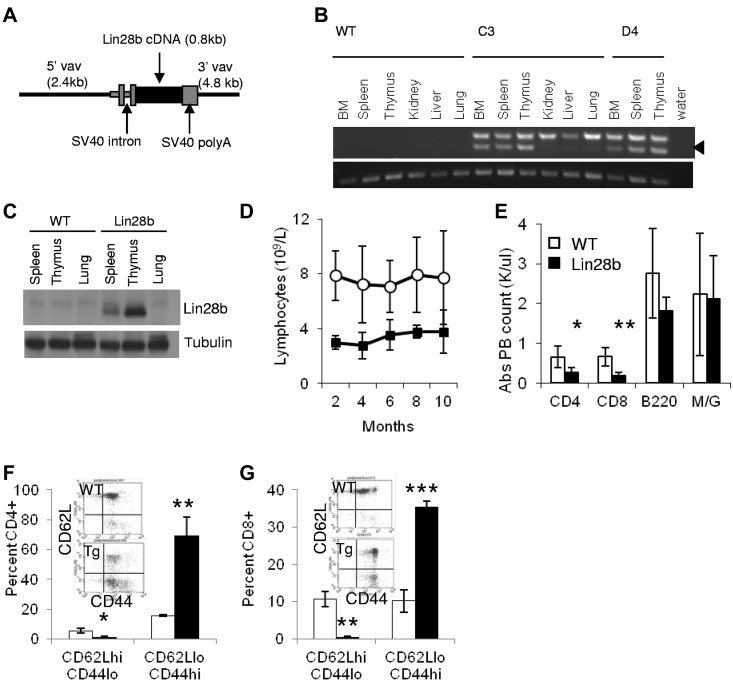
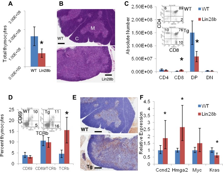
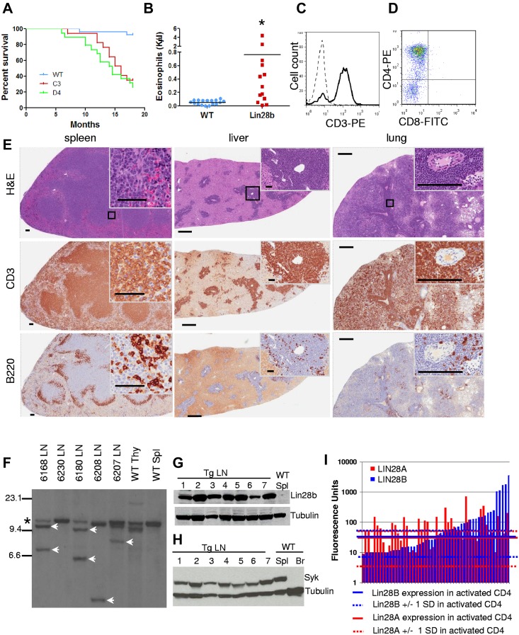
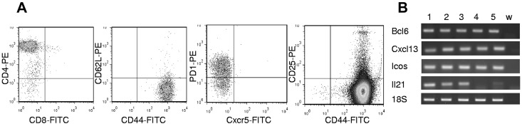
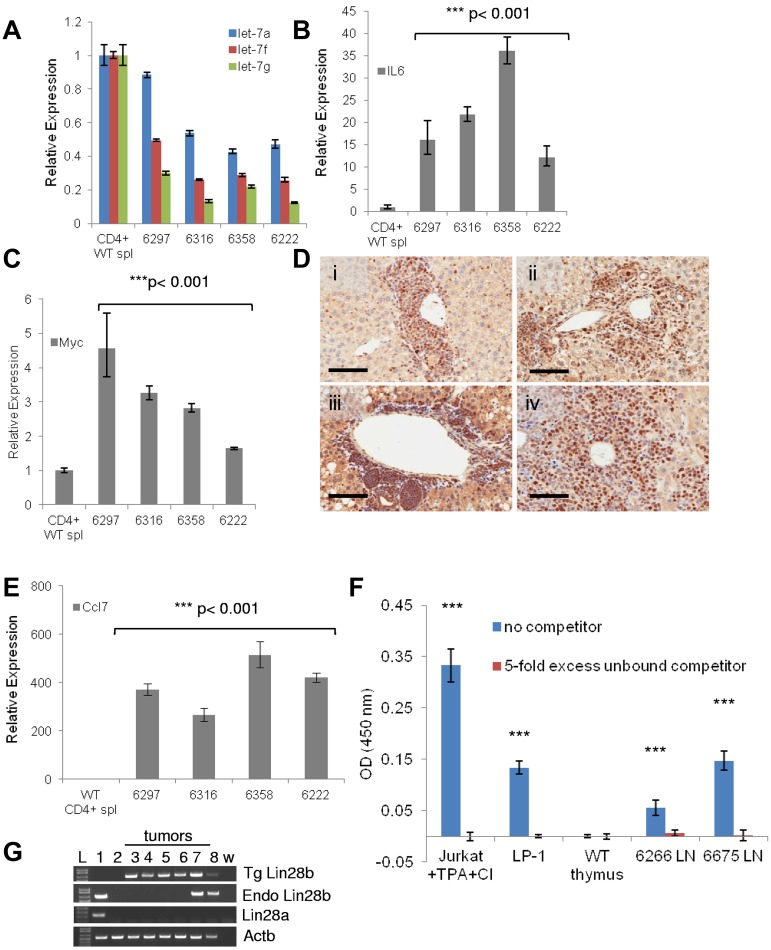
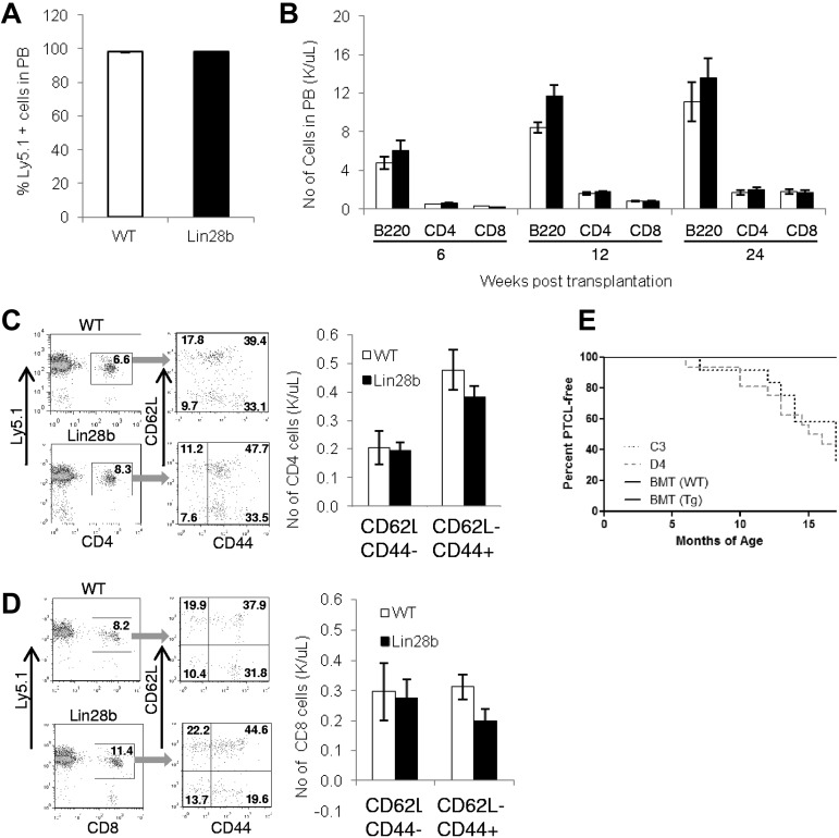
LIN28A and LIN28B, the mammalian homologs of lin-28, are implicated in malignant transformation in part because of their ability to promote degradation of the let-7 family of miRs. In the present study, we show that overexpression of Lin28b in vivo leads to an aggressive peripheral T-cell lymphoma (PTCL) characterized by widespread infiltration of parenchymal organs with malignant CD4(+) cells. Similar to patients with PTCL, Lin28b-transgenic mice show signs of inflammation such as eosinophilia, increased C-reactive protein, release of inflammatory cytokines, and pleural effusion. The PTCLs that develop in Lin28b mice are derived from activated T cells and show decreased let-7 expression, increased Il6 expression, activation of NF-κB, and infiltration of B cells, all resulting in an inflammatory microenvironment. In addition, LIN28B is overexpressed 7.5-fold in PTCL patient samples compared with activated CD4(+) cells. The results of the present study demonstrate for the first time that Lin28b can transform primary cells in vivo, identify a previously unsuspected link between Lin28b and PTCL, and provide a unique animal model for the study of PTCL biology and therapy.


Figures

Figure 1
Figure 1
Expression of Lin28b leads to lymphopenia in transgenic mice. (A) Schematic of Vav expression vector used to generate transgenic mice. (B) Expression of the Lin28b transgene. Top panel: spliced transgenic Lin28b mRNA (arrowhead); the upper band seen in all transgenic tissues is due to amplification of unspliced, contaminating genomic DNA. Bottom panel: Actb used as a loading control. (C) Western blot for Lin28b protein in hematopoietic tissues of Lin28b mice. (D) Serial lymphocyte counts in the peripheral blood of WT and Lin28b-transgenic mice (n = 11). P < .01 for each time point. (E-F) Leukocyte subsets in peripheral blood (E; n = 5) and spleen (F; n = 3). M/G indicates myeloid cells that stain for the antigens Mac1 and Gr-1. (G-H) Percent of naive (CD62L+CD44) and effector memory (CD62LCD44+) CD4 (G) and CD8 (H) cells in peripheral blood from WT or Lin28b mice (n = 3). *P < .05; **P < .01; ***P < .001. WT is indicated by open circles or white bars; Lin28b, black squares or bars.
Figure 2
Figure 2
Developmental abnormalities in the Lin28b thymus. (A) Total thymocyte number in WT (blue) or Lin28b (red) thymi (n = 4). (B) H&E staining of WT or Lin28b whole thymus. M indicates medulla; and C, cortex. Scale bar indicates 400 μm. (C) Absolute numbers from the thymus with representative FACS plots showing percentages (inset; n = 4). (D) Retention of mature CD69/Tcrb+ cells in the thymus (n = 3). Inset shows representative FACS. (E) B220 staining in the medulla of the thymus. Scale bar indicates 400 μm. *P < .05; ***P < .001. WT is indicated by blue; Lin28b, red. (F) Let-7 target gene expression in the thymi from mice 1-4 months of age (n = 6 per genotype). Samples were compared with the WT samples set at 1. *P < .05.
Figure 3
Figure 3
Lin28b-transgenic mice develop T-cell tumors. (A) Survival of Lin28b mice from C3 (n = 17) and D4 (n = 19) founders compared with WT littermate controls (n = 27). Median survival was 14.5 and 16 months for the D4 and C3 lines, respectively, P < .0001 compared with WT controls by log-rank test. (B) Eosinophil counts from PTCL mice with a mean age of 12 months compared with WT mice, also mean age 12 months. *P < .05. (C-D) CD3 staining of tumor cells (C; dashed line, isotype control; solid line, CD3) and CD4 positivity of tumor cells from mouse 6180 (D). (E) Immunohistochemistry of tissues from mouse 6180. Scale bars for the spleen indicate 500 μm (low magnification) and 50 μm (high magnification inset). Scale bars for the liver and lung indicate 500 μm (low magnification) and 200 μm (high magnification inset). Black square indicates area enlarged for inset. (F) Southern blot for Tcrb gene rearrangements. SstI-restricted genomic DNA, hybridized to a TCRb probe *Germline band; white arrows, nongermline-rearranged bands. (G) Immunoblot for Lin28b expression. Lanes 1-7, Lin28b tumor no. 6266, 6222, 6226, 6297, 6316, 6358, and 6549 with WT spleen as the negative control. (H) Immunoblot for Syk expression. Lanes 1 through 7, Lin28b tumors 6266, 6222, 6226, 6297, 6316, 6358, and 6569; lane 8, WT spleen (positive control); lane 9, WT brain (negative control). (I) LIN28A and LIN28B expression in patients with PTCL compared with mean values of activated CD4+ (aCD4) T cells (n = 4). Solid blue (LIN28B) and solid red (LIN28A) lines represent means for activated CD4+ T cells; dashed lines represents 1 SD from these means. Each bar represents 1 PTCL patient; data are presented on a logarithmic scale.
Figure 4
Figure 4
Malignant cells from Lin28b lymph nodes resemble TFH cells. (A) FACS plots describing tumor cell phenotype from mouse 6502. Analyses for CD44/CD62L, CXCR5/PD1, and CD44/CD25 were gated on the CD4+ population. (B) Expression of Bcl6, Cxcl13, Icos, and Il21 in Lin28b tumors. Lanes 1 through 6, WT CD4+ splenocytes, 6502 LN, 6222 LN, 6208 LN, 6297 LN, and w (water).
Figure 5
Figure 5
A Lin28b-regulated inflammatory pathway in vivo. (A) Expression of let-7a, let-7f, and let-7g (determined by TaqMan assays) in tumors from mice with PTCL compared with CD4+ WT splenocytes. Values shown are means ± SEM. All P values for the tumor samples were < .001 except 6297 for let-7a, which was P < .01. (B-C) Comparison as in panel A for Il6 (B) and Myc (C) mRNA. Values shown are means ± SEM. (D) Immunohistochemistry demonstrates nuclear NF-κB (p65) staining in the infiltrated livers of mice with PTCL: 6168 (i), 6226 (ii), 6297 (iii), and 6358 (iv). Scale bar indicates 100 μm. (E) Expression of the NF-κB target gene Ccl7 in PTCL tumors. Values shown are means ± SEM. For all RNA-expression experiments, the data were normalized to WT CD4+ splenocytes, which is set at 1. (F) Measurement of activated NF-κB in nuclear extracts from Lin28b tumors. Jurkat and LP-1 nuclear extracts were used as positive controls. The optical density 450 nm reflects binding of NF-κB p65 to the immobilized oligonucleotide (blue bars); the red bars reflect competition by a 5-fold excess of nonimmobilized oligonucleotide. TPA indicates 12-O-tetradecanoylphorbol 13-acetate; CI, calcium ionophore; and LN, lymph node. ***P < .001 compared with WT thymus. (G) Expression levels of Lin28a and Lin28b in tumors from transgenic Lin28b mice with PTCL. L-DNA ladder: mouse embryonic stem cell line (1); WT CD4+ splenocytes (2); 6222 (3); 6297 (4); 6316 (5); 6358 (6); 6502 (7); 6266 (8). w indicates water control; Tg, transgenic Lin28b; and Endo, endogenous Lin28b.
Figure 6
Figure 6
Transplantation with WT BM rescues lymphopenia. (A) Donor WT cells (Ly5.1) were transplanted into either WT or Lin28b mice and an engraftment assay was performed at 24 weeks after transplantation (n = 6 Lin28b recipients, n = 5 WT recipients). (B) Lymphocyte engraftment after transplantation at the indicated time points. White bars indicate WT; and black bars, Lin28b. (C) Quantification of naive (CD62L+CD44), central memory (CD62L+CD44+), and effector memory (CD62LCD44+) CD4+ cells in the peripheral circulation. (D) Quantification of naive, central memory, and effector memory CD8+ cells. White bars indicate WT; and black bars, Lin28b. (E) Percentage of WT (n = 3) or Lin28b (n = 3) recipients free of PTCL after transplantation of WT BM cells (C3, n = 12; D4, n = 16). Mice that died of causes other than PTCL were not included in this analysis (WT, n = 2; Lin28b, n = 3; C3, n = 5; D4, n = 3).

References

    1. Guo Y, Chen Y, Ito H, et al. Identification and characterization of lin-28 homolog B (LIN28B) in human hepatocellular carcinoma. Gene. 2006;384:51–61. - PubMed
    1. Moss EG, Lee RC, Ambros V. The cold shock domain protein LIN-28 controls developmental timing in C. elegans and is regulated by the lin-4 RNA. Cell. 1997;88(5):637–646. - PubMed
    1. Ambros V, Horvitz HR. Heterochronic mutants of the nematode Caenorhabditis elegans. Science. 1984;226(4673):409–416. - PubMed
    1. Yu J, Vodyanik MA, Smuga-Otto K, et al. Induced pluripotent stem cell lines derived from human somatic cells. Science. 2007;318(5858):1917–1920. - PubMed
    1. Viswanathan SR, Powers JT, Einhorn W, et al. Lin28 promotes transformation and is associated with advanced human malignancies. Nat Genet. 2009;41(7):843–848. - PMC - PubMed

Publication types

MeSH terms