. 2012 Jun 20;32(25):8480-90.
doi: 10.1523/JNEUROSCI.5895-11.2012.

Medial prefrontal cortex neuronal activation and synaptic alterations after stress-induced reinstatement of palatable food seeking: a study using c-fos-GFP transgenic female rats

Affiliations

Medial prefrontal cortex neuronal activation and synaptic alterations after stress-induced reinstatement of palatable food seeking: a study using c-fos-GFP transgenic female rats

Carlo Cifani et al. J Neurosci. .

Abstract

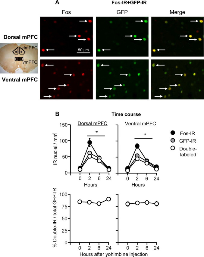
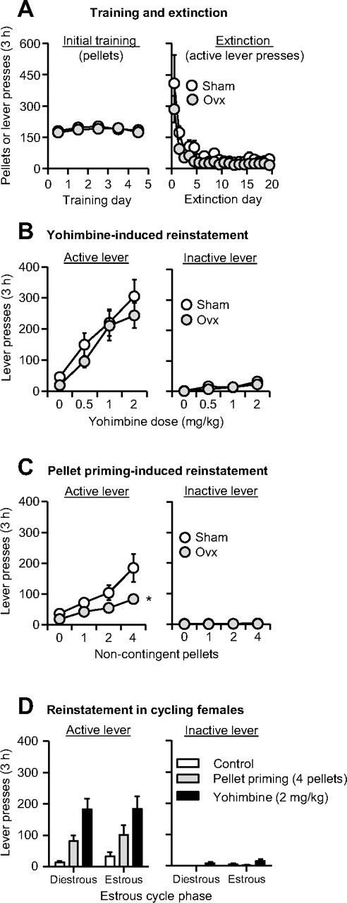
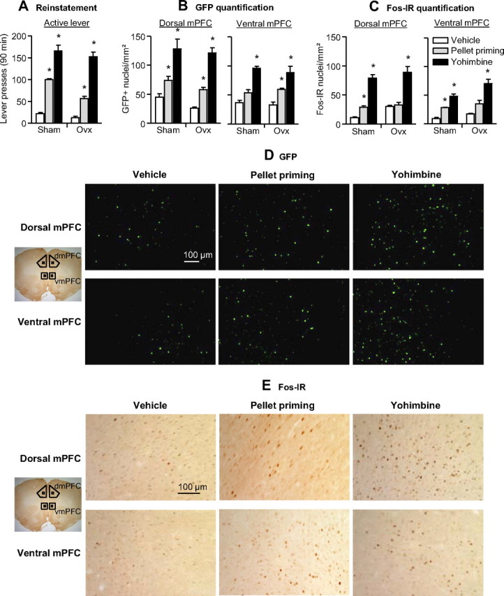
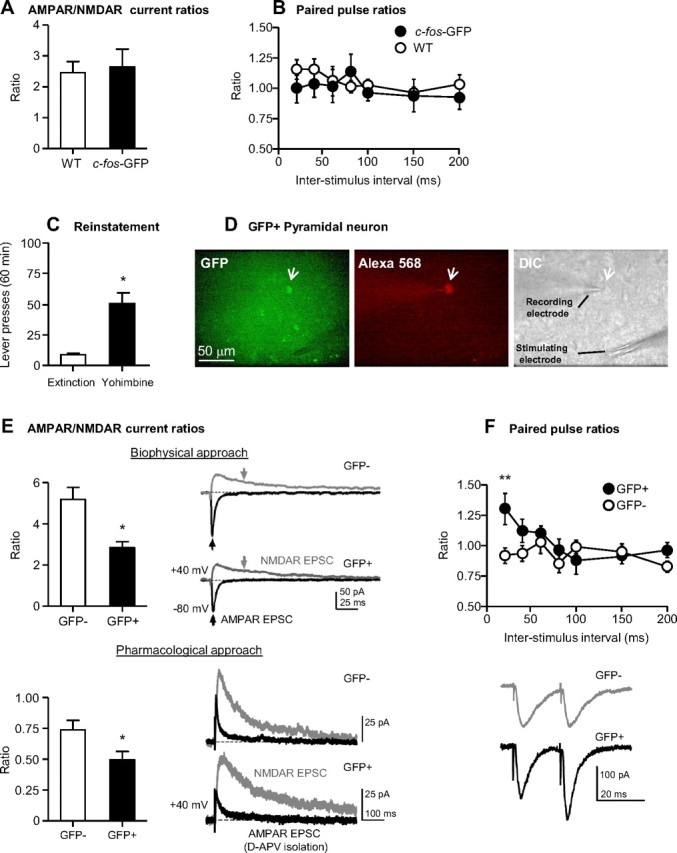
Relapse to maladaptive eating habits during dieting is often provoked by stress and there is evidence for a role of ovarian hormones in stress responses and feeding. We studied the role of these hormones in stress-induced reinstatement of food seeking and medial prefrontal cortex (mPFC) neuronal activation in c-fos-GFP transgenic female rats, which express GFP in strongly activated neurons. Food-restricted ovariectomized or sham-operated c-fos-GFP rats were trained to lever-press for palatable food pellets. Subsequently, lever-pressing was extinguished and reinstatement of food seeking and mPFC neuronal activation was assessed after injections of the pharmacological stressor yohimbine (0.5-2 mg/kg) or pellet priming (1-4 noncontingent pellets). Estrous cycle effects on reinstatement were also assessed in wild-type rats. Yohimbine- and pellet-priming-induced reinstatement was associated with Fos and GFP induction in mPFC; both reinstatement and neuronal activation were minimally affected by ovarian hormones in both c-fos-GFP and wild-type rats. c-fos-GFP transgenic rats were then used to assess glutamatergic synaptic alterations within activated GFP-positive and nonactivated GFP-negative mPFC neurons following yohimbine-induced reinstatement of food seeking. This reinstatement was associated with reduced AMPA receptor/NMDA receptor current ratios and increased paired-pulse facilitation in activated GFP-positive but not GFP-negative neurons. While ovarian hormones do not appear to play a role in stress-induced relapse of food seeking in our rat model, this reinstatement was associated with unique synaptic alterations in strongly activated mPFC neurons. Our paper introduces the c-fos-GFP transgenic rat as a new tool to study unique synaptic changes in activated neurons during behavior.


Figures

Figure 1.
Figure 1.
GFP-IR and Fos-IR overlap in mPFC neurons. A, Fos-IR and GFP-IR double labeling: representative photomicrographs of Fos and GFP labeling for dorsal and ventral mPFC (see small squares in the picture on the left of the photomicrographs for approximate areas). Data are from naive rats that were anesthetized and perfused 2 h after yohimbine (2 mg/kg, i.p) injections. B, Time course of Fos-IR, GFP-IR, and double-labeling after yohimbine (2 mg/kg, i.p.) injections. *Different from the 0 and 24 h time points for Fos-IR, GFP-IR, and double-labeling, p < 0.05, n = 3–4 per time point. dmPFC, Dorsal mPFC; vmPFC, ventral mPFC.
Figure 2.
Figure 2.
Effect of yohimbine and pellet priming on reinstatement of food seeking in ovariectomized and sham-operated rats and in cycling female rats. A, Mean ± SEM number of food pellets or previously active lever presses during the initial training sessions and the extinction sessions. B, C, Mean ± SEM number of active and inactive lever presses after yohimbine injections or after noncontingent pellet delivery in OVX and sham rats, n = 12 per group. D, Mean ± SEM number of active and inactive lever presses in female rats during the estrous and diestrous phases after water injections, yohimbine (2 mg/kg, i.p.) injections, or noncontingent delivery of 4 pellets at the beginning of the test session, n = 7. *Different from the Sham group, p < 0.01.
Figure 3.
Figure 3.
Effect of yohimbine- and pellet priming-induced reinstatement on GFP expression and Fos-immunoreactivity in the dorsal and ventral mPFC. A, Mean ± SEM number of active and inactive lever presses after water (vehicle) injections, yohimbine (2 mg/kg, i.p.) injections, and pellet priming (4 pellets). B, C, Mean ± SEM number of GFP-positive nuclei (per mm2) (B) or Fos-IR nuclei (per mm2) (C) in the dorsal (left) and ventral (right) mPFC 90 min after the test sessions. D, E, Representative pictures of GFP-positive (D) and Fos-IR (E) nuclei in the dorsal and ventral mPFC. *Different from the water (vehicle) condition, p < 0.05. dmPFC, Dorsal mPFC; vmPFC, ventral mPFC. n = 4 per experimental condition.
Figure 4.
Figure 4.
Electrophysiological properties of c-fos-GFP rat dorsal mPFC pyramidal neurons. A, B, Mean ± SEM of AMPAR/NMDAR current ratios (A) (n = 5 cells/3 rats for c-fos-GFP transgenic rats; n = 7 cells/4 rats for wild-type rats) and paired-pulse ratios from control c-fos-GFP and wild-type rats (B) (n = 6 cells/2 rats for c-fos-GFP rats; n = 10 cells/4 rats for wild-type rats) that did not undergo training, but were injected with yohimbine (2 mg/kg, i.p.) and killed 90 min later. C, Mean ± SEM number of active lever presses after water (vehicle) and yohimbine (2 mg/kg, i.p.) injections from c-fos-GFP rats used in Experiment 3. D, Representative micrograph of a GFP-positive pyramidal neuron indicated by a white arrow in the GFP panel. The same neuron was filled with Alexa Fluor 568 included in the patch pipette and indicated by a white arrow in the Alexa Fluor 568 panel. The DIC panel indicates the patched cell and the position of the stimulating and recording electrodes. E, Top, Mean ± SEM of AMPAR/NMDAR current ratios determined using the biophysical approach (n = 5 cells/4 rats for GFP-negative neurons; n = 4 cells/3 rats for GFP-positive neurons); the black and gray arrows indicate where the peak AMPAR and NMDAR-mediated EPSC, respectively, were obtained. Bottom, Mean ± SEM of AMPAR/NMDAR current ratios determined using d-APV application (n = 10 cells/6 rats for GFP-negative neurons; n = 6 cells/6 rats for GFP-positive neurons). Example traces of AMPAR and NMDAR-mediated EPSCs are indicated in black and gray, respectively. F, Top, Paired-pulse ratios from GFP-negative and GFP-positive neurons (n = 10 cells/6 rats for GFP-negative neurons; n = 9 cells/6 rats for GFP-positive neurons). Bottom, Representative EPSC pairs, evoked with 20 ms interstimulus intervals, are shown for single GFP-positive and GFP-negative pyramidal neurons.

References

    1. Anker JJ, Carroll ME. Sex differences in the effects of allopregnanolone on yohimbine-induced reinstatement of cocaine seeking in rats. Drug Alcohol Depend. 2010;107:264–267. - PMC - PubMed
    1. Anzalone CR, Hong LS, Lu JK, LaPolt PS. Influences of age and ovarian follicular reserve on estrous cycle patterns, ovulation, and hormone secretion in the Long-Evans rat. Biol Reprod. 2001;64:1056–1062. - PubMed
    1. Asarian L, Geary N. Modulation of appetite by gonadal steroid hormones. Philos Trans R Soc Lond B Biol Sci. 2006;361:1251–1263. - PMC - PubMed
    1. Badiani A, Belin D, Epstein D, Calu D, Shaham Y. Opiate versus psychostimulant addiction: the differences do matter. Nat Rev Neurosci. 2011;12:685–700. - PMC - PubMed
    1. Barth AL, Gerkin RC, Dean KL. Alteration of neuronal firing properties after in vivo experience in a FosGFP transgenic mouse. J Neurosci. 2004;24:6466–6475. - PMC - PubMed

Publication types

MeSH terms