. 2012 Jun 20;32(25):8703-15.
doi: 10.1523/JNEUROSCI.0204-12.2012.

Survival motor neuron protein in motor neurons determines synaptic integrity in spinal muscular atrophy

Affiliations

Survival motor neuron protein in motor neurons determines synaptic integrity in spinal muscular atrophy

Tara L Martinez et al. J Neurosci. .

Abstract

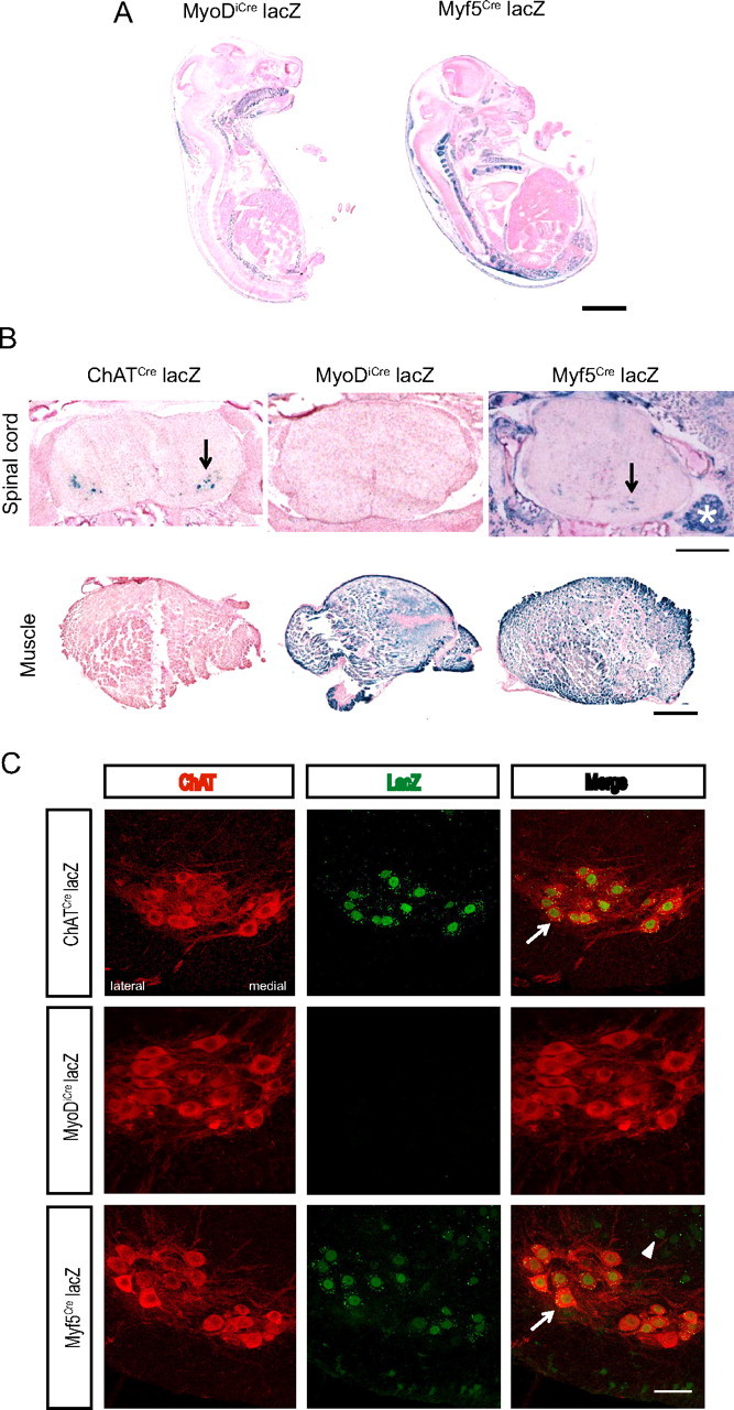
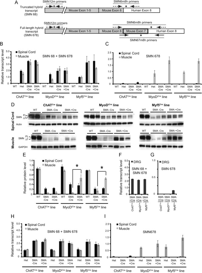
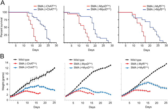
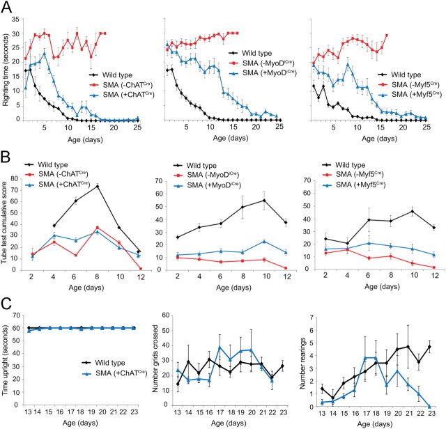
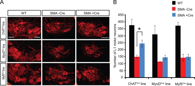
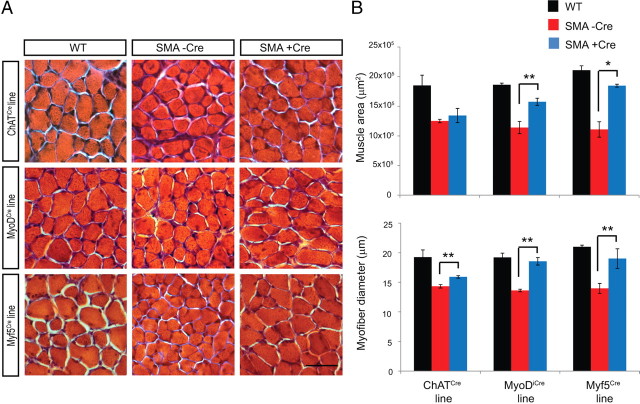
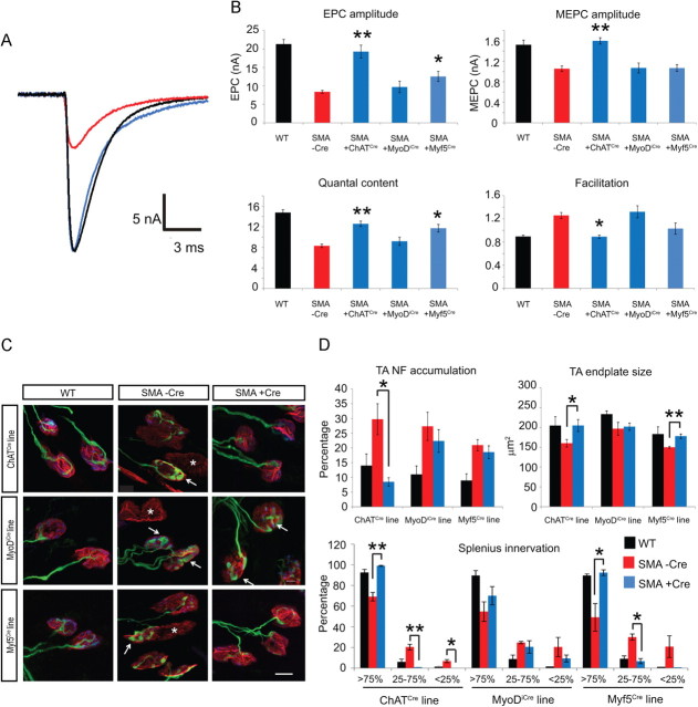
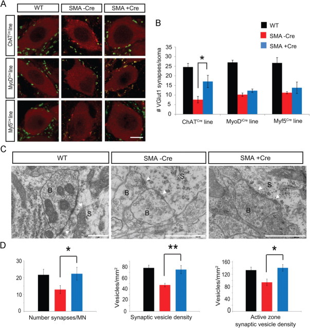
The inherited motor neuron disease spinal muscular atrophy (SMA) is caused by deficient expression of survival motor neuron (SMN) protein and results in severe muscle weakness. In SMA mice, synaptic dysfunction of both neuromuscular junctions (NMJs) and central sensorimotor synapses precedes motor neuron cell death. To address whether this synaptic dysfunction is due to SMN deficiency in motor neurons, muscle, or both, we generated three lines of conditional SMA mice with tissue-specific increases in SMN expression. All three lines of mice showed increased survival, weights, and improved motor behavior. While increased SMN expression in motor neurons prevented synaptic dysfunction at the NMJ and restored motor neuron somal synapses, increased SMN expression in muscle did not affect synaptic function although it did improve myofiber size. Together these data indicate that both peripheral and central synaptic integrity are dependent on motor neurons in SMA, but SMN may have variable roles in the maintenance of these different synapses. At the NMJ, it functions at the presynaptic terminal in a cell-autonomous fashion, but may be necessary for retrograde trophic signaling to presynaptic inputs onto motor neurons. Importantly, SMN also appears to function in muscle growth and/or maintenance independent of motor neurons. Our data suggest that SMN plays distinct roles in muscle, NMJs, and motor neuron somal synapses and that restored function of SMN at all three sites will be necessary for full recovery of muscle power.


Figures

Figure 1.
Figure 1.
Cre is expressed in tissue-specific patterns in ChATCre, MyoDiCre, and Myf5Cre mice. A, Representative images of lacZ staining of whole mount E15.5 embryos in MyoDiCre+ lacZ (n = 2) and Myf5Cre+ lacZ lines (n = 5). Scale bar, 2.5 mm. B, Representative images of lacZ staining of spinal cord (top row) and skeletal muscle cross sections (bottom row) from P7 ChATCre+ lacZ, MyoDiCre+ lacZ, and Myf5Cre+ lacZ mice (n = 2 each). *Indicates a DRG. Arrow indicates neurons in ventral horn of spinal cord. Scale bar, 500 μm. C, Representative images of ChAT/lacZ IHC in spinal cords from ChATCre+ lacZ (n = 3 lacZ-positive), MyoDiCre+ lacZ (n = 3 lacZ-positive, 3 lacZ-negative) and Myf5Cre+ lacZ mice (n = 4 lacZ-positive, 3 lacZ-negative examined). Punctate lacZ staining is evident in the cytoplasm and diffuse staining is visible in the nuclei of all motor neurons in ChATCre mice. LacZ staining is present in some, but not all motor neurons in Myf5Cre mice (indicated by arrows) and in other cells of the spinal cord (indicated by arrowheads). Scale bar, 50 μm.
Figure 2.
Figure 2.
SMN is expressed in a tissue-specific manner in conditional SMA mice. A, Schematic demonstrating the exonic structures of the possible transcript products of the SmnRes hybrid allele. In the absence of Cre recombinase, truncated hybrid transcript (SMN 68) is made containing upstream mouse Smn (exons 1–6), human SMN2 exon 8, and no exon 7 sequence. In the presence of Cre recombinase, a full-length hybrid transcript (SMN 678) containing mouse Smn exons 1–7 and human SMN2 exon 8 sequence may be made. The positions of qRT-PCR primers used are indicated with arrows indicating forward and reverse primers and asterisks indicating the positions of the probes. SMN6m8h primers detect both truncated and full-length hybrid transcripts, the SMN67m8h primers only detect full-length transcripts, and the SMN12m primers were used as an endogenous control. B, SMN6m8h primers detect hybrid transcripts in both spinal cord and muscle of P10 Cre+ Het, Cre− SMA, and Cre+ SMA mice, but not in WT mice. C, SMN67m8-h primers detect SMN 678 transcripts in spinal cord only in ChATCre+ and to a lesser extent, in Myf5Cre+ Het and SMA mice. SMN 678 transcripts are expressed in muscle only in MyoDiCre+ and Myf5Cre+ Het and SMA mice (n = 3–8 per genotype). D, SMN protein levels were determined in spinal cord (top row) and muscle (bottom row) tissues of P10 WT, Cre− SMA, and Cre+ SMA mice from each conditional SMA line. Both full-length (FL) and truncated (Δ7) SMN protein isoforms are visible and are compared with the endogenous controls, actin or GAPDH. E, Densitometry analysis of FL SMN protein expression shows reduced FL SMN expression in Cre− SMA mice compared with WT mice in all three lines in both spinal cord and muscle. FL-SMN expression is significantly increased in the muscles of MyoDiCre+ and Myf5Cre+ SMA mice compared with Cre− SMA mice. *p < 0.01. F, SMN6m8h primers detect hybrid transcripts in the DRGs of all lines of Cre+ SMA mice, but (G) SMN67m8 h primers detect SMN 678 transcripts only in DRGs of Myf5Cre+ SMA mice, not ChATCre+ or MyoDiCre+ SMA mice (n = 2–3 per genotype). H, SMN6m8-h primers and I, SMN67m8h primers detect hybrid transcripts that are expressed in the appropriate tissue-specific patterns at early developmental time points. (All tissues isolated from mice at P2 except 2 of 3 Myf5Cre+ SMA mice, which were isolated at P5). n = 2–4 per genotype.
Figure 3.
Figure 3.
Increased SMN expression in motor neurons or muscle increases survival and weights of SMA mice. A, Survival curves of ChATCre (n = 15 Cre− SMA, 28 Cre+ SMA mice), MyoDiCre (n = 16 Cre− SMA, 28 Cre+ SMA mice), and Myf5Cre (n = 13 Cre− SMA, 15 Cre+ SMA mice) mice indicate increased survival of Cre+ SMA mice in all 3 lines (log rank p < 0.0005). B, Weight curves are improved in all three lines, p < 0.001 (ChATCre: n = 11 WT, 12 Cre− SMA, and 27 Cre+ SMA, MyoDiCre: n = 11 WT, 12 Cre− SMA, and 18 Cre+ SMA, Myf5Cre: n = 10 WT, 14 Cre− SMA, and 15 Cre+ SMA mice).
Figure 4.
Figure 4.
Motor behavior is improved in conditional SMA mice. A, Righting latency is reduced in Cre+ SMA mice compared with Cre− SMA mice in all 3 lines of mice, p < 0.0001 (ChATCre: n = 11 WT, 12 Cre− SMA, and 27 Cre+ SMA, MyoDiCre: n = 11 WT, 12 Cre− SMA, and 18 Cre+ SMA, Myf5Cre: n = 10 WT, 14 Cre− SMA, and 15 Cre+ SMA mice). B, Cumulative tube test scores are increased in MyoDiCre+ SMA mice compared with Cre− SMA mice, p = 0.01 (ChATCre: n = 5 WT, 6 Cre− SMA, and 13 Cre+ SMA, MyoDiCre: n = 5 WT, 6 Cre− SMA, and 7 Cre+ SMA, Myf5Cre: n = 5 WT, 6 Cre− SMA, and 6 Cre+ SMA mice). C, Ambulation indices including time upright (left), numbers of grids crossed (middle), and number of rearings (right) is equivalent between ChATCre+ SMA mice and WT mice between P13 and P20 (n = 3–6 WT, 5–6 Cre+ SMA mice).
Figure 5.
Figure 5.
Increased SMN expression in motor neurons, but not muscle results in increased motor neuron number. A, Representative images of L1 spinal cord ventral horn and ChAT-positive motor neurons in ChATCre, MyoDiCre, and Myf5Cre WT, Cre− SMA, and Cre+ SMA mice. Scale bar, 50 μm. B, Quantification of total number of L1 motor neurons (ChATCre: n = 4 WT, 4 Cre− SMA, and 4 Cre+ SMA, MyoDiCre: n = 4 WT, 3 Cre− SMA, and 4 Cre+ SMA, Myf5Cre: n = 4 WT, 6 Cre− SMA, and 5 Cre+ SMA mice). *p = 0.01.
Figure 6.
Figure 6.
SMN expression in muscle restores myofiber size. A, Representative images of TA muscle cross sections from ChATCre, MyoDiCre, and Myf5Cre WT, Cre− SMA, and Cre+ SMA mice. Scale bar, 50 μm. B, Quantification of total muscle area and myofiber diameter (ChATCre: n = 3 WT, 3 Cre− SMA, and 3 Cre+ SMA, MyoDiCre: n = 3 WT, 4 Cre− SMA, and 4 Cre+ SMA, Myf5Cre: n = 3WT, 3 Cre− SMA, and 3 Cre + SMA mice). *p = 0.01, **p = 0.001.
Figure 7.
Figure 7.
NMJ physiology and structure is improved by expression of SMN in motor neurons. A, Representative EPC traces from ChATCre WT (black), Cre− SMA (red), and Cre+ SMA mice (blue). B, Quantification of EPC amplitudes, MEPC amplitudes, quantal content, and facilitation in the three lines of mice (n = 16 WT, 10 Cre− SMA, 6 ChATCre+ SMA, 6 MyoDiCre+ SMA, and 6 Myf5Cre+ SMA mice). *p ≤ 0.05, **p < 0.001. C, Representative images of NMJs from the splenius capitis muscle in ChATCre, MyoDiCre, and Myf5Cre WT, Cre− SMA, and Cre+ SMA mice (red, α-bungarotoxin; green, NF; blue, synaptophysin). Arrows indicate presynaptic terminals with NF accumulations. Asterisks indicate fully or partially denervated NMJs. Scale bar, 10 μm. D, Quantification of TA muscle presynaptic terminal NF accumulation, TA muscle endplate size, and innervation status of the splenius capitis muscle in the 3 lines of mice (ChATCre: n = 4 WT, 4 Cre− SMA, 4 Cre+ SMA, MyoDiCre: n = 3 WT, 4 Cre− SMA, and 4 Cre+ SMA, Myf5Cre: n = 3 WT, 3 Cre− SMA, 3 Cre+ SMA mice). *p < 0.05, **p < 0.001.
Figure 8.
Figure 8.
Motor neuron expression of SMN improves motor neuron somal synapses. A, Representative images of VGluT1+ synapses on L1 motor neurons in ChATCre, MyoDiCre, and Myf5Cre WT, Cre− SMA, and Cre+ SMA mice (red, ChAT; green, VGluT1). Scale bar, 10 μm. B, Quantification of VGluT1+ synapses in the three lines [ChATCre: n = 3 WT (17 neurons), 4 Cre− SMA (20 neurons), 3 Cre+ SMA (22 neurons), MyoDiCre: n = 3 WT (16 neurons), 4 Cre− SMA (17 neurons), and 3 Cre+ SMA (21 neurons), Myf5Cre: n = 3 WT (14 neurons), 3 Cre− SMA (21 neurons) and 4 Cre+ SMA (15 neurons)]. *p ≤ 0.5. C, Representative EM images of motor neuron somal synapses in ChATCre WT, Cre− SMA, and Cre+ SMA mice. S, soma; B, bouton; arrowheads indicate PSDs. Scale bar, 1 μm. D, Quantification of synapse number, PSD length, overall synaptic vesicle density, and active zone synaptic vesicle density in ChATCre WT, Cre− SMA, and Cre+ SMA mice (motor neuron#: n = 14 WT, 18 Cre− SMA, and 14 Cre+ SMA). *p < 0.05, **p < 0.001.

Similar articles

Cited by

References

    1. Avila AM, Burnett BG, Taye AA, Gabanella F, Knight MA, Hartenstein P, Cizman Z, Di Prospero NA, Pellizzoni L, Fischbeck KH, Sumner CJ. Trichostatin A increases SMN expression and survival in a mouse model of spinal muscular atrophy. J Clin Invest. 2007;117:659–671. - PMC - PubMed
    1. Biondi O, Grondard C, Lécolle S, Deforges S, Pariset C, Lopes P, Cifuentes-Diaz C, Li H, della Gaspera B, Chanoine C, Charbonnier F. Exercise-induced activation of NMDA receptor promotes motor unit development and survival in a type 2 spinal muscular atrophy model mouse. J Neurosci. 2008;28:953–962. - PMC - PubMed
    1. Bosch-Marcé M, Wee CD, Martinez TL, Lipkes CE, Choe DW, Kong L, Van Meerbeke JP, Musarò A, Sumner CJ. Increased IGF-1 in muscle modulates the phenotype of severe SMA mice. Hum Mol Genet. 2011;20:1844–1853. - PMC - PubMed
    1. Braun S, Croizat B, Lagrange MC, Warter JM, Poindron P. Constitutive muscular abnormalities in culture in spinal muscular atrophy. Lancet. 1995;345:694–695. - PubMed
    1. Burghes AH, Beattie CE. Spinal muscular atrophy: why do low levels of survival motor neuron protein make motor neurons sick? Nat Rev Neurosci. 2009;10:597–609. - PMC - PubMed

Publication types

MeSH terms