. 2012 Jun 20;32(25):8725-31.
doi: 10.1523/JNEUROSCI.1160-12.2012.

Wnt7A identifies embryonic γ-motor neurons and reveals early postnatal dependence of γ-motor neurons on a muscle spindle-derived signal

Affiliations

Wnt7A identifies embryonic γ-motor neurons and reveals early postnatal dependence of γ-motor neurons on a muscle spindle-derived signal

Soha Ashrafi et al. J Neurosci. .

Abstract

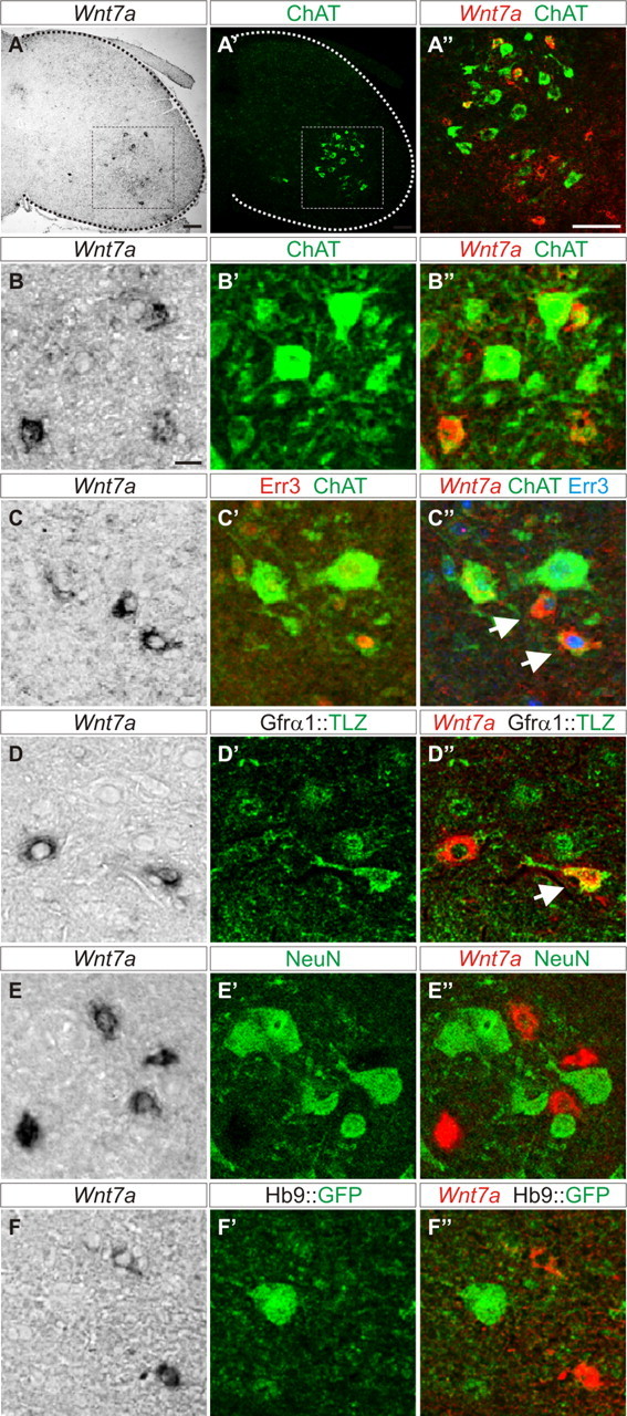
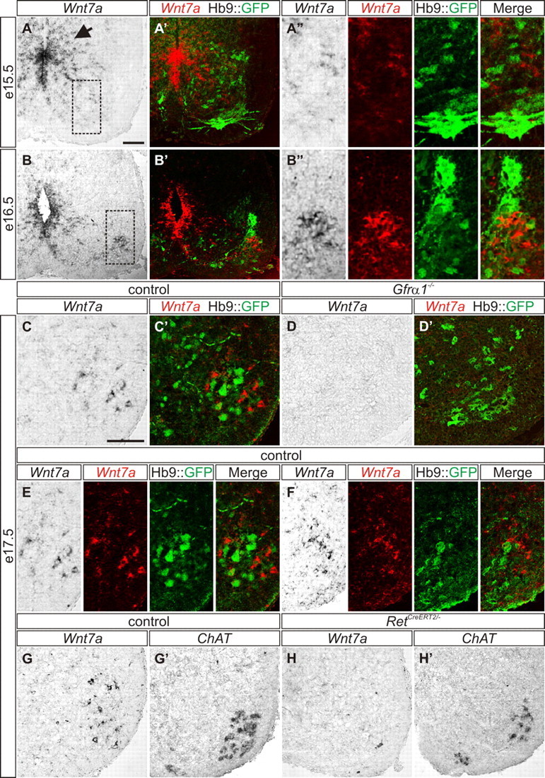
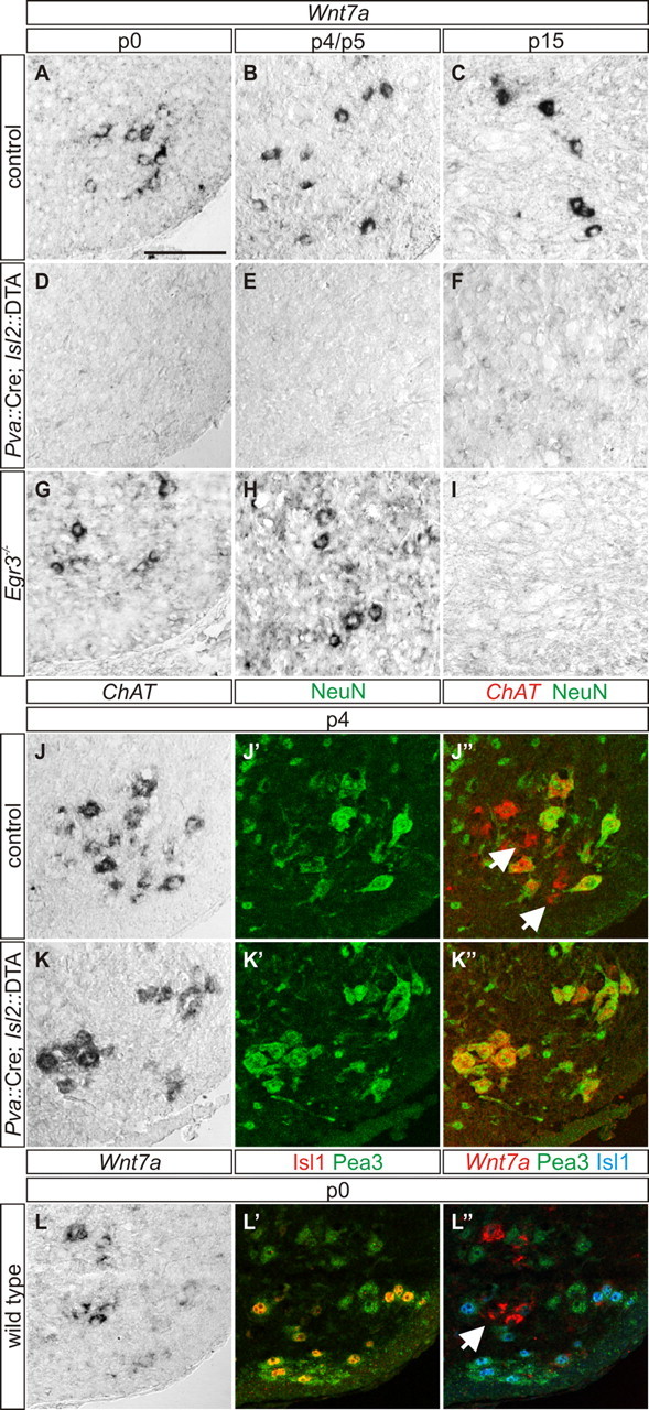
Motor pools comprise a heterogeneous population of motor neurons that innervate distinct intramuscular targets. While the organization of motor neurons into motor pools has been well described, the time course and mechanism of motor pool diversification into functionally distinct classes remains unclear. γ-Motor neurons (γ-MNs) and α-motor neurons (α-MNs) differ in size, molecular identity, synaptic input and peripheral target. While α-MNs innervate extrafusal skeletal muscle fibers to mediate muscle contraction, γ-MNs innervate intrafusal fibers of the muscle spindle, and regulate sensitivity of the muscle spindle in response to stretch. In this study, we find that the secreted signaling molecule Wnt7a is selectively expressed in γ-MNs in the mouse spinal cord by embryonic day 17.5 and continues to molecularly distinguish γ-from α-MNs into the third postnatal week. Our data demonstrate that Wnt7a is the earliest known γ-MN marker, supporting a model of developmental divergence between α- and γ-MNs at embryonic stages. Furthermore, using Wnt7a expression as an early marker of γ-MN identity, we demonstrate a previously unknown dependence of γ-MNs on a muscle spindle-derived, GDNF-independent signal during the first postnatal week.


Figures

Figure 1.
Figure 1.
Wnt7A is a γ-MN marker. A–A″, Wnt7aON cells (A) are located within the ChATON motor nucleus (A′) in the lateral ventral horn of P15 mouse spinal cord. A″, Magnification of boxed region in A and A′ showing colabeling of Wnt7a with a subset of ChATON cells. B–B″, Small ChATON/Wnt7aON cells are interspersed with large ChATON/Wnt7aOFF cells at P15. C–C″, Small ChATON/Wnt7aON cells express Err3 (arrows) at P15. D–D″, Small Wnt7aON cells express LacZ (arrow) in P21 Gfrα1::LacZ reporter mice. E–E″, Wnt7aON cells exclude NeuN at P21. F–F″, Wnt7aON cells exclude Hb9::GFP at P21. Split and merged channels shown for B–F″. Scale bars: A–A″, 100 μm; B–F″, 20 μm.
Figure 2.
Figure 2.
Wnt7A is expressed in embryonic γ-MNs. A–A″, Wnt7a is expressed in the ventricular zone (arrow) and is absent from the ventral motor neuron region in E15.5 Hb9::GFP mice (A, A′). A″, Magnified boxed region in A. B–B″, At E16.5, Hb9::GFPLOW/Wnt7aON cells are interspersed with Hb9::GFPON/Wnt7aOFF cells in the ventral spinal cord. Wnt7a labeling is also present in the ventricular zone (B, B′). B″, High magnification of boxed region in B. C, C′, E, F, Wnt7aON cells are localized throughout the motor neuron nucleus of E17.5 Hb9::GFP mice and are segregated from Hb9::GFPON cells. E, Split and merged channels of C′. D, D′, Wnt7aON cells are absent from the ventral spinal cord of E17.5 Gfrα1−/−; Hb9::GFP mice (D), while Hb9::GFPON/Wnt7aOFF cells persist (D′). G, H′, Wnt7aON cells in the motor nucleus of E17.5 control mice (G). RetCreERT2/− mice lack Wnt7aON cells at E17.5 (H). ChATON cells in control (G′) and RetCreERT2/− mice (H′). Split and merged channels shown for A″, B″, E, and F. Scale bars: A–H′, 100 μm.
Figure 3.
Figure 3.
A–C, A spindle-derived GDNF-independent signal is required for γ-MN development. Wnt7aON putative γ-MNs are localized within the motor neuron nucleus of P0 (A), P4 (B), and P15 (C) control mice. D–F, Wnt7a labeling is eliminated in P0 (D), P4 (E), and P15 (F) Pva::Cre; Isl2::DTA mutant mice. G–I, Wnt7aON γ-MNs are localized within the ventral spinal cord of P0 (G) and P5 (H) Egr3−/− mice, but are lost at P15 (I). J–J″, In control mice, NeuNOFF/ChATON γ-MNs (arrows in J″) intermingle with NeuNON/ChATON α-MNs at P4. K–K″, NeuNOFF/ChATON γ-MNs are absent in P4 Pva::Cre; Isl2::DTA mice. L–L″, In P0 wild type mice, Wnt7aON putative γ-MNs (arrow) are localized within the Cm motor pool, identified by coexpression of Islet1 and Pea3. Scale bar: A–L″, 100 μm.

Similar articles

Cited by

References

    1. Agalliu D, Takada S, Agalliu I, McMahon AP, Jessell TM. Motor neurons with axial muscle projections specified by Wnt4/5 signaling. Neuron. 2009;61:708–720. - PMC - PubMed
    1. Arber S, Han B, Mendelsohn M, Smith M, Jessell TM, Sockanathan S. Requirement for the homeobox gene Hb9 in the consolidation of motor neuron identity. Neuron. 1999;23:659–674. - PubMed
    1. Burke RE, Strick PL, Kanda K, Kim CC, Walmsley B. Anatomy of medial gastrocnemius and soleus motor nuclei in cat spinal cord. J Neurophysiol. 1977;40:667–680. - PubMed
    1. Chakkalakal JV, Nishimune H, Ruas JL, Spiegelman BM, Sanes JR. Retrograde influence of muscle fibers on their innervation revealed by a novel marker for slow motoneurons. Development. 2010;137:3489–3499. - PMC - PubMed
    1. Enjin A, Rabe N, Nakanishi ST, Vallstedt A, Gezelius H, Memic F, Lind M, Hjalt T, Tourtellotte WG, Bruder C, Eichele G, Whelan PJ, Kullander K. Identification of novel spinal cholinergic genetic subtypes disclose Chodl and Pitx2 as markers for fast motor neurons and partition cells. J Comp Neurol. 2010;518:2284–2304. - PubMed

Publication types

LinkOut - more resources