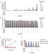
. 2012;10(6):e1001346.
doi: 10.1371/journal.pbio.1001346. Epub 2012 Jun 19.

Small heat shock proteins potentiate amyloid dissolution by protein disaggregases from yeast and humans

Affiliations

Small heat shock proteins potentiate amyloid dissolution by protein disaggregases from yeast and humans

Martin L Duennwald et al. PLoS Biol. 2012.

Abstract

How small heat shock proteins (sHsps) might empower proteostasis networks to control beneficial prions or disassemble pathological amyloid is unknown. Here, we establish that yeast sHsps, Hsp26 and Hsp42, inhibit prionogenesis by the [PSI+] prion protein, Sup35, via distinct and synergistic mechanisms. Hsp42 prevents conformational rearrangements within molten oligomers that enable de novo prionogenesis and collaborates with Hsp70 to attenuate self-templating. By contrast, Hsp26 inhibits self-templating upon binding assembled prions. sHsp binding destabilizes Sup35 prions and promotes their disaggregation by Hsp104, Hsp70, and Hsp40. In yeast, Hsp26 or Hsp42 overexpression prevents [PSI+] induction, cures [PSI+], and potentiates [PSI+]-curing by Hsp104 overexpression. In vitro, sHsps enhance Hsp104-catalyzed disaggregation of pathological amyloid forms of α-synuclein and polyglutamine. Unexpectedly, in the absence of Hsp104, sHsps promote an unprecedented, gradual depolymerization of Sup35 prions by Hsp110, Hsp70, and Hsp40. This unanticipated amyloid-depolymerase activity is conserved from yeast to humans, which lack Hsp104 orthologues. A human sHsp, HspB5, stimulates depolymerization of α-synuclein amyloid by human Hsp110, Hsp70, and Hsp40. Thus, we elucidate a heretofore-unrecognized human amyloid-depolymerase system that could have applications in various neurodegenerative disorders.


Conflict of interest statement

The authors have declared that no competing interests exist.

Figures

Figure 1
Figure 1. Mechanism of Sup35 prion assembly.
Sup35 is composed of a C-terminal GTPase domain (amino acids 254–685, black) that confers translation termination activity, a highly charged middle domain (M, amino acids 124–253, dark grey), and a prionogenic N-terminal domain (N, amino acids 1–123, light grey) enriched in glutamine, asparagine, tyrosine, and glycine. Together N and M (NM) confer all the properties needed to form a stable prion in yeast . Hence, NM is termed the prion domain. Within N, prion recognition elements termed the “Head” (red) and “Tail” (green), which flank a “Central Core” (blue), play important roles in prionogenesis. After a lag phase (steps 1–3), Sup35 prions assemble rapidly (steps 4 and 5). Prion recognition elements within N make homotypic intermolecular contacts such that Sup35 prions are maintained by an alternating sequence of Head-to-Head (red) and Tail-to-Tail (green) contacts. The Central Core (blue) is sequestered by intramolecular contacts. The amino acids that comprise the Head, Core, and Tail region when NM is assembled at 25°C are indicated. The steps antagonized by Hsp26 and Hsp42 are indicated.
Figure 2
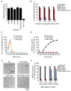
Figure 2. Hsp26 and Hsp42 synergize to inhibit spontaneous Sup35 prionogenesis.
(A, B) NM (5 µM) was incubated at 25°C with agitation for 6 h in the presence of increasing concentrations of BSA, Hsp26, Hsp42, or Hsp26 and Hsp42 (0–5 µM). For the mixture of Hsp26 and Hsp42, a 1∶1 ratio was employed. Thus, a concentration of 2 µM on the x-axis reflects 1 µM Hsp26 and 1 µM Hsp42. Fibrillization was measured by Thioflavin-T (ThT) fluorescence (A) or by determining the amount of SDS-resistant NM (B). Values represent means±SD (n = 3). (C) NM (5 µM) was assembled at 25°C with agitation for 6 h in the absence or presence of Hsp26 (0.6 µM or 3 µM), Hsp42 (0.6 µM or 3 µM), or Hsp26 and Hsp42 (0.3 µM or 1.5 µM of each). Reaction products were concentrated and transformed into [psi ] cells. No NM and soluble NM served as negative controls. The proportion of [PSI +] colonies was then determined. Values represent means±SD (n = 3). (D) Sup35 (5 µM) was incubated at 25°C with agitation for 6 h in the presence of increasing concentrations of BSA, Hsp26, Hsp42, or Hsp26 and Hsp42 (0–5 µM). For the mixture of Hsp26 and Hsp42, a 1∶1 ratio was employed. Thus, a concentration of 2 µM on the x-axis reflects 1 µM Hsp26 and 1 µM Hsp42. Fibrillization was measured by ThT fluorescence. Values represent means±SD (n = 3).
Figure 3
Figure 3. Hsp26 and Hsp42 inhibit de novo Sup35 prionogenesis by distinct mechanisms.
(A) NM (5 µM) was rotated for 5 min (80 rpm) in the absence or presence of Hsp26 (3 µM), Hsp42 (3 µM), or Ssa1 plus Ydj1 (3 µM). Oligomeric NM was recovered by centrifugation at 436,000 g for 30 min, resolved by SDS-PAGE, Coomassie stained, and the amount in the pellet fraction determined. Values represent means±SD (n = 3). (B) Fluorescence of NM-N21C-, Q38C-, G96C-, or Y106C-acrylodan (5 µM) after 15 min at 25°C in the absence or presence of BSA (3 µM), Hsp26 (3 µM), Hsp42 (3 µM), or Hsp26 (1.5 µM) and Hsp42 (1.5 µM). Values represent means±SD (n = 3). (C, D) NM (5 µM) was incubated at 25°C with agitation for 10 min at which point (arrow) buffer, Hsp26 (3 µM), or Hsp42 (3 µM) was added. The reaction was then continued at 25°C with agitation to 150 min. At the indicated times, the amount of A11-reactive species present was determined (C) or the amount of ThT-reactive species was determined (D). Datasets representative of three replicates are shown. (E) Electron microscopy of NM assembly at 25°C with agitation for 6 h in the absence or presence of Hsp26 (3 µM), Hsp42 (3 µM), or Hsp26 (1.5 µM) and Hsp42 (1.5 µM). Note the presence of small fibers in the presence of Hsp26 (arrow), the accumulation of oligomers in the presence of Hsp42 or Hsp26 and Hsp42. Bar, 0.5 µm. (F) NM cysteine variants were either left uncrosslinked or crosslinked under denaturing conditions with a flexible 11 Å BMB crosslink at position 25 or 31. The indicated NM protein (5 µM) was then assembled with agitation at 25°C in the absence or presence of BSA (3 µM), Hsp26 (3 µM), Hsp42 (3 µM), or Hsp26 and Hsp42 (1.5 µM of each). Fibrillization was measured by ThT fluorescence. Values represent means±SD (n = 3).
Figure 4
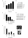
Figure 4. Hsp26 and Hsp42 antagonize Sup35 prion formation in vivo.
(A) Sup35 was overexpressed for 16 h at 30°C in wild-type, Δhsp26, Δhsp42, or Δhsp26Δhsp42 [psi ] [RNQ+] cells. Cells were plated on 25% YPD and the proportion of [PSI+] colonies was determined. Compared to wild-type cells there was significantly more [PSI+] induction in Δhsp26 cells (*p = 0.0264, two-tailed Student's t test), Δhsp42 cells (**p = 0.0059, two-tailed Student's t test), or Δhsp26Δhsp42 cells (***p = 0.0002, two-tailed Student's t test). Values represent means±SEM (n = 3). (B) Immunoblots demonstrating that neither Hsp104 nor Hsp70 (3A3 antibody that recognizes yeast Ssa1, Ssa2, Ssa3, and Ssa4 [95]) expression is affected in the Δhsp26, Δhsp42, or Δhsp26Δhsp42 background. Pgk1 is included as a loading control. (C) Sup35 was overexpressed for 16 h at 30°C in [psi ] [RNQ+] cells expressing elevated levels of Hsp26, Hsp42, Hsp26 plus Hsp42, or Hsp104. Cells were plated on 25% YPD and the proportion of [PSI+] colonies was determined. Compared to the vector control there was significantly less [PSI+] induction in cells expressing Hsp26 (*p = 0.0253, two-tailed Student's t test), Hsp42 (*p = 0.028, two-tailed Student's t test), Hsp26 and Hsp42 (**p = 0.0021, two-tailed Student's t test), or Hsp104 (***p<0.0001, two-tailed Student's t test). Values represent means±SEM (n = 3). (D) Immunoblots demonstrating that neither Hsp104 nor Hsp70 (3A3 antibody that recognizes yeast Ssa1, Ssa2, Ssa3, and Ssa4 [95]) expression is affected by elevated expression of Hsp26, Hsp42, or Hsp26 and Hsp42. Pgk1 is included as a loading control. (E) NM-YFP was transiently overexpressed for 4 h at 30°C in [psi ] [RNQ+] cells expressing elevated levels of Hsp26, Hsp42, or Hsp26 and Hsp42. Cells were processed for fluorescence microscopy. The proportion of cells with NM-YFP foci was then determined. (**p<0.01, ***p<0.001, two-tailed Student's t test). Values represent means±SEM (n = 3). Examples of cells with NM-YFP foci (“dots” and “ribbons” indicated by arrows) and cells with diffuse NM-YFP fluorescence are shown on the right.
Figure 5
Figure 5. Hsp26 and Hsp42 inhibit cross-seeding by Rnq1 fibers.
NM (2 µM) was incubated at 25°C for 16 h with or without Rnq1 fibers (10% or 20% wt/wt) without agitation in the absence or presence of BSA (3 µM), Hsp26 (3 µM), Hsp42 (3 µM), or Hsp26 (1.5 µM) and Hsp42 (1.5 µM). Fibrillization was measured by ThT fluorescence. Values represent means±SD (n = 3).
Figure 6
Figure 6. Hsp26 inhibits seeded assembly of Sup35 more potently than Hsp42 in a temperature-sensitive manner.
(A) NM (5 µM) was incubated at 25°C for 12 h in the presence of preformed NM fibers (5% wt/wt) plus increasing concentrations of either BSA, Hsp26, Hsp42, or Hsp26 and Hsp42 (0–24 µM). For the mixture of Hsp26 and Hsp42, a 1∶1 ratio was employed. Thus, a concentration of 2 µM on the x-axis reflects 1 µM Hsp26 and 1 µM Hsp42. For the Hsp26 alone condition, Hsp26 was pretreated at either 25°C or 45°C for 10 min. Fibrillization was measured by ThT fluorescence. Values represent means±SD (n = 3). (B) NM (5 µM) was incubated at 25°C for 12 h in the presence of preformed NM fibers (5% wt/wt) plus BSA, Hsp26, or Hsp42 (12 µM). Hsp26 was pretreated at either 25°C or 45°C for 10 min. Fibrillization was measured by determining the amount of SDS-resistant NM. Values represent means±SD (n = 3). (C) Sup35 (5 µM) was incubated at 25°C for 12 h in the presence of preformed Sup35 fibers (5% wt/wt) plus BSA, Hsp26, or Hsp42 (12 µM). Hsp26 was pretreated at either 25°C or 45°C for 10 min. Fibrillization was measured by ThT fluorescence. Values represent means±SD (n = 3). (D) Chemically denatured GDH or NM (5 µM) was incubated at 25°C for 4 h with agitation in the presence of Hsp26 (1–10 µM), which had been pretreated at either 25°C or 45°C for 10 min. GDH aggregation was assessed by turbidity and NM fibrillization by ThT fluorescence. Values represent means±SD (n = 3). (E) NM proteins (5 µM) carrying pyrene labels at the indicated single site were assembled at 25°C for 12 h in the presence of preformed NM fibers (5% wt/wt) plus either Hsp26 or Hsp42 (12 µM). Hsp26 was pretreated at either 25°C or 45°C for 10 min. The ratio of excimer to non-excimer fluorescence (I465 nm/I375 nm) was then determined as a measure of intermolecular contact formation. Soluble NM serves as a negative control.
Figure 7
Figure 7. Hsp26 interacts with NM fibers to prevent seeding.
(A) NM fibers (5 µM NM monomer) were incubated for 60 min at 25°C without or with BSA, Hsp26, or Hsp42 (10 µM). NM fibers were then recovered by centrifugation at 16,000 g, gently washed, resuspended in an equal volume of assembly buffer, and used to seed (2% wt/wt) assembly of NM (5 µM) for 12 h at 25°C. Fibrillization was measured by ThT fluorescence. Values represent means±SD (n = 3). (B) NM fibers were pretreated as in (A) and used to seed (2% wt/wt) assembly of NM proteins (5 µM) carrying pyrene labels at the indicated single site. The ratio of excimer to non-excimer fluorescence (I465 nm/I375 nm) was then determined as a measure of intermolecular contact formation. (C) Preassembled NM fibers (5 µM NM monomer) were incubated for 60 min at 25°C without or with Hsp26 or Hsp42 (10 µM). NM fibers were then recovered by centrifugation at 16,000 g for 30 min. Pellet fractions with or without a wash step were processed for SDS-PAGE and Coomassie stained. Note the presence of Hsp26 and Hsp42 in the pellet and washed pellet fractions. (D) NM-his (5 µM) was incubated for 60 min at 25°C without or with Hsp26 or Hsp42 (10 µM). NM-his was recovered with Ni-NTA agarose, washed, and eluted. Eluates were processed for SDS-PAGE and Coomassie stained. Note the presence of Hsp42 but not Hsp26 in the eluted fractions. (E) NM-his (5 µM) that had been pretreated with buffer, BSA, Hsp26, or Hsp42 (10 µM) was incubated with NM fibers (5% wt/wt) for 12 h. Fibrillization was measured by ThT fluorescence. Values represent means±SD (n = 3). (F) Ex vivo Sup35 prions (2% wt/wt) were used to seed the assembly of soluble full-length Sup35 (5 µM) for 12 h at 25°C in the absence or presence of BSA, Hsp26, or Hsp42 (10 µM). Fibrillization was measured by ThT fluorescence. Values represent means±SD (n = 3).
Figure 8
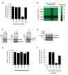
Figure 8. Elevated expression of Hsp26 and Hsp42 effectively cures [PSI +].
Hsp26, Hsp42, Hsp26 plus Hsp42, or Hsp104 were overexpressed in [PSI+] cells in liquid for 6 h at 30°C. Empty vector served as the control. Cells were plated on 25% YPD and the proportion of [PSI+] colonies was determined. Compared to the vector control there was significantly more [PSI+] curing in cells expressing Hsp26 (**p = 0.00249, two-tailed Student's t test), Hsp42 (*p = 0.0387, two-tailed Student's t test), Hsp26 and Hsp42 (***p = 0.000187, two-tailed Student's t test), or Hsp104 (**p = 0.00148, two-tailed Student's t test). Values represent means±SD (n = 3).
Figure 9
Figure 9. Hsp42 synergizes with Ssa1:Sis1 and Ssa1:Ydj1 to inhibit seeded assembly of NM.
(A–E) NM fibers (5 µM NM monomer) were incubated for 60 min at 25°C with buffer or Hsp42 (10 µM). NM fibers were then recovered by centrifugation at 16,000 g, gently washed, resuspended in an equal volume of assembly buffer, and used to seed (2% wt/wt) assembly of NM (5 µM) for 12 h at 25°C in the presence of ATP (5 mM) and increasing concentrations of Ssa1 (A), Sis1 (B), Ydj1 (C), Ssa1:Sis1 (D), or Ssa1:Ydj1 (E). Fibrillization was monitored by ThT fluorescence. Values represent means±SD (n = 3). (F) NM fibers (5 µM or 1.25 µM) were sonicated and transformed into [psi ] cells overexpressing the indicated combination of Hsp26, Hsp42, Sis1, and Ydj1. The proportion of [PSI +] colonies was then determined. Values represent means±SD (n = 5).
Figure 10
Figure 10. Hsp26 or Hsp42 binding destabilizes NM fibers.
(A) NM fibers (5 µM NM monomer) were incubated for 60 min at 25°C without or with Hsp26 (10 µM), Hsp42 (10 µM), or Ssa1:Sis1 (10 µM of each) in the presence of ATP (5 mM). The stability of the various NM fibers was then determined by SDS–PAGE and quantitative immunoblot. The amount of SDS-soluble NM, which reflects susceptibility of NM fibers to thermal solubilization, was plotted against temperature and fitted to a sigmoidal function. Values represent means±SD (n = 3). (B) NM proteins (5 µM) carrying pyrene labels at the indicated single site were assembled at 25°C with agitation for 12 h. Assembled NM fibers (5 µM NM monomer) were then incubated for 60 min at 25°C without or with buffer, Hsp26, Hsp42, or Ssa1:Sis1 (10 µM) in the presence of ATP (5 mM). The ratio of excimer to non-excimer fluorescence (I465 nm/I375 nm) was then determined.
Figure 11
Figure 11. Hsp26 or Hsp42 promotes rapid amyloid disassembly by Hsp104.
(A, B) NM fibers (2.5 µM monomer) were pretreated for 1 h at 25°C with either buffer, Hsp26 (10 µM), or Hsp42 (10 µM). Hsp104 (0–0.5 µM) was then added in the absence (A) or presence (B) of Ssa1:Sis1 (2.5 µM each) and reactions were incubated for a further 60 min at 25°C. Fiber integrity was then assessed by ThT fluorescence. Values represent means±SD (n = 3). (C) NM fibers (2.5 µM monomer) were pretreated for 1 h at 25°C with either buffer, Hsp26 (10 µM), or Hsp42 (10 µM). Hsp104 (0.15 µM) was then added in the absence or presence of Ssa1:Sis1 (2.5 µM each) and reactions were incubated for a further 60 min at 25°C. Reaction products were concentrated, sonicated, and transformed into [psi ] cells. The proportion of [PSI +] colonies was then determined. Values represent means±SD (n = 3). (D) Hsp104, Hsp26, Hsp42, or the indicated combination were overexpressed in [PSI+] cells in liquid for 6 h at 30°C. Cells were plated on 25% YPD and the proportion of [psi ] colonies was determined. Values represent means±SD (n = 3). (E) α-Syn fibers (0.5 µM monomer, green bars) or Q62 fibers (1 µM monomer, black bars) were pretreated for 1 h at 25°C with either buffer, Hsp26 (10 µM), or Hsp42 (10 µM). Hsp104 (10 µM) was then added in the absence or presence of Ssa1:Sis1 (10 µM each) and reactions were incubated for a further 60 min at 25°C (for Q62) or 37°C (for α-syn). Fiber integrity was then assessed by ThT fluorescence. Values represent means±SD (n = 3). (F) Luciferase (0.1 µM) was aggregated for 15 min at 45°C in the presence of the indicated concentration of sHsp (Hsp26 or Hsp42). Protein aggregates were diluted 20-fold into chaperone mixtures containing Hsp104 (1 µM), Ssa1 (1 µM), and Ydj1 (1 µM) plus ATP (5 mM) and incubated for 90 min at 25°C. Native luciferase activity at the same concentration was set to 100%. Note that Hsp26 stimulates luciferase reactivation, whereas Hsp42 does not. Note that Hsp104 alone or Ssa1 and Ydj1 alone do not promote luciferase reactivation under these conditions .
Figure 12
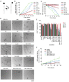
Figure 12. Gradual disassembly of Sup35 prions by Hsp26 or Hsp110, Hsp70, and Hsp40.
(A) Molecular recycling model. Monomers at the fiber ends are constantly dissociating and reassociating. An example monomer is shown in black. k off is the rate constant for the dissociation of a monomer from a fiber end and k on is the rate constant for the association of a monomer with a fiber end. k on is several orders of magnitude greater than k off. (B–E) NM fibers (2.5 µM) were sonicated and then incubated at 25°C for 0–28 d in the absence or presence of Hsp26 (5 µM), Hsp42 (5 µM), Sis1 (5 µM), Ydj1 (5 µM), Ssa1 (5 µM), Sse1 (5 µM), Sse1:Ssa1, Sse1:Sis1, Sse1:Ydj1, Ssa1:Sis1 (2.5 µM each), Ssa1:Ydj1 (2.5 µM each), Sse1:Ssa1:Sis1 (1.67 µM each), or Sse1:Ssa1:Ydj1 (1.67 µM each). At the indicated times, reactions were centrifuged at 436,000 g for 30 min. The amount of SDS-soluble NM in the supernatant was then determined by quantitative immunoblot (B). Values represent means±SD (n = 3). Alternatively, at the indicated times, fiber integrity was assessed by ThT fluorescence (C). Values represent means±SD (n = 3). After 28 d, fiber integrity was assessed by electron microscopy (D). Sonicated NM fibers at the start of the reaction (t = 0) are shown at the top left for comparison. Note the absence of fibers in the presence of Sse1:Ssa1:Sis1, Sse1:Ssa1:Ydj1, and Ssa1:Sis1 and a collection of shorter forms in the presence of Hsp26 or Ssa1:Ydj1. Bar, 0.5 µm (D). Alternatively, at the indicated times, reaction products were concentrated and transformed into [psi ] cells. The proportion of [PSI +] colonies was then determined (E). Values represent means±SD (n = 3). (F) NM fibers (2.5 µM) were not sonicated and then incubated at 25°C for 0–28 d in the absence or presence of Hsp26 (5 µM), Ssa1:Sis1 (2.5 µM each), Ssa1:Ydj1 (2.5 µM each), Sse1:Ssa1:Ydj1 (1.67 µM each), or Sse1:Ssa1:Sis1 (1.67 µM each). At the indicated times, reactions were centrifuged at 436,000 g for 30 min. The amount of SDS-soluble NM in the supernatant was then determined by quantitative immunoblot. Values represent means±SD (n = 3). Note the extent of disassembly is not as extensive as when NM fibers are sonicated as in (B).
Figure 13
Figure 13. sHsps promote gradual depolymerization of Sup35 prions by Hsp110, Hsp70, and Hsp40.
(A) Schematic illustrating the concept of NM-his capped NM fibers (left) or NM capped NM-his fibers (right). Cyan circles depict NM-his and white circles depict NM. (B, C) NM fibers with NM-his caps (2.5 µM monomer) were not sonicated and incubated at 25°C for 0–28 d in the absence or presence of Hsp26 (5 µM), Ssa1:Sis1 (2.5 µM each), Ssa1:Ydj1 (2.5 µM each), Sse1:Ssa1:Ydj1 (1.67 µM each), or Sse1:Ssa1:Sis1 (1.67 µM each). At the indicated times, reactions were centrifuged at 436,000 g for 30 min. The amount of SDS-soluble untagged NM (B) or NM-his (C) in the supernatant was then determined by quantitative immunoblot. Values represent means±SD (n = 3). (D, E) NM-his fibers with NM caps (2.5 µM monomer) were not sonicated and incubated at 25°C for 0–28 d in the absence or presence of Hsp26 (5 µM), Ssa1:Sis1 (2.5 µM each), Ssa1:Ydj1 (2.5 µM each), Sse1:Ssa1:Ydj1 (1.67 µM each), or Sse1:Ssa1:Sis1 (1.67 µM each). At the indicated times, reactions were centrifuged at 436,000 g for 30 min. The amount of SDS-soluble untagged NM (D) or NM-his (E) in the supernatant was then determined by quantitative immunoblot. Values represent means±SD (n = 3). (F, G) NM-his fibers with NM caps (2.5 µM monomer) were sonicated and incubated at 25°C for 0–28 d in the absence or presence of Hsp26 (5 µM), Ssa1:Sis1 (2.5 µM each), Ssa1:Ydj1 (2.5 µM each), Sse1:Ssa1:Ydj1 (1.67 µM each), or Sse1:Ssa1:Sis1 (1.67 µM each). At the indicated times, reactions were centrifuged at 436,000 g for 30 min. The amount of SDS-soluble untagged NM (F) or NM-his (G) in the supernatant was then determined by quantitative immunoblot. Values represent means±SD (n = 3). (H, I) NM fibers (2.5 µM) were sonicated and then incubated for 1 h at 25°C with buffer, Hsp26, or Hsp42 (10 µM). (H) Sse1:Ssa1:Sis1 (1.67 µM each) or (I) Sse1:Ssa1:Ydj1 (1.67 µM each) was then added and fibers were incubated for 0–28 d at 25°C. At the indicated times, reactions were centrifuged at 436,000 g for 30 min. The amount of SDS-soluble NM in the supernatant was then determined by quantitative immunoblot. Values represent means±SD (n = 3).
Figure 14
Figure 14. Human HspB5 promotes gradual depolymerization of α-syn fibers by human Hsp110, Hsp70, and Hsp40.
(A, B) α-Syn fibers (2.5 µM monomer) were sonicated and then incubated for 0–30 d at 25°C with either buffer, Hsc70 (10 µM), Hdj1 (10 µM), Apg-2 (10 µM), HspB5 (10 µM), Hsc70:Hdj1 (5 µM of each), Hsc70:Apg-2 (5 µM of each), Hsc70:HspB5 (5 µM of each), Hdj1:Apg-2 (5 µM of each), Hdj1:HspB5 (5 µM of each), Apg-2:HspB5 (5 µM of each), Hsc70:Hdj1:Apg-2 (3.3 µM of each), Hsc70:Apg-2:HspB5, Hsc70:Hdj1:HspB5, Hdj1:Apg-2:HspB5 (3.3 µM of each), or Hsc70:Hdj1:Apg-2:HspB5 (2.5 µM of each). At the indicated times, reactions were centrifuged at 436,000 g for 30 min (A). The amount of α-syn in the supernatant was then determined by quantitative immunoblot (A). Alternatively, fiber integrity was assessed by ThT fluorescence (B). Values represent means±SD (n = 3). (C) α-Syn fibers with his-α-syn caps (2.5 µM monomer) were not sonicated and incubated at 25°C for 0–30 d in the presence of Hsc70:Hdj1:Apg-2:HspB5 (2.5 µM of each). At the indicated times, reactions were centrifuged at 436,000 g for 30 min. The amount of his-α-syn (blue) or untagged α-syn (red) in the supernatant was then determined by quantitative immunoblot. Values represent means±SD (n = 3). (D) Preformed amyloid fibers composed of α-syn (0.5 µM monomer) were incubated for 6 h at 37°C in buffer plus ATP (5 mM) and ATP-regeneration system without or with the indicated combination of HspB5 (10 µM), Hsc70 (10 µM), Hdj1 (10 µM), and Apg-2 (10 µM) in the absence (blue bars) or presence of Hsp104 (2.5 µM, red bars; or 10 µM, green bars). Fiber integrity was then determined by sedimentation analysis and quantitative immunoblot. Values represent means±SD (n = 3).

References

    1. Cushman M, Johnson B. S, King O. D, Gitler A. D, Shorter J. Prion-like disorders: blurring the divide between transmissibility and infectivity. J Cell Sci. 2010;123:1191–1201. - PMC - PubMed
    1. Goldschmidt L, Teng P. K, Riek R, Eisenberg D. Identifying the amylome, proteins capable of forming amyloid-like fibrils. Proc Natl Acad Sci U S A. 2010;107:3487–3492. - PMC - PubMed
    1. Chiti F, Dobson C. M. Protein misfolding, functional amyloid, and human disease. Annu Rev Biochem. 2006;75:333–366. - PubMed
    1. Fowler D. M, Koulov A. V, Alory-Jost C, Marks M. S, Balch W. E, et al. Functional amyloid formation within mammalian tissue. PLoS Biol. 2006;4:e6. doi: 10.1371/journal.pbio.0040006. - DOI - PMC - PubMed
    1. Watt B, van Niel G, Fowler D. M, Hurbain I, Luk K. C, et al. N-terminal domains elicit formation of functional Pmel17 amyloid fibrils. J Biol Chem. 2009;284:35543–35555. - PMC - PubMed

Publication types

MeSH terms