. 2012;7(6):e38760.
doi: 10.1371/journal.pone.0038760. Epub 2012 Jun 19.

Imatinib enhances functional outcome after spinal cord injury

Affiliations

Imatinib enhances functional outcome after spinal cord injury

Mathew B Abrams et al. PLoS One. 2012.

Abstract

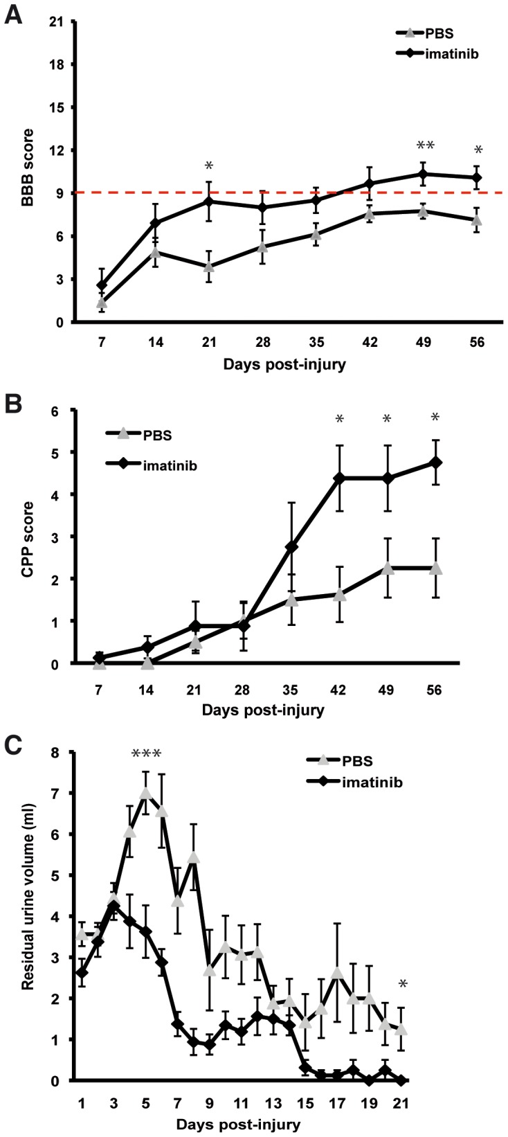
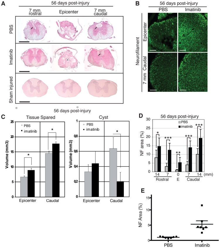
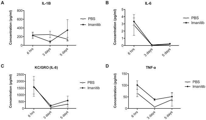
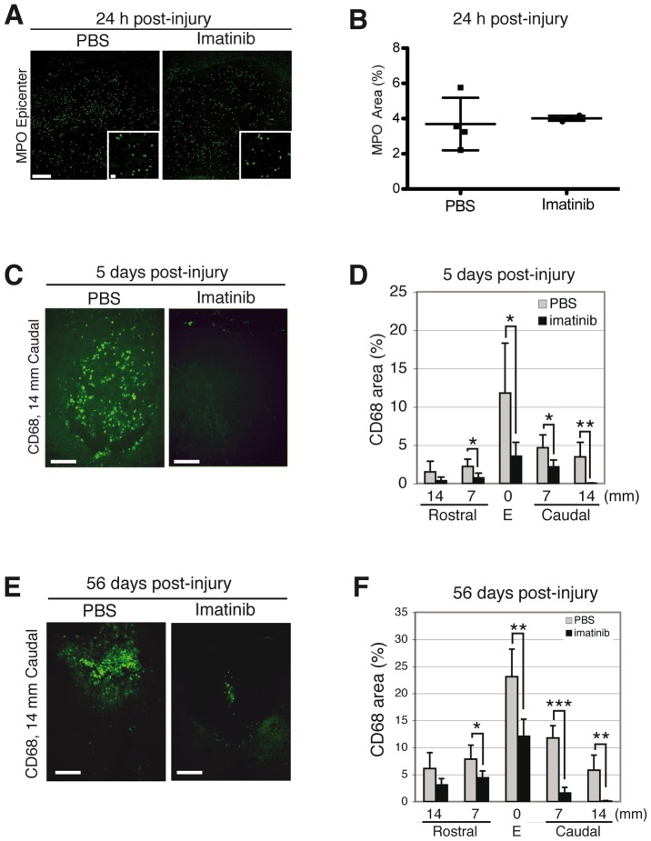
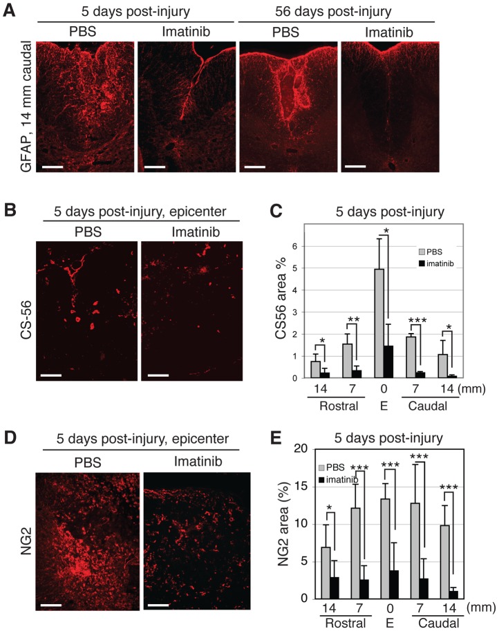
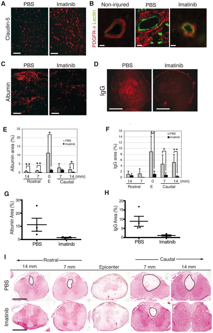
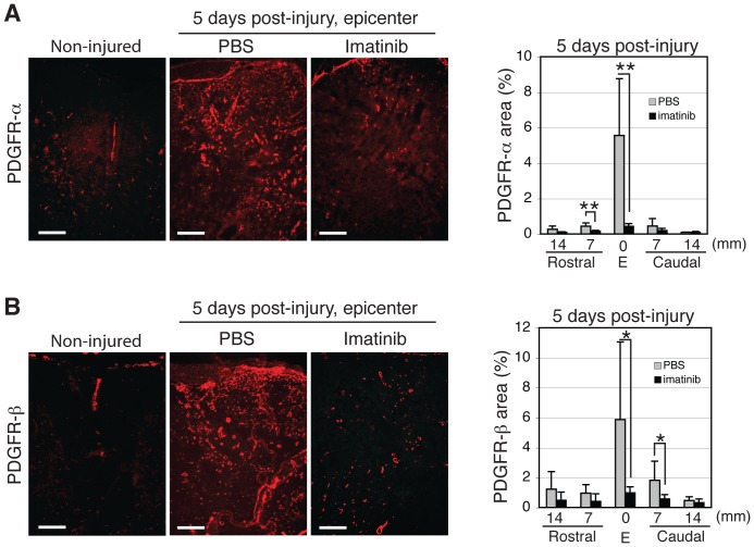
We investigated whether imatinib (Gleevec®, Novartis), a tyrosine kinase inhibitor, could improve functional outcome in experimental spinal cord injury. Rats subjected to contusion spinal cord injury were treated orally with imatinib for 5 days beginning 30 minutes after injury. We found that imatinib significantly enhanced blood-spinal cord-barrier integrity, hindlimb locomotor function, sensorimotor integration, and bladder function, as well as attenuated astrogliosis and deposition of chondroitin sulfate proteoglycans, and increased tissue preservation. These improvements were associated with enhanced vascular integrity and reduced inflammation. Our results show that imatinib improves recovery in spinal cord injury by preserving axons and other spinal cord tissue components. The rapid time course of these beneficial effects suggests that the effects of imatinib are neuroprotective rather than neurorestorative. The positive effects on experimental spinal cord injury, obtained by oral delivery of a clinically used drug, makes imatinib an interesting candidate drug for clinical trials in spinal cord injury.


Conflict of interest statement

Competing Interests: The authors have read the journal’s policy and have the following conflict: Ulf Eriksson and Ludwig Institute of Cancer Research (LICR) have submitted a patent application on the use of imatinib in CNS injuries (US patent 20070265203, filed 17 Apr 2007, published 15 Nov 2007). Lars Olson has received financial support from AFA Insurance company. Neither alters the authors' adherence to all the PLoS ONE policies on sharing data and materials.

Figures

Figure 1
Figure 1. Imatinib enhances functional outcome.
(A) Hindlimb locomotor function assessed by the BBB locomotor rating scale. The red line indicates BBB score 9, when weight-support begins. Scores above 9 represent walking, while scores below 9 represent no walking. (B) Contact plantar placement (CPP) test of sensorimotor function. (C) Bladder residual urine volumes. Imatinib (N = 10) compared to PBS (N = 10). Data presented as mean ± SEM: *P<0.05, **P<0.01, and ***P<0.001.
Figure 2
Figure 2. Imatinib enhances tissue preservation.
H&E staining was used to visualize tissue and spinal cysts within the epicenter and in spinal segments 7 and 14 mm rostral and caudal to the injury. (A) Representative micrographs of tissue preservation and spinal cysts at day 56 post-injury in the injury site and in spinal segments 7 mm rostral and caudal to the injury. Arrows indicate spinal cysts. Asterisk indicates possible enlargement of the central canal. (B) Representative micrographs of NF immunoreactivity within the injury site at day 56 post-injury. (C) Quantification of tissue preservation and spinal cyst volumes within the injury site and 7 mm caudal to the epicenter at day 56 post-injury (imatinib n = 10; PBS n = 10). (D) Quantification of NF immunoreactivity within the injury site and in spinal segments 7 and 14 mm rostral and caudal to the injury at day 56 post-injury. (E) Scatter plot of NF immunoreativity within the injury site. Imatinib (N = 7) compared to PBS (N = 7). Data presented as mean ± SD: *P<0.05, **P<0.01, and ***P<0.001. Scale bars: (A) 1 mm, (B) 100 µm.
Figure 3
Figure 3. Impact of imatinib on plasma concentrations of pro-inflammatory cytokines.
(A) IL-1β plasma concentrations. (B) IL-6 plasma concentrations. (C) IL-8 plasma concentrations. (D). TNF-α plasma concentrations. Imatinib (N = 4) compared to PBS (N = 4). Data presented as the mean ± SD. ***P<0.001.
Figure 4
Figure 4. Imatinib attenuates acute and chronic inflammation.
Effects of 30 minute delayed imatinib treatment on MPO immunoreactive neutrophils within the injury site at 24 hours post-injury and CD68 immunoreactive activated macrophages/microglia at day 5 post-injury. (A) Representative micrographs of MPO immunoreactivity at 24 hours post-injury. (B) Quantification of MPO immunoreactivity within the injury site at 24 hours post-injury (imatinib, N = 4; PBS, N = 4). (C) Representative micrographs of CD68 immunoreactivity at day 5 post-injury. (D) Quantification of CD68 immunoreactivity within the spinal cord at day 5 post-injury (imatinib, N = 4; PBS, N = 4). (E) Representative micrographs of CD68 immunoreactivity for activated macrophages/microglia within the injury site 14 mm caudal of the epicenter at day 56 post-injury. (F) Quantification of the relative area occupied by CD68 immunoreactivity within the injury site and in spinal segments 7 and 14 mm rostral and caudal to the injury at day 56 post-injury (imatinib, N = 6; PBS, N = 6). Data presented as mean ± SD: *P<0.05, **P<0.01, and ***P<0.001. Scale bars: 100 µm, inset: 10µm.
Figure 5
Figure 5. Imatinib attenuates astrogliosis and CSPG deposition.
(A) Representative micrographs of dorsomedial GFAP immunoreactivity 14 mm caudal to the injury site at 5 and 56 days post-injury. (B) Representative micrographs of CS56 immunoreactivity within the injury site at day 5 post-injury. (C) Quantification of CS56 immunoreactivity within the injury site and spinal segments 7 and 14 mm rostral and caudal to the injury at day 5. (D) Representative micrographs of NG2 immunoreactivity within the injury site at day 5 post-injury. (E) Quantification of NG2 immunoreactivity within the injury site and in spinal segments 7 to 14 mm rostral and caudal to the injury site at day 5. Imatinib (day 5, N = 4; day 56, N = 6) compared to PBS (day 5, N = 4; day 56, N = 6). Data presented as mean ± SD: *P<0.05, **P<0.01, and ***P<0.001. Scale bars: 100 µm.
Figure 6
Figure 6. Imatinib enhances vascular integrity.
(A) Tight junction protein, claudin-5, immunoreactivity in endothelial cells in the dorsal horn at 5 days post-injury is stronger in imatinib than in PBS-treated rats. This indicates better preservation of the BSCB integrity. (B) Representative micrographs of PDGFR-β/lectin immunoreactivity at day 5 post-injury in the spinal cords of non-injured, PBS-treated, and imatinib-treated rats. (C) Representative micrographs of albumin immunoreactivity within the injury site at day 5 post-injury. (D) Representative micrographs of IgG immunoreactivity within the injury site at day 5 post-injury. (E) Quantification of albumin immunoreactivity within the spinal cord at day 5 post-injury. (F) Quantification of IgG immunoreactivity within the spinal cord at day 5 post-injury. (G) Scatter plot of the area of albumin immunoreactivity within the injury site at day 5 post-injury. (H) Scatter plot of the area of IgG immunoreactivity within the injury site at day 5 post-injury. (I) H and E staining of the spinal cord at day 5 post-injury. Circles indicate areas without H and E staining. Imatinib (day 5, N = 4) compared to PBS (day 5, N = 4). Data presented as mean ± SD: *P<0.05, **P<0.01, and ***P<0.001. Scale bars: (A,C,E) 100 µm,(G,I) 1 mm, (B) 10 µm.
Figure 7
Figure 7. Effects of 30 minute delayed, 5-day imatinib treatment on PDGFR expression at day 5 after contusion spinal cord injury.
(A) Representative micrographs of PDGFR-α immunoreactivity within the injury site and quantification of PDGFR-α immunoreactivity within the injury site and in spinal segments rostral and caudal to the injury in non-injured, PBS-treated, and imatinib-treated rats. (B) Representative micrographs of PDGFR-β expression within the injury site and quantification of PDGFR-β expression within the injury site and spinal segments rostral and caudal to the injury in non-injured, PBS-treated, and imatinib-treated rats. Imatinib (N = 4) compared to PBS (N = 4). Data presented as mean ± SD: *P<0.05. Scale bars: 100 µm.

References

    1. Tator CH, Fehlings MG. Review of the secondary injury theory of acute spinal cord trauma with emphasis on vascular mechanisms. J Neurosurg. 1991;75:15–26. - PubMed
    1. Norenberg MD, Smith J, Marcillo A. The pathology of human spinal cord injury: defining the problems. J Neurotrauma. 2004;21:429–440. - PubMed
    1. Nakajima K, Kohsaka S. Microglia: activation and their significance in the central nervous system. J Biochem. 2001;130:169–175. - PubMed
    1. Neumann H, Wekerle H. Neuronal control of the immune response in the central nervous system: linking brain immunity to neurodegeneration. J Neuropathol Exp Neurol. 1998;57:1–9. - PubMed
    1. Paniagua RT, Sharpe O, Ho PP, Chan SM, Chang A, et al. Selective tyrosine kinase inhibition by imatinib mesylate for the treatment of autoimmune arthritis. J Clin Invest. 2006;116:2633–2642. - PMC - PubMed

Publication types

MeSH terms